Introduction
Leishmaniases are neglected diseases caused by protozoan parasites of genus Leishmania, which infect cells of mononuclear phagocytic system of the mammalian hosts (WHO, 2012). This disease complex is mainly represented by tegumentary leishmaniasis (TL), which causes since self-limiting lesions until ulcerous scars leading to the patients morbidity, and visceral leishmaniasis (VL), which can be fatal if acute and left non-treated (Torres-Guerrero et al., Reference Torres-Guerrero, Quintanilla-Cedillo, Ruiz-Esmenjaud and Arenas2017). Besides the high prevalence of VL in several countries worldwide, co-infection cases involving the human immunodeficiency virus (HIV) have also been reported (WHO, 2019).
The VL/HIV co-infection can cause a decrease in the number of CD4+ T cells in the patients, which is dangerous for them, since both pathologies accelerate the development of clinical symptoms and, if uncontrolled, cause the patients' death (Lindoso et al., Reference Lindoso, Moreira, Cunha and Queiroz2018). The impact of co-infection is observed in the occurrence of severe clinical manifestations and in the poor diagnosis and therapeutic response against both diseases (Alvar et al., Reference Alvar, Aparicio, Aseffa, Den Boer, Canavate, Dedet, Gradoni, Ter Horst, Lopez-Velez and Moreno2008; Cota et al., Reference Cota, Sousa and Rabello2011). In this context, rapid, sensitive and specific diagnosis of VL/HIV co-infection could significantly increase the probability for the patients to be promptly treated, contributing to the improvement of their life quality (Russo et al., Reference Russo, Laguna, Lopez-Velez, Medrano, Rosenthal, Cacopardo and Nigro2003; Hot et al., Reference Hot, Schmulewitz, Viard and Lortholary2007).
The parasitological methods remain as a gold standard to VL diagnose. However, these tests present variable sensitivity, since parasite or its content will be present in the collected samples. As a consequence, false-negative results can be found. In addition, in the VL/HIV co-infection, patients can present less parasites; thus making more difficult to perform the parasitological diagnosis (Srivastava et al., Reference Srivastava, Dayama, Mehrotra and Sundar2011). Immunological methods, such as direct agglutination tests (DAT), enzyme-linked immunosorbent assay (ELISA) and Western-blotting have shown good efficacy for the VL diagnosis, when immunocompetent patients are evaluated (Srividya et al., Reference Srividya, Kulshrestha, Singh and Salotra2012). However, their performance has been hampered, when VL/HIV co-infection cases are investigated (Silva et al., Reference Silva, Brandão, Colovati, Sousa, Lima, Dorta, Ribeiro-Dias, Costa, Costa and Oliveira2018). In this context, and due to the scarce of sensitive antigens to be applied in these cases, new studies should be performed to identify more suitable antigenic markers (Santos-Gomes et al., Reference Santos-Gomes, Gomes-Pereira, Campino, Araújo and Abranches2000; Kumar et al., Reference Kumar, Pai, Tripathi, Pandey and Sundar2002; Cota et al., Reference Cota, Sousa, Demarqui and Rabello2012). In fact, proteins that stimulate the humoral response in the infected hosts and present high reactivity as antigen in ELISA assays against specific samples; should help for the development of more accurate diagnostic tests.
Immunoproteomics is a biotechnological tool also used to identify antigens with a diagnostic application for canine and human VL (Lage et al., Reference Lage, Ludolf, Silveira, Machado, Ramos, Dias, Ribeiro, Costa, Vale, Tavares, Martins, Chávez-Fumagalli, Caligiorne, Chaves, Gonçalves, Rocha, Duarte and Coelho2019; Santos et al., Reference Santos, Cardoso, Machado, Siqueira, Ramos, Oliveira-da-Silva, Tavares, Lage, Costa, Freitas, Martins, Bandeira, Chávez-Fumagalli, Lyon, Moreira, Magalhães-Soares, Silveira, Tupinambás, Caligiorne, Chaves, Rocha, Fujiwara and Coelho2019; Vale et al., Reference Vale, Dias, Machado, Ribeiro, Lage, Costa, Steiner, Tavares, Ramos, Martínez-Rodrigo, Chávez-Fumagalli, Caligiorne, Magalhães-Soares, Silveira, Machado-de-Ávila, Teixeira and Coelho2019). In the present work, an immunoproteomics approach was performed in Leishmania infantum antigenic extracts using VL and VL/HIV co-infected patients sera; aiming to identify new candidates to serologically diagnose both patients classes. Results in the immunoblottings showed the identification of 26 proteins by mass spectrometry, which reacted with VL and VL/HIV patients sera, but not with those collected of HIV patients and healthy subjects. In addition, one of the identified proteins, β-tubulin, was expressed, purified and tested in ELISA experiments as a proof of concept for validation of our immunoproteomics findings.
Materials and methods
Human sera
Samples of VL patients (n = 30, including 16 males and 14 females with ages ranging from 29 to 63 years) living in an endemic region of disease (Belo Horizonte) were used. They were diagnosed by clinical evaluation and demonstration of L. infantum kDNA in spleen or bone marrow aspirates by polymerase chain reaction (PCR) technique. Sera of HIV patients (n = 25, including 16 males and 9 females with ages ranging from 22 to 48 years) were also used, as well as those of VL/HIV co-infected patients (n = 28, including 18 males and 10 females with ages ranging from 23 to 55 years). In both cases, patients were submitted to medical evaluation, and their blood samples were collected for laboratory tests for CD4+ T cell count and determination of the viral load. Inclusion criteria were determined after the clinical exam and VL confirmation by parasitological methods to identify L. infantum kDNA (Cota et al., Reference Cota, Sousa, Freitas-Nogueira, Gomes, Oliveira, Assis, Mendonça, Pinto, Saliba and Rabello2013). None of the patients had been treated before sample collection. Sera were also collected from healthy individuals living in an endemic area of VL (n = 25, including 15 males and 10 females with ages ranging from 21 to 50 years), which did not present clinical signal and showed negative serological results. Samples of Chagas disease patients (n = 10, including 3 males and 7 females, with ages ranging from 24 to 52 years), which were diagnosed by hemoculture, Chagatest® recombinant ELISA v.4.0 kit and/or Chagatest® hemmaglutination inhibition (Wiener lab., Rosario, Argentina), as well as those of leprosy (n = 10, with 6 males and 4 females, with ages ranging from 23 to 49 years), tuberculosis (n = 10, 6 males and 4 females with ages ranging from 38 to 67 years) and malaria (n = 10, 7 males and 3 females with ages ranging from 20 to 46 years) patients, were also used.
Parasite and prepare of the Leishmania antigenic extract
Leishmania infantum (MHOM/BR/1970/BH46) strain was used. Stationary promastigotes were cultured at 24°C in complete Schneider's medium (Sigma-Aldrich, USA), which was composed by the medium plus 20% inactivated fetal bovine serum (FBS; Sigma-Aldrich), 20 mm L-glutamine, 200 U mL−1 penicillin and 100 μg mL−1 streptomycin at pH 7.4 (Coelho et al., Reference Coelho, Tavares, Carvalho, Chaves, Teixeira, Rodrigues, Charest, Matlashewski, Gazzinelli and Fernandes2003). The soluble Leishmania antigenic extract (SLA) was prepared from 2 × 10⁸ stationary promastigotes as described (Martins et al., Reference Martins, Duarte, Lage, Costa, Carvalho, Mendes, Roatt, Menezes-Souza, Soto and Coelho2017). For the immunoblottings, stationary promastigotes (1 × 10⁸ cells) were washed in cold PBS 1x, and their morphology was evaluated after staining by the Giemsa method in an optical microscope. The protein extraction for bidimensional electrophoresis was performed following the 2-D Electrophoresis Principles and Methods handbook (GE Healthcare). Briefly, parasite pellet was resuspended in DeStreak rehydration solution (GE Healthcare) containing protease inhibitor cocktail. After homogenization, samples were disrupted by six cycles of ultrasonication (30 s each, at 38 MHz) in ice bath, and centrifuged at 20 000 × g for 7 min at 4°C. The supernatant was collected and protein content was estimated by Bradford method (Bradford, Reference Bradford1976).
Isoeletric focusing (IEF) and 12% sodium dodecyl sulfate polyacrylamide gel (SDS-PAGE)
For the first-dimension electrophoresis, 150 μg protein extract was added to 125 μL DeStreak rehydration solution (GE Healthcare) including 1% immobilized pH gradient buffer, pH 4–7, for each IPG strip. Samples were applied to IPG strips (7 cm, pH 4–7; GE Healthcare) for passive rehydration for 16 h at room temperature. After gel rehydration for 12 h, isoelectric focusing was performed in a six-step run: (1) 300 V for 200 V h−1, (2) 1000 V for 300 V h−1, (3) 5,000 V for 4,800 V h−1, (4) 5,000 V for 3000 V h−1, (5) 300 V for 18 h and (6) 5,000 V for 1 h; by using the Ettan IPGphor III electrophoresis unit (GE Healthcare). Each strip was then equilibrated in reducing buffer (containing 6 M Urea, 30% glycerol, 2% odium dodecyl sulfate (SDS), 75 mm Tris-HCl pH 8.8, 0.001% bromophenol blue and 130 mm DTT) for 15 min and in an alkylating buffer (containing 135 mm iodoacetamide) for 15 min. Strips and molecular weight standard were placed on 12% SDS-PAGE gels and sealed with agarose solution. The Multicolor Broad Range Protein Ladder (Thermo Fisher, USA) was used. The second-dimension electrophoresis was carried out using Mini-Protean II system (BioRad) in a Tris/glycine/SDS buffer, under 100 V until the dye front had reached the gel bottom. For each experiment, three distinct gels were performed, being one to be used in the immunoblottings and two to be stained by Colloidal Coomassie Blue G-250.
Immunoblottings
To perform the immunoblottings, sera of VL patients (n = 14, including 8 males and 6 females with ages ranging from 32 to 59 years), as well as those of HIV (n = 14, including 9 males and 5 females with ages ranging from 24 to 44 years) and VL/HIV co-infected (n = 14, including 8 males and 6 females with ages ranging from 25 to 50 years) patients were used. Also, healthy subjects samples (n = 14, including 8 males and 6 females with ages ranging from 23 to 44 years) were employed. Bidimensional gels were transferred to cellulose membrane (Schleicher and Schull, Dassel, Germany) by using a Trans-Blot Electrophoretic Transfer Cell (Bio-Rad), at 100 V (3 mA cm−1) for 120 min with transfer buffer (containing 25 mm Tris-Base, 192 mm glycine and 20% methanol). Membranes were blocked with 5% (w/v) low-fat dried milk in PBS 1x plus 0.05% Tween 20, for 16 h at room temperature. Next, they were washed 2 times (10 min each) with blocking buffer and incubated with VL, VL/HIV and HIV patients' pooled sera, as well as with healthy individuals' sera pool. Samples were 1:100 diluted in PBS 1x and 0.05% Tween 20, and the incubation was performed for 2 h at room temperature. Afterwards, membranes were washed 3 times (15 min each) and incubated with peroxidase-conjugated goat anti-human IgG secondary antibody (1: 10 000 diluted in PBS 1x plus 0.05% Tween 20) for 2 h at room temperature. After washing 3 times with PBS 1x plus 0.05% Tween 20, immunoblottings were revealed by the addition of a solution composed by chloronaphtol, diaminobenzidine and H2O2 30 vol. Reactions were then stopped by the addition of distilled water.
Protein digestion, peptide extraction and spot handling
The immunoblottings and their corresponding Colloidal Coomassie Blue-stained gels were overlapped. The interest spots were excised of corresponding gels for identification by mass spectrometry. Firstly, they were washed in milli-Q water and destained for 15 min by using 50% acetonitrile (ACN) and 25 mm ammonium bicarbonate (AB) at pH 8.0. Gel fragments were dried in 100% ACN for 5 min, followed by rehydration in 100 mm AB for 5 min and addition of the same volume of ACN. After removing ACN, spots were dried in a speed vac for 20 min. The dried spots were re-swollen in 20 μg mL−1 Sequencing Grade Modified Trypsin (Promega) and 25 mm AB for 10 min. The protein digestion was performed for 16 h at 37°C. After, the supernatant was transferred to a clean tube and 30 μL 5% formic acid (FA) plus 60% ACN were added for the peptide extraction. The procedure was performed 2 times for 30 min and under constant agitation. The supernatant was pooled to the respective tube containing the initial solution, which was dried in a speed vac, and peptide fragments were resuspended in 8 μL 0.1% trifluoracetic acid (TFA) solution.
Protein identification by mass spectrometry and database search
Reverse-phase nanochromatography and high-resolution nanoelectrospray were used to identify tryptic peptides. Firstly, each peptide was purified on a 2 cm trap column (100 μm internal diameter), followed by separation on a 15 cm separation column (75 μm internal diameter), both packed with 3 μm 120 A Reprosil-Pur C18 AQ matrix, which was home-made using a laser puller P 2000 Sutter Instrument (USA). The chromatography was carried on a UPLC EASY-nLC 1200 instrument (Thermo Scientific, USA), and samples were loaded (2000 nL per min) and the chromatographic separation occurred at 200 nL min−1. The mobile phase A consisted of 0.1% (v/v) formic acid diluted in distillated water, whereas the mobile phase B consisted of 0.1% (v/v) formic acid diluted in 80% ACN. The gradient conditions were 2 to 50% B for 58 min, and up to 100% B for 2 min. The eluted peptides were introduced to a Q-Exactive HF-X (Thermo, USA) for analysis. The voltage source was set to 1.9 kV with the capillary temperature at 250°C. MS1 spectra were acquired on the Orbitrap analyzer (300–1500 m z−1), at a 120 000 resolution (for m z−1 445.1200). For each spectrum, the seven most intense ions were submitted to higher-energy collisional (HCD) fragmentation, with isolation windows of 2.0 m z−1, isolation off set 0.5 m z−1, and maximum IT 50 ms, followed by mass spectrometry (MS) acquisition on the Orbitrap. The peptide mass profile for each spot was submitted to MS/MS ion search, by using Peaks Studio X (Bioinformatics Solutions Inc.), in order to search in the Uniprot Leishmania database. The following parameters were used: allowance of two tryptic miss cleavages, peptide error tolerance of 15 ppm, MS/MS error tolerance of ± 0.1 Da, variable modification of methionine (oxidation) and fixed modification of cysteine (carbamidomethylation). To avoid random matches, only ions with a high score or extensive homology (P < 0.05) were accepted.
Preparation of recombinant β-tubulin protein
The β-tubulin amino acid sequence present in L. infantum (SUZ41687.1 and SUZ45083.1) and L. braziliensis (XP_001567862.1) species was aligned using the Multialin server (Corpet, Reference Corpet1988). The L. braziliensis protein was previously cloned in our laboratory (Duarte et al., Reference Duarte, Pimenta, Menezes-Souza, Magalhães, Diniz, Costa, Chávez-Fumagalli, Lage, Bartholomeu, Alves, Fernandes, Soto, Tavares, Gonçalves, Rocha and Coelho2015), and it was used in serological assays as a proof of concept for the validation of our immunoproteomics findings. Briefly, the protein-codifying gene was amplified by PCR using L. braziliensis kDNA as template, and it was cloned in the pET28a-TEV vector in Escherichia coli BL21 cells (Agilent Technologies). The protein expression was induced with isopropyl β-D-1-thiogalactopyranoside (IPTG; 1.0 mm), with incubation at 150 × g for 24 h at 12°C. Cells were ruptured by six cycles of ultrasonication (30 s each, at 38 MHz), followed by six cycles of freezing and thawing. They were then centrifuged at 20 000 × g for 7 min at 4°C, and cellular debris was removed by centrifugation. The recombinant β-tubulin (rβ-tubulin) protein was purified by Ni-IDA affinity chromatography and passed through a polymyxin-agarose column (Sigma-Aldrich, USA) to remove any residual endotoxin content.
ELISA experiments
Previous titration curves were performed to determine the most appropriate antigen concentration and antibody dilution to be used. Flexible microtiter immunoassay plates (Jetbiofil®, Belo Horizonte) were coated with rβ-tubulin or L. infantum SLA (0.25 and 1.0 μg per well, respectively), which were diluted in 100 μL coating buffer (50 mm carbonate buffer) at pH 9.6, for 16 h at 4°C. Next, free binding sites were blocked using 250 μL PBS 1x and Tween 20 0.05% (PBS-T) added with 5% non-fat dry milk, for 1 h at 37°C. After washing the plates five times with PBS-T, sera were individually added (1:200 diluted in PBS-T), where samples of VL (n = 30), HIV (n = 25), VL/HIV (n = 28), Chagas disease (n = 10), leprosy (n = 10), tuberculosis (n = 10) and malaria (n = 10) patients, as well as of healthy individuals (n = 25) were used. Plates were incubated for 1 h at 37°C, and later they were washed five times in PBS-T. A peroxidase-conjugated goat anti-human IgG antibody was added (1: 20 000 diluted in PBS-T; catalogue SAB3701282, Sigma-Aldrich, USA), and the incubation was performed for 1 h at 37°C. Plates were then washed five times in PBS-T, and reactions were developed by incubation with solution composed by ortho-phenylenediamine, H2O2 and citrate-phosphate buffer at pH 5.0, for 30 min and in the dark. They were then stopped by adding 2 N H2SO4, and optical density (OD) values were read in an ELISA microplate spectrophotometer (Molecular Devices, Spectra Max Plus, Canada) at 492 nm.
Statistical analysis
Results were entered into Microsoft Excel (version 10.0) spreadsheets and analyzed using GraphPad PrismTM (version 6.0 for Windows). The receiver operating characteristic (ROC) curves were constructed to obtain the cut-off values, as well as sensitivity (Se), specificity (Sp) and area under the curve (AUC) of the antigens. The one-way analysis of variance (ANOVA) followed by the Bonferroni's post-test, were used for comparisons between groups. Differences were significant with P < 0.05.
Results
Immunoblottings using leishmania infantum protein extracts and VL, HIV and VL/HIV co-infected patients sera
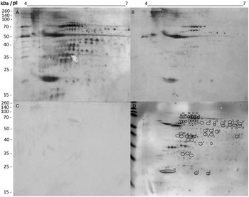
Bidimensional gels were stained with Colloidal Coomassie Blue G-250 and results showed several identified spots, which were distributed between 20 and 70 kDa, with pH varying between 4.5 and 7.0 (Fig. 1). VL patients sera recognized about 200 spots (Fig. 1A), while VL/HIV co-infected patients sera identified about 50 spots in the immunoblottings (Fig. 1B). The HIV-infected patients sera (Fig. 1C), as well as those of healthy subjects (data not shown), showed low reactivity against the parasite antigenic extract. The immunoblottings and respective Colloidal Coomassie Blue-stained gels were overlapped, and a total of 43 spots were identified by both patients classes. They were excised of the gels when 37 valid sequences corresponding to 26 distinct proteins were then identified (Table 1).
Bidimensional gels and immunoblottings performed in Leishmania infantum antigenic extracts. Bidimensional gels were obtained after separation of promastigote extracts (150 μg) using the first dimension: 7 cm IEF pH range 4–7, and in the second dimension: 12% SDS-PAGE. Immunoblots were developed after incubation of membranes with pooled sera from VL (A), VL/HIV (B) and HIV-infected (C) patients, all 1:100 diluted in PBS 1x plus 0.05% Tween 20. Bound antibodies were detected using a peroxidase-conjugated goat anti-human IgG secondary antibody (1: 10 000 diluted in PBS 1x plus 0.05% Tween 20). The x-axis represents the isoelectric point (pI) and y-axis represents the molecular weight (kDa) indicated by a commercial marker (Spectra Multicolor Broad Range Protein Ladder). The identified spots by antibodies in both VL and VL/HIV sera were marked in the bidimensional gels, which were stained with Coomassie Brilliant Blue G-250 (D).

Identified proteins in Leishmania infantum extracts by VL and VL/HIV patients' sera

a Spot number identified in the bidimensional gel.
b Name of the identified protein.
c Accession numbers according to NCBI.
d Predicted molecular weight (Mr, in KDa).
e Predicted isoelectric point (pI).
Leishmania proteins identified by MS/MS and database search
Among the 26 identified proteins by both VL and VL/HIV sera, two hypothetical and 24 known proteins were described. Between them, housekeeping proteins, such as tubulins and heat shock proteins were identified. Some of these antigens were already showed with a diagnostic role and/or as vaccine candidates against leishmaniasis, such as paraflagellar rod component (Maharana et al., Reference Maharana, Tewari and Veer2015) and HSP-83 (Menezes-Souza et al., Reference Menezes-Souza, Mendes, Gomes, Reis-Cunha, Nagem, Carneiro, Coelho, Galvão, Fujiwara and Bartholomeu2014); while others were showed to present an immunotherapeutic action, such as cytochrome c oxidase VII (Dey et al., Reference Dey, Meneses, Salotra, Kamhawi, Nakhasi and Duncan2010), s-adenosylhomocysteine hydrolase (Yang and Borchardt, Reference Yang and Borchardt2000) and dihydrolipoamide dehydrogenase (Chiranjivi and Dubey, Reference Chiranjivi and Dubey2018).
β-tubulin protein used to validate the immunoproteomics findings
One of the proteins identified in the immunoblottings by VL and VL/HIV patients sera, β-tubulin, was tested as a proof of concept to validate our immunoproteomics findings. The protein used here was cloned from L. braziliensis kDNA, since it presents high amino acid sequence similarity (higher than 99.0%) with L. infantum (SUZ41687.1 and SUZ45083.1) proteins (Fig. 2). In addition, due to the high degree of conservation in the amino acid sequences between such species, it is expected that the most B cell epitopes be conserved between them. For the serological assays, sera samples of VL, HIV and VL/HIV patients, as well as of Chagas disease, leprosy, tuberculosis,\ and malaria patients were evaluated. Results showed that both VL and VL/HIV patients sera recognized with high sensitivity and specificity the recombinant protein, with individual DO values above of the cut-off, while the other sera classes presented low reactivity with it (Fig. 3). ROC curves were constructed and results showed sensitivity and specificity values of 100% for rβ-tubulin; while with SLA used as an antigen, the values were of 96.4% and 44.4%, respectively (Table 2).
Multiple alignment of β-tubulin amino acid sequences between L. infantum and L. braziliensis species. The L. infantum (SUZ41687.1 and SUZ45083.1) and L. braziliensis (XP_001567862.1) β-tubulin amino acid sequences were aligned and results are shown. The letter ‘a’ indicates difference between the amino acid sequences of the SUZ41687.1 and XP_001567862 proteins; while the letter ‘b’ indicates the difference between the amino acid sequences of the SUZ45083.1 and XP_001567862 proteins.

Diagnostic assays for VL and VL/HIV co-infection using the rβ-tubulin protein and L. infantum SLA. ELISA assays were performed using the β-tubulin protein and L. infantum SLA. Sera samples from visceral leishmaniasis (VL; n = 30), VL/HIV co-infected (n = 28) and HIV (n = 25) patients were used. In addition, samples from healthy individuals living in endemic area (CT; n = 25) of disease, as well as from patients with Chagas Disease (CD; n = 10), leprosy (LE; n = 10), tuberculosis (TB; n = 10) and malaria (MA; n = 10) were used. The cut-off values were calculated by Receiver Operator Curves (ROC). Results showing the individual optical density (OD) values, as well as the mean of the groups against β-tubulin and SLA are shown. ROC curves were used to determine sensitivity (95% CI), specificity (95% CI), and area under the curve (AUC) for each antigen.

Diagnostic evaluation of the rβ-tubulin protein in the VL and VL/HIV co-infection

A human serological panel was used in ELISA experiments against rβ-tubulin protein and L. infantum SLA. Receiver operating characteristic (ROC) curves were constructed to obtain sensitivity (Se), specificity (Sp) and area under the curve (AUC).
Discussion
The VL/HIV co-infection is endemic in several countries in the world (WHO, 2019). Diagnostic tests applied to identify VL usually are not suitable to detect VL/HIV cases, since lower sensitivity is found (Cota et al., Reference Cota, Sousa, Demarqui and Rabello2012; Silva et al., Reference Silva, Brandão, Colovati, Sousa, Lima, Dorta, Ribeiro-Dias, Costa, Costa and Oliveira2018). In this context, there is the necessity to identify more sensitive antigens able to diagnose both VL and VL/HIV. Immunoproteomics approach based on the combination of bidimensional gels, immunoblottings and mass spectrometry has been developed to identify new biomarkers for VL (Coelho et al., Reference Coelho, Oliveira, Valadares, Chávez-Fumagalli, Duarte, Lage, Soto, Santoro, Tavares, Fernandes and Coelho2012; Lage et al., Reference Lage, Ludolf, Silveira, Machado, Ramos, Dias, Ribeiro, Costa, Vale, Tavares, Martins, Chávez-Fumagalli, Caligiorne, Chaves, Gonçalves, Rocha, Duarte and Coelho2019). In this context, in the present study, an immunoproteomics assay was performed in L. infantum antigenic extracts by using VL and VL/HIV co-infected patients sera, aiming to identify new targets able to diagnose both patients classes. Parasite extracts were solved in bidimensional gels, which were transferred to nitrocellulose membranes and reacted against VL, VL/HIV and HIV patients and healthy subjects sera. The immune reactive spots only for VL and VL/HIV sera were selected, and 26 proteins were identified.
On the basis of the identified proteins, some of them were already showed to be antigenic in the VL, such as tubulins and heat shock proteins; which are parasite housekeeping proteins. These antigens are known to induce cellular and/or humoral response in the course of Leishmania infection in mammalian hosts, and they have shown a diagnostic value for VL (Menezes-Souza et al., Reference Menezes-Souza, Mendes, Gomes, Reis-Cunha, Nagem, Carneiro, Coelho, Galvão, Fujiwara and Bartholomeu2014; Costa et al., Reference Costa, Alves, Carneiro, Dias, Fujimura, Araujo, Tavares, Ramos, Duarte, Menezes-Souza, Briza, Briza, Coelho and Goulart2019; Humbert et al., Reference Humbert, Costa, Katis, Ramos, Machado, Sones, Coelho and Christodoulides2019). In this context, to partially validate our immunoproteomics findings, we selected the β-tubulin protein to perform ELISA experiments against our serological panel composed by VL, HIV, VL/HIV, Chagas disease, leprosy, tuberculosis and malaria patients sera; as well as healthy subjects samples. This protein was shown also to be antigenic in previous immunoproteomics studies developed by our group, such as when L. infantum extracts were reacted against canine (Coelho et al., Reference Coelho, Oliveira, Valadares, Chávez-Fumagalli, Duarte, Lage, Soto, Santoro, Tavares, Fernandes and Coelho2012) and human (Lage et al., Reference Lage, Ludolf, Silveira, Machado, Ramos, Dias, Ribeiro, Costa, Vale, Tavares, Martins, Chávez-Fumagalli, Caligiorne, Chaves, Gonçalves, Rocha, Duarte and Coelho2019) VL sera, as well as when L. braziliensis antigenic preparations were reacted against TL patients sera (Duarte et al., Reference Duarte, Pimenta, Menezes-Souza, Magalhães, Diniz, Costa, Chávez-Fumagalli, Lage, Bartholomeu, Alves, Fernandes, Soto, Tavares, Gonçalves, Rocha and Coelho2015). In addition, when β-tubulin was applied in a recombinant version in ELISA assays, it showed satisfactory diagnostic value to detect TL (Duarte et al., Reference Duarte, Pimenta, Menezes-Souza, Magalhães, Diniz, Costa, Chávez-Fumagalli, Lage, Bartholomeu, Alves, Fernandes, Soto, Tavares, Gonçalves, Rocha and Coelho2015; Lima et al., Reference Lima, Costa, Lage, Dias, Ribeiro, Machado, Ramos, Salles, Fagundes, Carvalho, Franklin, Chávez-Fumagalli, Machado-de-Ávila, Menezes-Souza, Duarte, Teixeira, Gonçalves and Coelho2018) and VL (Costa et al., Reference Costa, Alves, Carneiro, Dias, Fujimura, Araujo, Tavares, Ramos, Duarte, Menezes-Souza, Briza, Briza, Coelho and Goulart2019) cases.
Leishmania proteins usually present a high degree of evolutionary conservation between Trypanosomatides. It is estimated that these parasites have approximately 8,000 genes, which are able to codify proteins and that about 99% of them maintain synteny, with 94% being constitutively expressed (Vasconcelos et al., Reference Vasconcelos, Terrão, Ruiz, Vêncio and Cruz2012). In this context, the β-tubulin protein used in our study was cloned from L. braziliensis kDNA. It was expressed, purified and tested in ELISA experiments, and high sensitivity and specificity values were found to diagnose VL and VL/HIV co-infection. Such facts demonstrate the partial validation of our immunoproteomics findings, and can be considered as a proof of concept of our study; thus opening the possibility to test other identified proteins as diagnostic biomarkers for VL and VL/HIV, which could well be applied in other immunodiagnostic platforms, as well as by using higher serological panel and field conditions to validate their diagnostic potential.
The Leishmania β-tubulin amino acid sequence presents similarity with its homologous in humans, with homology values near 85.0%, such as NP_001060.1 and NP_821080.1 sequences. This fact can be considered as limiting for use of this protein in a specific diagnosis of VL, mainly due to the occurrence of cross-reactivity in the immunological assays. However, the sensitivity and specificity values found here demonstrate that even a conservation degree is described; tubulins can present variations in their amino acid sequences, such as those related to specific B cell epitopes (Kaur and Kaur, Reference Kaur and Kaur2013; Costa et al., Reference Costa, Alves, Carneiro, Dias, Fujimura, Araujo, Tavares, Ramos, Duarte, Menezes-Souza, Briza, Briza, Coelho and Goulart2019); which could justify the serological distinction found here, mainly between the VL and VL/HIV patients sera, which presented distinct reactivity as compared to that found using healthy subjects samples.
Immunoproteomics studies have shown potential to identify new biological targets on leishmaniasis in mammalian hosts (Duarte et al., Reference Duarte, Pimenta, Menezes-Souza, Magalhães, Diniz, Costa, Chávez-Fumagalli, Lage, Bartholomeu, Alves, Fernandes, Soto, Tavares, Gonçalves, Rocha and Coelho2015; Lage et al., Reference Lage, Ludolf, Silveira, Machado, Ramos, Dias, Ribeiro, Costa, Vale, Tavares, Martins, Chávez-Fumagalli, Caligiorne, Chaves, Gonçalves, Rocha, Duarte and Coelho2019). Differential protein expression models in promastigotes and amastigotes have shown a protein conservation degree between such parasite stages, with few antigens being characterized as specific-amastigote, such as A2 (Charest and Matlashewski, Reference Charest and Matlashewski1994), LiHyp1 (Martins et al., 2013), LdA1 (Avishek et al., Reference Avishek, Ahuja, Pradhan, Gannavaram, Selvapandiyan, Nakhasi and Salotra2018), among others; as well as preferentially expressed in the promastigote stage, such as lipophosphoglycans (Kelleher et al., Reference Kelleher, Curtis, Sacks, Handman and Bacic1994), zinc metalloproteinase GP63 (McKean et al., Reference McKean, Denny, Knuepfer, Keen and Smith2001), promastigote surface antigen (PSA) (Petitdidier et al., Reference Petitdidier, Pagniez, Pissarra, Holzmuller, Papierok, Vincendeau, Lemesre and Bras-Gonçalves2019), among others. However, due to the fact that amastigotes are found parasitizing the mammalian cells a few hours after infection, their antigens should also be considered suitable to be used as diagnostic markers for active disease.
However, and like described above, since the majority of Leishmania proteins are commonly expressed in both parasite life stages; they could be also considered interesting since could indicate the initial and/or late infection in the mammalian hosts. As an example, kinesin-derived proteins have been employed as recombinant antigens for VL diagnosis, and satisfactory results for detection of the active disease have been obtained, with such molecules being found expressed in both parasite stages (Sivakumar et al., Reference Sivakumar, Sharma, Chang and Singh2006; Dhom-Lemos et al., Reference Dhom-Lemos, Viana, Cunha, Cardoso, Mendes, Pinheiro, Siqueira, Lobo, Teles, Bueno, Guimarães-Carvalho, Bartholomeu and Fujiwara2019). Certainly, in the present study, we consider that the absence of evaluation of an amastigote protein extract against VL and VL/HIV sera is one drawback of the work; and new experiments should be performed to respond this question, mainly with the purpose of identifying candidates expressed in both Leishmania stages, for future diagnostic applications.
This work used a low and controlled serological panel, and TL patients samples were not tested. Other identified proteins in the immunoblottings were also not used to validate our immunoproteomics findings, and such facts can be considered limitations of the study. However, the identified proteins showed here and the diagnostic action of one of them indicate to the possibility to test these antigens in future works by means of distinct immunological techniques, making possible the improvement of the sensitivity for the diagnosis of VL and VL/HIV co-infection.
Acknowledgements
The authors would like to thank CAPES, CNPq and FAPEMIG for the scholarships.
Financial support
This work was supported by grants from CNPq (APQ-408675/2018-7).
Conflict of interest
None.
Ethical standards
The study was approved by the Human Research Ethics Committee from Federal University of Minas Gerais (UFMG; Belo Horizonte, Minas Gerais, Brazil), with protocol number CAAE–32343114.9.0000.5149. The work was also approved by the Ethical Review Boards of FHEMIG (Belo Horizonte) and Centro de Pesquisa from René Rachou (Fundação Oswaldo Cruz, Belo Horizonte). All patients agreed to participate in the study, and those who did not want to participate had the same attention and medical care as compared to the enrolled participants, and all of them received the same treatment, follow-up and necessary care.