Case report
A 15-year-old boy with simple transposition of the great arteries underwent arterial switch operation during the 1st week of life. He also had severe scoliosis requiring nocturnal non-invasive positive-pressure ventilation and surgical repair. The long-term follow-up after arterial switch operation was complicated by complex obstruction of the right ventricular outflow tract involving the origin of both pulmonary arteries and a severe proximal stenosis of the right coronary artery. Myocardial scintigraphy demonstrated reversible ischaemia, and the boy was treated with aspirin and β-blockers.
Before undergoing orthopaedic surgery, the patient underwent cardiac catheterisation in order to evaluate the feasibility of right coronary artery recanalisation and relief of right ventricular outflow tract obstruction.
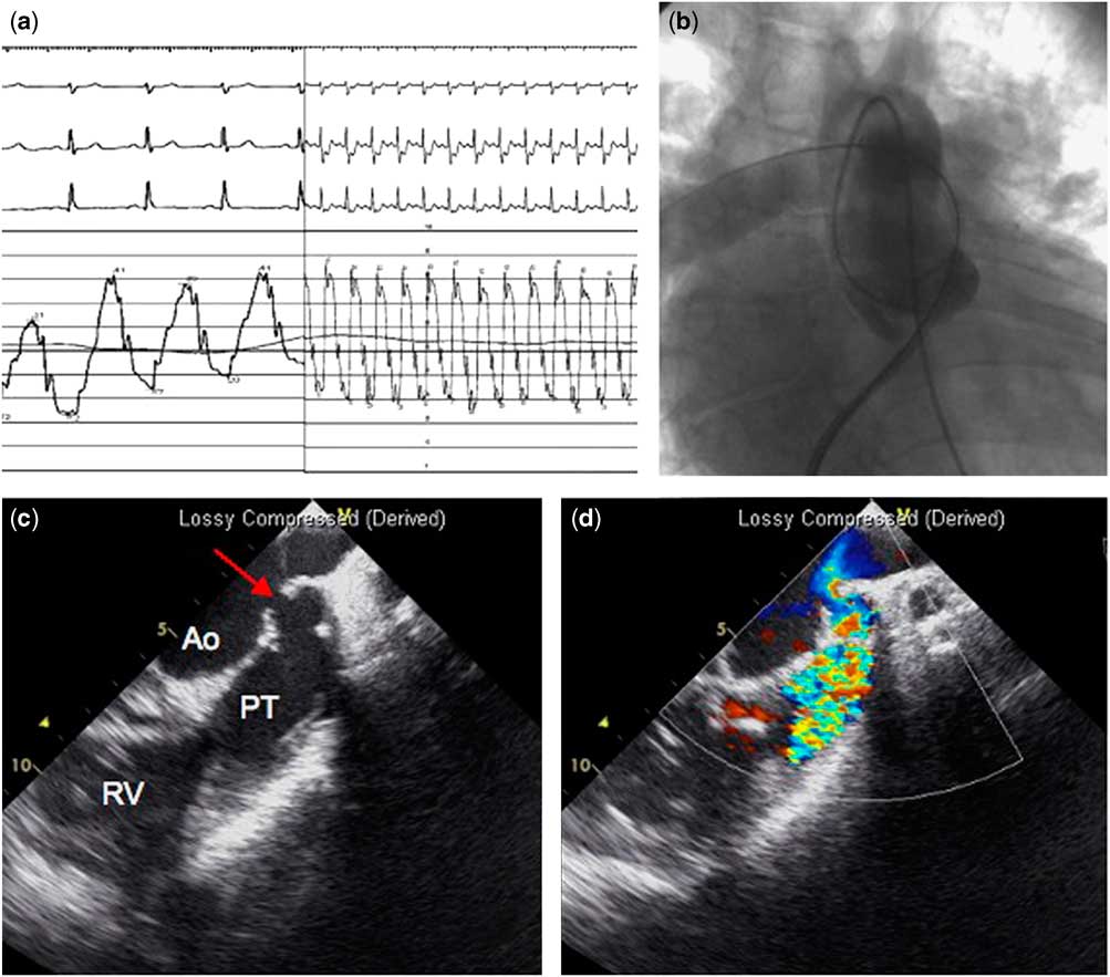
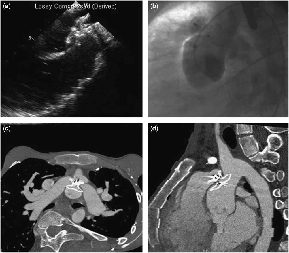
Cardiac catheterisation was performed under conscious sedation. Systolic right ventricular pressure was isosystemic. Immediately after hand-inflation of a 12-mm diameter and 3-cm long Mullins-XTM balloon (NuMED, Inc., Hopkinton, New York, United States of America), ischaemic changes on the electrocardiogram and increasing pulmonary pressures occurred (Fig 1a). Aortography was repeated to rule out coronary complications and revealed an aortopulmonary shunt with visualisation of the pulmonary arteries (Fig 1b, Supplementary movie 1). The patient was then intubated, and transoesophageal echocardiography detected a 10-mm “aortopulmonary window” with a mobile flap between the posterior wall of the pulmonary trunk and the anterior wall of the ascending aorta (Fig 1c and d). The lesion was closed via an arteriovenous circuit with a 13-mm Amplatzer Septal Occluder (St. Jude Medical Inc., St. Paul, Minnesota, United States of America) (Fig 2a). Immediately after closure, electrocardiogram changes regressed and pulmonary pressures normalised. A moderate residual shunt through the device persisted immediately after the procedure (Fig 2b, Supplementary movie 2). The peak gradient between the right ventricle and the pulmonary arteries remained unchanged. The patient was hospitalised for 15 days due to a transient haemolysis and deteriorated ventilation requiring non-invasive positive pressure and oxygen. CT scan confirmed the correct position of the device (Fig 2c and d).
Electrocardiogram (ECG) and pulmonary artery pressures before (left panel) and after (right panel) balloon angioplasty ( a ). After balloon angioplasty, the ECG showed ischaemic changes, and the pulmonary arterial pressure increased from 40/17/25 to 83/28/35 mmHg. Ascending aortography visualised the pulmonary arteries revealing a left-to-right shunt ( b ). Transoesophageal echocardiography detected a communication between the aorta and the pulmonary artery ( c ) with a left-to-right shunt ( d ). Ao=aorta; PT=pulmonary trunk; RV=right ventricle.

The aortopulmonary window was closed with a 13-mm Atrial Septal Occluder ( a ). Final angiography showed a trivial residual shunt ( b ). CT scan in the axial view ( c ) and oblique view ( d ) demonstrated the device correctly positioned between the aorta and the pulmonary bifurcation.

After 6 months, the patient underwent successful surgical repair including closure of the aortopulmonary window with a Dacron patch, right and left pulmonary artery plasty using a pericardial patch, and right coronary artery bypass grafting with right internal mammary artery. Surgical treatment of the scoliosis is now considered possible and is scheduled for in the near future.
Pulmonary artery disruption with aortopulmonary shunt is a rare, potentially lethal complication of pulmonary angioplasty. When it occurs in the pulmonary trunk or at the proximal segment of the pulmonary arteries, covered stent implantation may be an option.Reference Preminger, Lock and Perry 1 In this case, mimicking a traumatic aortopulmonary window, the lesion occurred in the posterior wall of the pulmonary trunk at the level of the pulmonary bifurcation where the use of a covered stent is not possible; however, emergency closure of the aortopulmonary window using an Amplatzer Septal Occluder was effective for clinical stabilisation before proceeding to surgical repair.
To note, the iatrogenic aortopulmonary window was discovered because of the ischaemic changes in the electrocardiogram, likely due to a steal phenomenon in the patient with an already-known severe stenosis of the right coronary artery; however, this complication might also be asymptomatic and misdiagnosed for a long time.Reference Vida, Biffanti, Stellin and Milanesi 2 Thus, in patients with transposition of great arteries corrected with arterial switch operation, the occurrence of this life-threatening complication and this possible rescue procedure should be always kept in mind during pulmonary angioplasty.
Acknowledgements
We thank Dr. Roberto Bordese for providing transesophageal echocardiography
Financial Support
This research received no specific grant from any funding agency, commercial, or not-for-profit sectors.
Conflicts of Interest
None.
Supplementary Material
To view supplementary material for this article, please visit http://dx.doi.org/10.1017/S104795111500164X