The residues found on medical devices after cleaning vary according to the method of cleaning and the cleaning agents used, as well as the constitution and structure of these devices. Organic and inorganic residues, in addition to harboring microorganisms, may cause pyrogenic reactions or infection. 1 Bacterial residues and other organic matter have been detected even after cleaning and steam sterilization and has been linked to outbreaks of postoperative infections.Reference Parada, Grassbaugh, Devine and Arrington 2 – Reference Tosh, Disbot and Duffy 4
Standard operating procedures need to be developed and validated to ensure the effective and safe cleaning of medical devices. All processing steps should follow the manufacturer’s recommendations, particularly for medical devices of complex design (eg, those with narrow or long lumens, hinges, cracks, inaccessible slits, or complex geometries).Reference Cowperthwaite and Holm 5 , Reference Lucas, Nagaraja, Gordon and Hitchins 6 However, organizations, international societies, and experts recognize that there are limitations and even omissions in the development and validation of product reprocessing instructions. 7
Functionality is the main concern of manufacturers in the design of medical devices, and the same attention tends to not be paid to the disassembly of the complex designs to ensure efficient cleaning. This occasionally requires healthcare professionals to contact manufacturers to request instructions for appropriate reprocessing, as disassembly may be challenging to perform.
For the reprocessing of complex medical devices, there is no consensus in the literatureReference Vassey, Budge and Poolman 8 , Reference Alfa, Nemes, Olson and Mulaire 9 to indicate that performing manual cleaning followed by automated cleaning is more effective than performing manual cleaning alone. Taking this into account, this study aimed to compare the efficacy of cleaning methods in the removal of organic residues, such as those present on laparoscopic instruments.
METHODS
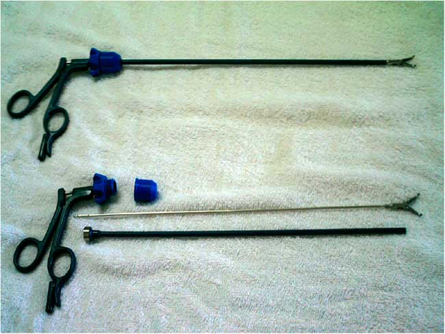
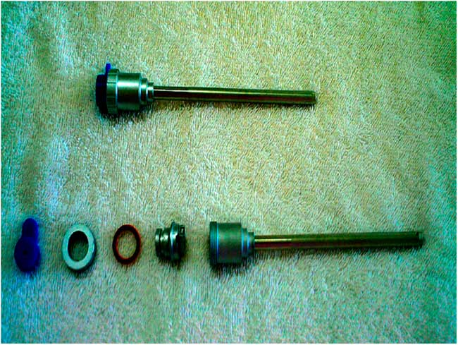
An experimental laboratory study was performed using 2 complex laparoscopic instruments: a trocar screw-window valve and laparoscopic dissection forceps. The trocar had a 5-mm lumen, consisted of 5 pieces, and measured 11 cm in length, with a total area of 81 cm2. The laparoscopic forceps consisted of 4 pieces, with a toothed end and a 30-cm-long lumen with a 3-mm internal diameter, and had a total surface area of 156 cm2 (Figure 1).
Laparoscopic dissection forceps.

Intentional Contamination and Cleaning Methods
The laparoscopic instruments were subjected to intentional contamination to simulate the use of laparoscopic instruments in clinical practice. Human placental blood (1 mL) was used to contaminate the external surface of each instrument, and an additional 1 mL was flushed into the lumen of each trocar. This procedure was performed using an aseptic technique, and the instruments were kept in contact with the source of contamination for 1 hour. The positive-control group, which was used to validate the contamination, contained high concentrations of carbohydrate, hemoglobin, and proteins. Visually, the volume of blood used was sufficient to contaminate the entire internal and external surfaces of the instruments.
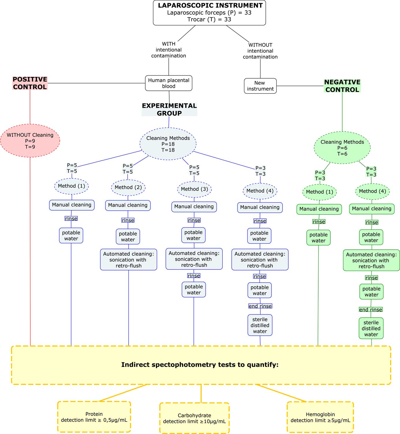
The experimental groups were defined according to various cleaning methods commonly implemented in the Central Sterile Supply Department, as detailed below:
Cleaning method 1: Manual-only cleaning and rinsing with potable tap water.
Cleaning method 2: Manual cleaning and rinsing with potable tap water, followed by ultrasonic cleaning without rinsing.
Cleaning method 3: Manual cleaning and rinsing with potable tap water, followed by ultrasonic cleaning and rinsing with potable tap water.
Cleaning method 4: Manual cleaning and rinsing with potable tap water, followed by ultrasonic cleaning, a rinse with potable tap water, and a final rinse with sterile distilled water.
Positive control group: The instruments were subjected to intentional contamination, and no cleaning method was applied.
Negative control groups: New laparoscopic instruments were subjected to the same cleaning as that in groups 1 and 4, without intentional contamination.
The experimental and control groups, as well as the sample sizes, are described in Figure 3.
Trocar screw-window valve.

Characterization of the Cleaning Methods
For manual cleaning, the laparoscopic instruments were disassembled, and all pieces were immersed in potable tap water (19–23°C) for 10 minutes to ensure moisture during processing. The samples were then rinsed with tap water and immersed in a multienzymatic detergent solution (Endozime AW Plus APA, Ruhof Healthcare, New York, NY) for 2 minutes, in accordance with the manufacturer’s instructions. The solution was additionally flushed into the laparoscopic lumens with a syringe. Soft- and firm-bristle brushes were used to decontaminate all external surfaces, with special attention to the recesses, rack, joints, and grooves. The lumen was washed using disposable 60-cm-long brushes with 2- and 5-mm diameters. The instruments were rinsed with tap water using a high-pressure water sprayer, which was particularly directed toward the internal structures of the lumens. The samples were cleaned until they were visually clean, varying for each sample and method used.
Automated cleaning was performed in an ultrasonic cleaner with a retro-flush pulsating pump system (H. Strattner, Sao Paulo, Brazil) with a 2-kg capacity and an ultrasonic frequency of 35 kHz, 127 V, 50/60 Hz. We used a 10-minute cycle at 45°C and included the same enzymatic detergent used for manual cleaning. The ultrasonic cleaner did not include a consecutive rinsing stage; therefore, the instruments were rinsed manually with potable tap water with the aid of a high-pressure water sprayer directed toward the internal structure of the parts (except in cleaning method 2). In cleaning method 4, rinsing was supplemented with an additional sterile distilled water rinse. After cleaning, all instrument pieces were placed vertically on white fabric for natural drying and visual inspection of possible residual debris.
Analysis of Organic Residues
To remove contaminant residues, each disassembled instrument was individually placed inside a sterile plastic bag using aseptic technique, and 250 mL of sterile distilled water was added before sealing. The instruments were then sonicated 3 times for 10 seconds each (USC-2800, Enge Solutions, Sao Paulo, Brazil) and subjected to orbital shaking (Agitator 255-B, Fanem, Brazil) at 160 rpm for 10 minutes.
Inside a biological safety cabinet (VLFS, Veco, Campinas, Brazil), 50 mL of the extraction solution was transferred to a conical tube. Of this solution, 1 mL was used in indirect testing for the detection of organic residues (proteins, carbohydrates, and hemoglobin). The values obtained from the analysis were multiplied by 250 to estimate the total organic residue levels present on the instruments.
Spectrophotometry (ELx800, Biotek, Winooski, VT) was used to compare the radiation absorbed by solutions containing unknown amounts of the contaminant residues to known amounts. Graphs of absorbance versus the concentration of standard solutions of protein standard solution, D-(+)- glucose, and lyophilized human hemoglobin (Sigma-Aldrich, St Louis, MO), were constructed to determine the concentrations of the unknown solutions.
Protein quantification was performed by indirect testing using the QuantiPro Bicinchoninic acid kit (Sigma-Aldrich), which detects low protein concentrations in small solution volumes. This results in a linear response from 0.5 to 30 μg/mL of protein.
Carbohydrate residue was quantified through indirect testing as well.Reference Liu, Lau, Chau and Pacepavicius 10 For each 250 µL of analyzed solution, 50 μL of 5% phenol was added with immediate vortexing (Genie 2, SI-A236, Scientific Industries, Bohemia, NY). Sulfuric acid (1 mL of 95%–98%) was then added and immediately vortexed. A portion of each sample (200 μL) was subsequently transferred to a microplate and incubated at room temperature for 30 minutes. The absorbance at 490 nm was then analyzed using a spectrophotometer (ELx800, Biotek). The detection limit of this test was ≥10 μg/mL.
For hemoglobin quantification, TMB One Solution (Promega, Madison, WI) was used. For each 50 μL of solution analyzed, 150 μL of TMB One was added, with slight shaking. The solution was then incubated at 37°C for 30 minutes and the absorbance at 650 nm was measured using a spectrophotometer. The detection limit of this test was ≥5 μg/mL.
The average values of organic residue levels are presented as a ratio of the area of the instruments (μg/cm2); that is, the amount of residue in µg obtained per material, divided by the total area of the instruments. Inferential statistics were performed using the Kruskal-Wallis nonparametric test to compare the average amounts of residue between the study groups; P<.05 was considered statistically significant.
RESULTS
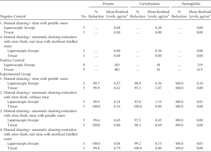
The average organic residue levels in the experimental groups were significantly decreased compared with those in the positive controls, with 95.1%–100% reduction in residual organic matter observed with different methods. However, there were no significant differences (P>.05) in the levels of organic residues between different cleaning methods. Moreover, there was no significant difference between rinsing with only potable tap water or with sterile distilled water. Unexpectedly, protein and carbohydrate residues were detected after cleaning of new, not-contaminated forceps and trocars as well (Table 1).
Mean Levels of Organic Residues of Protein, Carbohydrate, and Hemoglobin of Laparoscopic Instruments in the Various Study Groups, and the Percentage of Reduction Compared With the Respective Positive Controls

DISCUSSION
This study demonstrates that complex laparoscopic instruments may be adequately cleaned by either manual or manual/ultrasonic means, thus ensuring one of the essential requirements for successful sterilization. These 2 methods are the most highly recommended practices for the processing of laparoscopic instruments.
The FDA does not recommend the use of spores or other microbial tracers to determine the effectiveness of medical device cleaning; the focus is on the removal of organic and inorganic residues and not microbial reduction. 11 The contaminant challenge used in the present study produced satisfactory residue values compared with other soiling simulators, indicating the high degree of challenge imposed to the cleaning process and the robustness of the employed methods.Reference Alfa, DeGagne and Olson 12
Currently, there is no consensus on the reference values or maximum acceptable limits of various residues for a material to be considered satisfactorily clean.Reference Verjat, Prognon and Darbord 13 Some authorsReference Kruger 14 consider that a given material may not be accepted as clean when residual protein levels are >20 μg/cm2. In another study, a reference level of <0.1 μg/device was considered satisfactory. In the present study, the average protein residue levels found in forceps and trocars cleaned using method 1 were 0.42 and 0.57 μg/cm2, respectively.
The unexpected detection of protein and carbohydrates after cleaning new forceps and trocars suggests that even when not intentionally contaminated with organic matter, it is still possible to detect organic residues on medical devices. This finding stresses the need to establish acceptable safe limits of organic residues on medical devices in repeated use, especially since the limits supported by the human body without an inflammatory reaction remain unknown.
Comparisons between manual and manual/automated cleaning, and the residues left following their application, have been performed in other studies. In one study,Reference Alfa and Nemes 15 residues from ported and nonported disposable instruments were compared. The authors concluded that automated ultrasonic cleaning with retro-flushing of narrow lumens was more effective than manual cleaning, which is contrary to the results of this study; this discrepancy may be attributed to the fact that the application of direct friction was not possible in some instruments in the former study.
Automated ultrasonic cleaning, with and without an intermittent flush adapter, has also been compared with the manual cleaning of new, intentionally contaminated biopsy forcepsReference Alfa, Nemes, Olson and Mulaire 9 of similar length and diameter to those used in this study (240 cm×3.3 mm). Automated cleaning with an intermittent flush adapter was more effective than manual cleaning alone. However, organic residue levels after automated cleaning, without the use of the adapter, were similar to those observed following manual cleaning alone. Notably, in this study, brushing of the forceps’ lumen was not performed when manual cleaning alone was carried out.
A disadvantage of manual cleaning is the degree of subjectivity of the technique, which makes it difficult to guarantee the reproducibility of the method. Moreover, the type of detergent, water temperature, brush, and number and type of brushes used, 1 , Reference Vassey, Budge and Poolman 8 also affects the efficiency of the procedure. Likewise, the efficiency of automated cleaning is also influenced by several factors such as the chemical agents used, water quality, physical energy type (ultrasonic wave power or water jet pressure associated with unclogged sprinklers), functionality tests and equipment validation. 1 , Reference Vassey, Budge and Poolman 8
Our study has some limitations. Other important residues, such as endotoxins, that may recontaminate clean medical devices when rinsing is performed with clean water were not investigated. Our results demonstrate that manual-only cleaning and rinsing with potable tap water, followed by ultrasonic cleaning without rinsing, showed satisfactory results, but only against proteins, carbohydrates, and hemoglobin.
Water treated using methods, such as deionization, distillation, or reverse osmosis, is indicated for the final rinse after cleaning the products. 1 , Reference Cowperthwaite and Holm 5 Although the use of tap water has not been ruled out, the difficulty in controlling organic and inorganic contaminants has been recognized; this concern is mainly related to geographic and seasonal variations as well as possible formation of biofilms in the pipes.Reference Souza, Gonçalves, Ikeda, Cruz and Graziano 16
The findings obtained in this study may be extrapolated to other complex surgical instruments, as long as it is possible to exert direct friction on all internal and external surfaces. Furthermore, the results contradict the logical reasoning that greater technological investment in cleaning processes automatically implies better results in terms of the reduction of residues.
Flowchart describing the sample sizes, experimental group, and control group.

ACKNOWLEDGMENTS
We are grateful to the University Hospital of the University of São Paulo for access to the obstetrical center to obtain placental blood and to the staff of the Obstetric Center, the Biomaterials laboratory, and the Pontifical Catholic University of São Paulo (PUC-SP) School of Medical Sciences and Health Sorocaba campus.
Financial support: This research was funded by the Foundation for Research Support of the State of São Paulo, Brazil, the Support and Incentive to Teaching Research Program of the Pontifical Catholic University of São Paulo (PUC-SP), and the Teaching Capacity modality of the Teaching Research and Extension Council.
Potential conflicts of interest: The authors declare that there are no conflicts of interest related to this article.