Complex pulmonary and systemic venoatrial connections are known to be a major feature of patients with visceral heterotaxy.Reference Cohen1, Reference Pierpont2 We describe an infant with asplenia syndrome, and isomerism of the right atrial appendages, who also had a common atrium and atrioventricular valve, pulmonary atresia with multiple systemic-to-pulmonary collateral arteries, the right-sided aorta arising from the morphologically right ventricle, and infradiaphragmatic totally anomalous pulmonary venous connection. Following a unifocalization procedure, construction of a systemic-to-pulmonary arterial shunt, and repair of the anomalous pulmonary veins, the patient suffered ischemic necrosis of the right lobe of the liver. Subsequent noninvasive imaging, and anatomical inspection, suggested dependent portal venous flow from the anomalous pulmonary venous drainage. Noninvasive identification of separate portal venous systems may herald this complication.
Case report
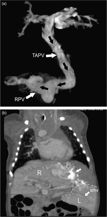
A full-term female newborn, diagnosed prenatally as having complex congenital cardiac malformations, was transported to our medical center on the first day of life following the initiation of an infusion of prostaglandin E1. A transthoracic echocardiogram confirmed the prenatal findings of isomerism of the right atrial appendages, with a common atrium and atrioventricular valve, the aorta arising from the morphologically right ventricle, pulmonary atresia with multiple systemic-to-pulmonary collateral arteries, a right sided aortic arch, and totally anomalous pulmonary venous connection. The pulmonary venous drainage was primarily to a descending transdiaphragmatic vein that entered the right-sided portal venous system. An additional small vertical vein draining into the retroaortic brachiocephalic vein was also identified. A preoperative computerized tomographic scan of the chest following administration of 6 millilitres of contrast via an umbilical venous catheter by slow hand injection further delineated the anomalous pulmonary venous connections (Fig. 1). The descending vein from the pulmonary confluence drained to the right lobe of the liver, while the superior mesenteric vein drained to the left lobe. Additionally, there were bilateral inferior caval veins that became confluent at the level of the liver. Other pertinent non-cardiac diagnoses included a midline liver, absence of the spleen, and an umbilical cord containing only two vessels. The patient weaned from mechanical ventilation, remained haemodynamically stable, and was fed enterally prior to surgery.
Preoperative helical computed tomographic scan of the chest (a) with coronal maximum intensity projection reconstruction of the totally anomalous pulmonary venous (TAPV) connection below the diaphragm supplying a right sided portal vein (RPV). (b) Coronal image showing large midline liver with greater enhancement of the left lobe, which was supplied by a separate superior mesenteric vein (SMV). Note dense contrast stain (upper arrow) following injection of the left umbilical vein.

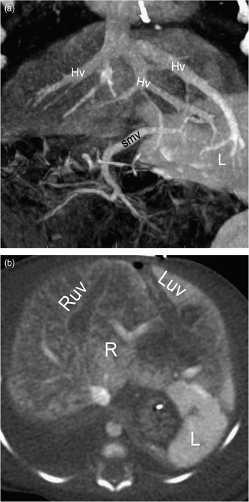
On the seventh day of life, the patient underwent unifocalization of the multiple systemic-to-pulmonary collateral and intrapericardial pulmonary arteries using a 3.0 millimetre central shunt, along with repair of the anomalous pulmonary venous connection. Cardiopulmonary bypass lasted for 180 minutes, with a period of aortic cross-clamping of 63 minutes. The patient separated from cardiopulmonary bypass without difficulty, and was taken to the cardiac intensive care on a continuous infusion of milrinone. Analysis of blood at the time of admission had demonstrated a lactic acidosis, at 4.6 millimoles per litre, and coagulapathy, with a prothrombin time of 22.8 seconds and activated partial thromboplastin time of greater than 250 seconds. These were treated. Escalation of the inotropic and vasopressor support, with addition of infusions of dopamine, epinephrine, and vasopressin, became necessary due to hypotension and persistent lactic acidosis. On the second postoperative day, hepatic transaminases were markedly elevated, with normal indirect and direct levels of bilirubin in the serum. By the third postoperative day, the liver had increased in size, and the abdomen was distended. Abdominal radiographs demonstrated no peritoneal free air or bowel pneumatosis. An abdominal ultrasound revealed a hepatic artery with increased resistive indexes. There was also a thrombosed vessel coursing cranially within the liver, which did not appear to be part of the hepatic venous or portal system, and was thought to likely be an anomalous vein. There was further elevation of the hepatic transaminases and the coagulopathy persisted. By the fifth postoperative day, the abdominal ultrasound revealed abnormal echogenicity of the right lobe of the liver, suggesting infarction as well as indeterminate portal venous flow. The patient failed to recover. Between the sixth and twelfth postoperative days, the metabolic acidosis continued to progress, with levels of lactate in excess of 37 millimoles per litre. Growing evidence for hepatic synthetic dysfunction, with increasing prothrombin times and decreased levels of fibrinogen, was observed. A repeated abdominal computed tomographic scan (Fig. 2a) on the twelfth postoperative day revealed an infarcted right lobe of the liver. Arterial supply to the right lobe could not be identified, and the hepatic artery to left lobe was small. By this time, there was evidence of intracranial haemorrhage, and at the request of the parents, the infant was removed from mechanical support. A postmortem examination confirmed the abdominal and cardiac findings. The right and left portal venous systems were separate (Fig. 2b). The right branch of the hepatic portal vein drained the gastric vein, which joined the anomalous common pulmonary vein. A right hepatic artery was present, but hypoplastic. The mesenteric vein drained into the left portal venous system, and there was a separate left hepatic artery. There was marked centrilobular necrosis seen in both lobes of the liver.
Postoperative computed tomography scan of the abdomen (a) with coronal maximum intensity projection reconstruction shows a necrotic right lobe and residual enhancing left lobe (L) supplied by left superior mesenteric vein (SMV). Note three draining hepatic veins (Hv) and (b) a right and left umbilical vein (RUV, LUV). The right (R) and left (L) lobes of the liver are indicated.

Discussion
Anomalies of the hepatic portal system are known to exist in the setting of visceral heterotaxy, as are anomalous forms of pulmonary venous connection.Reference Freedom, Jaeggi, Lim and Anderson3 Yet, this is the first report of which we are aware, describing hepatic infarction subsequent to repair of totally anomalous pulmonary venous connection that appears to be related specifically to the anomalous pulmonary venous connection to the portal system. In our patient, the portal venous systems of the left and right lobes of the liver were separate, with the primary source of portal venous flow to the right lobe originating from the anomalous pulmonary venous drainage. We speculate that the marginal arterial supply through the hypoplastic right hepatic artery made the right lobe of the liver dependent upon portal venous flow for viability. Once the anomalous connection from the pulmonary veins to the right portal system was ligated, the only flow to the right lobe originated from the gastric vein, creating an ischaemic environment. Abdominal computed tomographic scan proved accurate in the postoperative diagnosis of this previously unreported complication. Physicians and nurses who care for these patients in the immediate postoperative period should be aware of this rare complication.