Introduction
Leprosy is a chronic infectious disease caused by Mycobacterium leprae, which typically affects the skin, peripheral nerves and upper respiratory tract.Reference Kiris, Karlidag, Kocakoc, Bozgeyik and Sarsilmaz1 New cases of leprosy are extremely rare in Japan, where the mean age of ex-leprosy patients (i.e. leprosy patients who have been treated and cured) is now over 80 years. However, elsewhere leprosy is still common in endemic regions; in 2010, the number of registered patients with leprosy was approximately 210 000 worldwide.Reference Kiris, Karlidag, Kocakoc, Bozgeyik and Sarsilmaz1–4 The eradication of leprosy continues, but many new cases still occur.
The quality of life of ex-leprosy patients is extremely important.Reference Chen, Chu and Wang5 Nasal and paranasal lesions occur in the early stages of leprosy, and as many as 94 per cent of patients develop nasal symptoms.Reference Barton6 Leprosy also causes after-effects such as atrophic rhinitis and saddle nose deformity, complications which can permanently reduce patients' quality of life.Reference Soni2
The nasal mucosa is a very important initial focus of M leprae infection. The paranasal sinus can also be affected and then act as a reservoir of the bacilli.Reference Srinivasan, Nehru, Mann, Sharma, Bapuraj and Das7 Chronic granulomatous infection finally causes septal perforation, saddle nose deformity and secondary atrophic rhinitis.Reference Soni2 Previous studies of sinonasal morbidity in leprosy have usually targeted untreated leprosy patients.Reference Kiris, Karlidag, Kocakoc, Bozgeyik and Sarsilmaz1, Reference Srinivasan, Nehru, Mann, Sharma, Bapuraj and Das7–Reference Barton10 One study of sinonasal findings in treated lepromatous leprosy patients mainly focused on persistent leprosy infection of the paranasal sinuses, and did not address subsequent atrophic rhinitis and chronic rhinosinusitis.Reference Kiris, Karlidag, Kocakoc, Bozgeyik and Sarsilmaz1
This study investigated rhino-sinus abnormalities in ex-lepromatous leprosy patients with secondary atrophic rhinitis.
Material and methods
Between October 2008 and September 2009, 16 of 136 ex-leprosy patients in our sanatorium (National Sanatorium Tohoku Sinseien, Tome, Miyagi, Japan) were treated for atrophic rhinitis in the sanatorium otolaryngology department and the National Sendai Medical Center. Thirteen patients had lepromatous leprosy, two had borderline leprosy and one had tuberculoid leprosy. The current study included the 13 ex-lepromatous leprosy patients: 7 men and 6 women aged between 71 and 93 years (mean, 81 years). These patients had been aged between 13 and 33 years (mean, 19.5 years) when originally admitted to our sanatorium. These patients' treatment had been completed decades ago and all were clinically and serologically disease-free.
Informed consent to study inclusion was obtained from all patients.
Patients were interviewed about their sinonasal symptoms and also underwent flexible endoscopy and nasal swab culture. Computed tomography (CT) was performed in 12 of the 13 patients to evaluate the bony structures surrounding the paranasal sinus, nasal bones and nasal septum.
Endoscopic sinus surgery (ESS) had been performed for the treatment of chronic rhinosinusitis in 3 of the 13 patients. We evaluated these patients' clinical course and past CT findings (using the Lund–Mackay system).Reference Lund and Mackay11 Inflammation of the paranasal sinuses was still found in these patients, so forceps biopsies of the sinus mucosa were taken under local anaesthesia for analysis.
Results
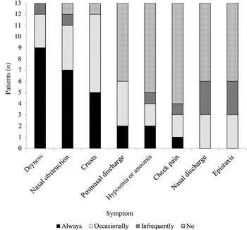
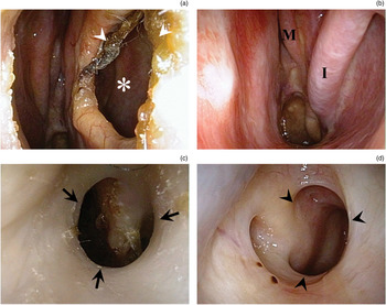
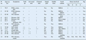
Table I summarises the clinical characteristics of the 13 patients. Sinonasal symptoms were present in all patients. A feeling of dryness, nasal obstruction and crusts were the most common symptoms noted (Figure 1). All patients had various pathological findings on nasal endoscopy (Figure 2). Atrophy of the inferior and/or middle turbinates was found in all patients. Anterior nasal stenosis and choanal stenosis were observed in five (38.5 per cent) and six patients (46.2 per cent), respectively. Septal perforation was identified in six patients (46.2 per cent). Methicillin-resistant Staphylococcus aureus was detected in six patients (46.2 per cent). Typical causative organisms of atrophic rhinitis (e.g. Klebsiella ozaenae and Coccobacillus foetidus ozaenae) were not detected.Reference Dutt and Kameswaran12 Computed tomography demonstrated soft tissue density shadows in the paranasal sinuses of 9 of the 12 patients examined (75 per cent). The most commonly encountered findings were maxillary sinus involvement (in eight patients; 66.7 per cent) and ethmoidal sinus involvement (five patients; 41.7 per cent).
Sinonasal symptoms in study patients.

Representative nasal endoscopic findings: (a) septal perforation (*) and crusts (arrowheads) seen in patient 8; (b) atrophy of the inferior (I) and middle (M) turbinates seen in patient 6; (c) anterior nasal stenosis (arrows) in patient 11; and (d) choanal stenosis (arrowheads) in patient 9.

Patient data

Pt no = patient number; y = years; Turb atr = turbinate atrophy; Ant = anterior; sten = stenosis; perf = perforation; Ma = maxillary; E = ethmoidal; Fr = frontal; S = sphenoidal; Srg = surgery; F = female; PND = postnasal discharge; NO = nasal obstruction; ++ = moderate to severe change; −= negative finding; += mild change; MRSA = methicillin-resistant Staphylococcus aureus; M = male; MSSA = methicillin-sensitive S aureus; α-strep = α-streptococcus; CB = corynebacterium species; AB = acinetobacter species; NA = no information available
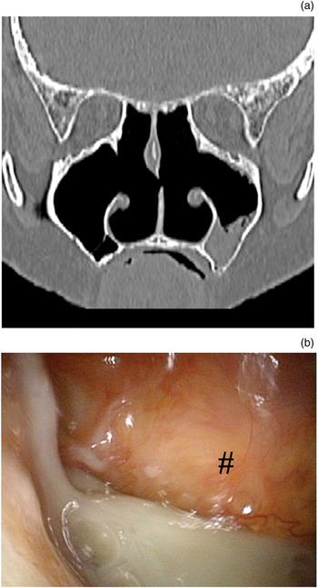
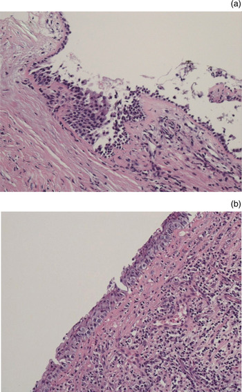
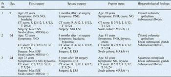
Table II shows the characteristics of the three patients with chronic rhinosinusitis treated with ESS. The first ESS procedure had been performed at an age of 69–73 years. All three patients experienced relapse of chronic rhinosinusitis, followed by a second ESS procedure seven months to six years later. After the second procedure, the patients had been treated with conservative measures. All three patients continued to have unpleasant symptoms, including a feeling of dryness (patients two and three), postnasal discharge (patients one and two), nasal obstruction (patients one and three) and crusts (patients one and two). Post-operative CT findings showed obvious improvement in patients one and two, but there was still persistent maxillary sinusitis with mucopus (Figure 3). Submucosal fibrosis, squamous metaplasia and a decreased number of submucosal glands were observed in the biopsy specimens (Figure 4).
Evidence of persistent maxillary sinusitis and mucopus after endoscopic sinus surgery in patient one: (a) coronal computed tomography image of paranasal sinuses: (b) nasal endoscopic view of the maxillary sinus (#).

Photomicrographs of sinus biopsies. (a) Patient two: submucosa shows thick fibrosis and a decreased number of submucosal glands (H&E; original magnification ×100). (b) Patient three: ciliated respiratory epithelium undergoing squamous metaplasia in places (H&E; original magnification ×200).

Extra data for ess patients

ESS = endoscopic sinus surgery; Pt no = patient number; F = female; PND = postnasal discharge; NO = nasal obstruction; CT = computed tomography; R = right; L = left; T = total; bilat = bilateral; MRSA = methicillin-resistant Staphylococcus aureus; (+) = positive; M = male; ND = nasal discharge; (−) = negative
Discussion
More than 90 per cent of our ex-lepromatous leprosy patients with atrophic rhinitis reported feelings of dryness, nasal obstruction and crusts. A previous study of treated lepromatous leprosy patients reported a lower prevalence of crusting (57.9 per cent) and nasal obstruction (18.4 per cent).Reference Kiris, Karlidag, Kocakoc, Bozgeyik and Sarsilmaz1 Our study included only ex-lepromatous leprosy patients with atrophic rhinitis. Our results suggest that atrophic rhinitis is an important source of prolonged nasal symptoms such as dryness, stiffness and crusts.
Nasal endoscopy demonstrated turbinate atrophy in all patients and nasal stenosis in approximately 40 per cent. Lepromatous leprosy is the severest type of leprosy, so we presumed that massive chronic granulomatous infection had caused these scar formations. Septal perforation was present in 46.2 per cent of patients (6 of 13). Previous descriptions of untreated lepromatous leprosy have reported septal perforation prevalences of 12 per cent (3 of 25), 20 per cent (2 of 10) and 12 per cent (3 of 25), variously.Reference Soni2, Reference Srinivasan, Nehru, Mann, Sharma, Bapuraj and Das7, Reference Srinivasan, Nehru, Bapuraj, Sharma and Mann8 On the other hand, a previous study of treated lepromatous leprosy patients found septal perforation in 47.3 per cent (18 of 38), suggesting that, in cases of persistent infection, cartilage destruction may continue even after the treatment of leprosy.Reference Kiris, Karlidag, Kocakoc, Bozgeyik and Sarsilmaz1 In addition, we suggest that treated lepromatous leprosy patients have a high prevalence of septal perforation as a result of repeated septal damage. In a study of intranasal sensation in lepromatous leprosy patients, 68 per cent of patients had partial or complete intranasal anaesthesia, leading to repeated trauma due to ‘picking’ of the nose.Reference Soni13 Lepromatous leprosy patient should be instructed in care of the nose, and otolaryngologists should be involved in the treatment and regular follow-up examination of these patients.
The findings of our patients' nasal swab cultures suggested that Staphylococcus aureus was the causative bacteria of rhinosinusitis. Ex-leprosy patients are sometimes exposed to antibiotics for management of the complications of leprosy. Otolaryngologists should pay attention to the results of bacterial testing, and select appropriate antibiotics with which to treat rhinosinusitis in ex-leprosy patients.
Follow-up CT studies of untreated leprosy patients have indicated that the ethmoidal sinuses are the most affected, being involved in 80 per cent of cases.Reference Srinivasan, Nehru, Mann, Sharma, Bapuraj and Das7, Reference Srinivasan, Nehru, Bapuraj, Sharma and Mann8 Similarly, the ethmoidal sinuses have been reported to be the most commonly involved sinuses (55.2 per cent) in treated lepromatous leprosy patients; furthermore, 33.3 per cent of these patients have had persistent acid-fast bacilli infection.Reference Kiris, Karlidag, Kocakoc, Bozgeyik and Sarsilmaz1 These results suggest that the ethmoidal sinuses are the main site of paranasal sinus lesions in leprosy. However, fewer of our patients had ethmoidal sinus involvement (41.7 per cent), compared with previous reports, with the maxillary sinuses being the most affected (66.7 per cent). Maxillary sinuses are the most affected sinuses in cases of chronic rhinosinusitis in ex-leprosy patients with atrophic rhinitis.
Endoscopic sinus surgery is currently regarded as the ‘gold standard’ surgery for chronic rhinosinusitis.Reference Chiu and Kennedy14 Unfortunately, we could not find any reports evaluating the use of ESS for chronic rhinosinusitis in ex-leprosy patients with secondary atrophic rhinitis; however, a few studies have reported that ESS can improve primary atrophic rhinitis.Reference Dutt and Kameswaran12, Reference Jiang, Hsu, Chen, Jan and Jang15, Reference Fang and Jin16
Three of our ex-leprosy patients with secondary atrophic rhinitis had undergone ESS for the treatment of chronic rhinosinusitis. Interestingly, all these patients experienced relapse and underwent a second ESS procedure; furthermore, despite continuous conservative treatment thereafter, all these patients continued to have unpleasant symptoms. Patient three had bronchial asthma, with histopathological analysis showing massive submucosal invasion by eosinophils; thus, this patient's condition might have been partly caused by allergic factors.
Although patients one and two gained improved CT scores following their first ESS procedure, their symptoms did not improve sufficiently. We speculate that two factors were responsible: atrophic rhinitis and continuous inflammation of the maxillary sinus. Atrophic rhinitis causes nasal mucosa anaesthesia, large crusts and an eddy current pattern of airflow, resulting in nasal obstruction and a feeling of dryness.Reference Dutt and Kameswaran12 Therefore, these latter two symptoms are difficult to relieve even after chronic rhinosinusitis treatment. Continuous maxillary sinusitis and mucopus were present in patients one and two despite wide opening of the maxillary ostia. These findings suggest that dysfunction of mucociliary transport, not closure of the ostium, caused chronic maxillary sinusitis in these patients. In a study of 21 ex-leprosy patients in Japan, the saccharin test indicated that mucociliary function had been severely impaired in half the patients.Reference Nakabayashi, Suzuki, Furukawa and Sasaki17 Impaired ciliary function is known to improve significantly after ESS; however, we speculate that ciliary function is slower to recover in ex-leprosy patients with atrophic rhinitis.Reference Uren, Psaltis and Wormald18–Reference Lund and Scadding20
• Leprosy has been eradicated in most regions
• However, many new cases still occur in endemic regions
• Ex-lepromatous leprosy patients with atrophic rhinitis have various rhino-sinus abnormalities and symptoms
• Otolaryngologists should regularly examine leprosy patients, both infectious and cured
Maxillary sinus irrigation was performed in patients one and two after confirming their remaining symptoms and the presence of maxillary mucopus. Maxillary sinus irrigation was easy to perform because of the widely opened maxillary ostium left after ESS, and was effective in relieving symptoms such as postnasal discharge and crusting. Nasal irrigation is known to be effective in improving the symptoms and health status of patients with sinonasal disease.Reference Tomooka, Murphy and Davidson21 A combination of ESS and maxillary sinus irrigation may reduce the nasal symptoms of ex-leprosy patients with chronic rhinosinusitis and atrophic rhinitis, and thus improve their QoL.
Conclusion
In this study, ex-lepromatous leprosy patients with atrophic rhinitis had various rhino-sinus abnormalities and persistent symptoms. These patients had underlying mucociliary dysfunction, so ESS was not sufficient for the treatment of chronic rhinosinusitis; thus, nasal obstruction and a feeling of dryness were hard to treat, whereas postnasal discharge and crusting could be reduced by a combination of ESS and nasal irrigation. Otolaryngologists should be involved in the serial observation of leprosy patients, not only in the infectious state but also in the treated and cured state.
Acknowledgements
We thank Dr Takashi Yokota (National Sanatorium Tohoku Sinseien) for his helpful advice, and Drs Daisuke Saito and Iori Takata (National Sanatorium Tohoku Sinseien) for their assistance in data collection.