Introduction
Malignant otitis externa is a potentially fatal infection of the external ear canal, skull base and adjacent soft tissues. Osteomyelitis of the temporal bone associated with necrotising otitis externa was described in the literature as early as 1838.Reference Toulmouche1 In 1959, Meltzer and Keleman reported a case of acute skull base osteomyelitis in a diabetic patient with auricular necrosis and purulent discharge.Reference Meltzer and Keleman2 The organism Bacillus pyocyaneus (known today as Pseudomonas aeruginosa) was identified in 1959.Reference Meltzer and Keleman2 The term ‘malignant’ was first used in 1968 by Chandler to reflect the condition's grim outcome (13 patients were reviewed, six of whom died).Reference Chandler3 Some prefer to use the terms ‘invasive’ or ‘necrotising’ otitis externa, in order to convey the fact that malignant otitis externa is not a neoplastic condition.Reference Nadol4
Patients with malignant otitis externa usually present with extreme otalgia and otorrhoea. The pain is classically worse at night-time. Diagnosis requires a high level of suspicion in a patient whose otitis externa is not responding to conventional management. Immunocompromised patients (e.g. diabetic, human immunodeficiency virus positive and transplant patients) are prone to this condition, but malignant otitis externa can also present in immunocompetent patients. The facial nerve is the most commonly involved cranial nerve; however, involvement of different cranial nerves at the jugular foramen and the petrous apex gives rise to a variety of clinical pictures and presentations (e.g. dysphonia and dysphagia). Malignant otitis externa is usually unilateral, but bilateral cases have also been reported in the literature.Reference Rowlands, Lekakis and Hinton5 Clinical examination reveals signs similar to ordinary otitis externa; however, granulation tissue on the floor of the ear canal at the osseocartilaginous junction is the tell-tale sign of underlying osteitis. The tympanic membrane, if visualised, is almost always intact.Reference Amorosa, Modugno and Pirodda6
Whilst surgical debridement was the treatment of choice in the early days, it is currently reserved for tissue sampling and occasionally for debridement. At present, long term systemic antibiotics are the mainstay of treatment. Appropriate and aggressive treatment has reduced the morbidity and mortality considerably over the past few decades.Reference Bhandary, Karki and Sinha7–Reference Carfrae and Kesser9
In light of current concerns regarding the emergence of ciprofloxacin resistance and the changing face of the disease, we reviewed our malignant otitis externa cases to evaluate the current epidemiology of the condition, and also to assess the status of drug resistance in our patient population.Reference Rubin Grandis, Branstetter and Yu10
Methods
We retrospectively identified all cases of malignant otitis externa diagnosed and treated in our otolaryngology department (the only otolaryngology department in a tertiary hospital in the north-east of England, with a catchment area of approximately one million people). All patients diagnosed with malignant otitis externa between January 2000 and April 2009 (i.e. nine years and four months) were identified and included in the study.
Diagnosis of malignant otitis externa was based on combined clinical and radiological criteria. The diagnosis was suspected in patients presenting with otorrhoea and deep-seated otalgia who did not respond to standard otitis externa management, and it was confirmed by computed tomography (CT) scanning demonstrating signs of bone involvement (i.e. osteitis). No other clinical signs or laboratory or microbiology results were included in the diagnostic criteria.
All relevant data (i.e. patient demographics, risk factors, and radiology and laboratory results) were retrieved from the hospital notes and the imaging archive system.
This study was approved by the local audit and clinical governance body.
Results
We identified 41 patients with a diagnosis of malignant otitis externa. Four patients were excluded because of insufficient information in their hospital notes, leaving 37 patients. These patients comprised 30 men (81 per cent) and seven women (19 per cent), aged 52 to 101 years (mean, 77 years; median, 81 years).
The interval between the first clinical consultation in our department and the establishment of the diagnosis varied. The diagnosis was usually made after a number of consultations and trials of conventional treatment. Diagnosis was expedited by the presence of cranial nerve palsies. On average, the diagnosis of malignant otitis externa was established and treatment commenced 30 days after the first otolaryngology consultation.
Clinical findings
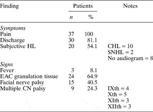
The presenting symptoms did not differ from ordinary otitis externa. Otalgia was the universal symptom at presentation. A summarised list of clinical findings at presentation is shown in Table I.
Clinical findings at presentation in 37 patients with malignant otitis externa

HL = hearing loss; CHL = conductive hearing loss; SNHL = sensorineural hearing loss; EAC = external auditory canal; CN = cranial nerve
Predisposing factors
A number of potential risk factors were surveyed. Half of the patients were diabetic (19/37; 51 per cent). One patient was iatrogenically immunosuppressed following a cardiac transplant. Four patients were receiving regular steroid therapy (11 per cent). Twelve patients were anaemic (32 per cent). A review of clinical documentation revealed no record of trauma to the affected ear, syringing or significant water exposure prior to symptom development. However, six patients (16 per cent) had regularly used hearing aids in the affected ear.
Inflammatory markers
Laboratory blood test results at the time of diagnosis were reviewed for all patients. The C-reactive protein concentration was measured in 30 patients, and was found to be elevated above the reference value in 28 patients (93 per cent), with a mean value of 41 mg/l. The erythrocyte sedimentation rate was tested in eight patients, seven of whom had an elevated value (mean, 88 mm/hour). A white blood cell count was performed for 33 patients, and was well within the normal reference range in the majority, being elevated in only seven patients (21 per cent; mean, 8.6 × 10⁹/l).
Microbiology results
Identification of the infective micro-organism was attempted in all 37 patients. Pseudomonas aeruginosa was identified in 20 patients (54 per cent), staphylococcal species in five (14 per cent), other organisms in seven (20 per cent) and no growth (after the standard inoculation time) in five (14 per cent). The miscellaneous organisms grown in the seven cases were Proteus mirabilis, candida, Corynebacterium striatum, Klebsiella pneumoniae and Aspergillus flavus.
Antimicrobial sensitivities were established for all positive samples. All pseudomonal isolates were found to be sensitive to ciprofloxacin, meropenem and Tazocin® (n = 20), whereas only 18 (18/20, 90 per cent) were sensitive to gentamicin.
Treatment protocols
When reviewing antimicrobial therapy and outcome, 10 patients had to be excluded because of insufficient data, insufficient documentation or failure to attend the follow-up clinic. Of the 27 remaining patients who received antibiotic treatment (as advised by the microbiologists), eight received monotherapy only (ciprofloxacin for seven and ceftazidime for one), while the rest received two or more antimicrobial agents. The most popular combinations were ciprofloxacin plus meropenem (n = 12) and ciprofloxacin plus Tazocin (n = 5). Two patients received triple therapy, in the form of ciprofloxacin plus meropenem plus linezolid, or ciprofloxacin plus meropenem plus Tazocin. Variconazole was added to the protocol if fungal growth was observed on microbiological culture. Topical antibiotic preparations were also used in 20 patients (usually for the initial two to three weeks); Gentisone® HC aural drops was most commonly used.
The duration of treatment varied but six weeks was the minimum course, and also the most commonly completed (n = 11/27). Some patients required a longer initial course (six to eight weeks) or a further course of treatment; thus, the mean duration of therapy was 8.7 weeks.
Response to treatment
We noted how soon after initiation of treatment the patient reported resolution of pain, as documented during clinical consultations. In general, all patients reported that their pain and discharge resolved prior to clinical consideration of treatment cessation. Overall, it took an average of five weeks for pain to resolve, in all 27 patients who received antibiotics and whose clinical notes and data were complete. Patients receiving monotherapy recovered more quickly than those receiving dual therapy (3.3 versus 5.8 weeks, respectively).
There was no objective, unified assessment of cranial nerve palsies at follow-up reviews; thus, valid statistical comparison was not possible. Of 14 patients with cranial nerve palsies and sufficient follow-up data, four reported resolution of their palsies, five showed improvement, four noticed no change at three to six month follow up, and one patient noticed worsening at four weeks and required further admission to hospital.
Three patients relapsed after treatment (three of 27; 11 per cent) and required a further course of antibiotics. The first patient initially received monotherapy (ciprofloxacin), but pain redeveloped following seven weeks of treatment; dual therapy was therefore prescribed for a further six weeks, with satisfactory results. The second patient received dual therapy with ciprofloxacin plus meropenem, but symptoms redeveloped three months after treatment. This patient made a good recovery on a repeated six-week course of the initial antibiotic regimen. The third patient died in hospital of an acute cardiac event not attributed to malignant otitis externa.
Discussion
Our series revealed a number of characteristics of malignant otitis externa, some in agreement with previous reports and some in contrast.
Diagnosis and clinical findings
We used similar criteria to previous studies in diagnosing malignant otitis externa.Reference Carfrae and Kesser9, Reference Mani, Sudhoff, Rajagopal, Moffat and Axon11 The diagnosis was based on combined clinical and radiological criteria. Malignant otitis externa was suspected in patients with a discharging ear and deep-seated pain who did not respond to medical treatment and aural toileting; the clinical suspicion was then confirmed by radiological findings of bone involvement on CT scan of the temporal bone. No other clinical findings (e.g. facial nerve palsy) or para-clinical findings (e.g. microbiological growth of P aeruginosa from the ear discharge specimen) were required to make the diagnosis, as previous studies had shown them not to be present in all cases.Reference Carfrae and Kesser9, Reference Rubin Grandis, Branstetter and Yu10 The clinical findings of granulation tissue on the floor of the ear canal at the osseocartilaginous junction and/or cranial nerve palsy (mainly facial nerve palsy) expedited the diagnosis.
In cases of malignant otitis externa, facial nerve palsy is usually the first neurological abnormality to develop.Reference Salit, McNeely and Chait12 Facial nerve involvement has been described in about 25 per cent of reported cases; however, a higher rate of 40 per cent was found in our cohort.Reference Carfrae and Kesser9 Although facial nerve involvement is a sign of malignant otitis externa progression and extension, it does not worsen the prognosis per se.Reference Soudry, Joshua, Sulkes and Nageris13 This was confirmed in our series, as the majority of patients with facial nerve involvement showed improvement or full recovery.
There was no mortality related to malignant otitis externa in our series.
Involvement of other cranial nerves (i.e. IXth, Xth, XIth and XIIth) has been reported if malignant otitis externa involves the jugular foramen and the petrous apex.Reference Chandler14 We found multiple nerve palsies in 24 per cent of our cases, a clinical feature which has not previously been clearly reported (but which was thought to be less common).Reference Carfrae and Kesser9 Involvement of these cranial nerves can broaden the spectrum of presenting or developing symptoms (e.g. causing dysphonia and dysphagia).
Hearing loss in our patients was mainly of the conductive type, similar to previous reports.Reference Carfrae and Kesser9, Reference Rubin Grandis, Branstetter and Yu10 This is usually secondary to soft tissue blocking the external canal. Only a few of our patients suffered sensorineural hearing loss. This is due to the fact that the middle-ear structures are usually preserved until late in the disease process.Reference Nadol4, Reference Kohut and Lindsay15
Predisposing factors
The pathogenesis of malignant otitis externa is not fully understood. The condition generally commences as a soft tissue infection of the external auditory canal. Pseudomonas aeruginosa is not a normal inhabitant of the healthy external ear canal. The shift from normal skin flora to proliferating Gram-negative bacilli (including P aeruginosa), and the subsequent tissue invasion, are thought to be triggered by three predisposing factors.
The first two factors are excessive moisture and ear canal trauma – the major predisposing factors described in the literature. Trauma to the ear canal can be caused by syringing, hearing aid use or other trivial insult.Reference Babiatzki and Sadé16 Although exposure to water and trauma (e.g. aural irrigation for wax removal) have been two of the most frequently reported predisposing factors, we did not encounter any such cases in our series.Reference Rubin Grandis, Branstetter and Yu10 This may be due to increasing reluctance to perform aural syringing within general practice. Approximately 16 per cent of our patients wore a hearing aid, which may have been responsible for a damp and humid aural environment as well as micro-trauma.
The third predisposing factor is immunocompromise. Immunocompromised individuals are the patient group most vulnerable to malignant otitis externa, with elderly diabetics being most at risk. However, other immunocompromised patients, and some healthy individuals, can also develop the condition. Cases of malignant otitis externa in patients with acquired immunodeficiency syndrome, myelodysplasia and neoplasia, and in transplant patients (iatrogenically immunocompromised), have been reported.Reference Carfrae and Kesser9, Reference Ress, Luntz, Telischi, Balkany and Whiteman17 It is not well established why diabetic individuals are most at risk. Diabetic microangiopathy has been identified as a predisposing factor causing ear canal tissue hypoperfusion. However, no significant increase in P aeruginosa colonisation has been found in the external ear canal of diabetic patients.Reference Salit, Miller, Wigmore and Smith18 Although the incidence of diabetes amongst malignant otitis externa patients has been reported as 65–100 per cent, diabetic patients formed only half (51 per cent) of our patient cohort.Reference Carfrae and Kesser9, Reference Rubin Grandis, Branstetter and Yu10
Microbiology
A wide range of different organisms has been cultured in malignant otitis externa cases; however, it is uncertain whether they are true pathogens.
Pseudomonas aeruginosa was grown from 54 per cent of our patients' samples, whereas it was grown in a clear majority of the cases reported in the literature.Reference Carfrae and Kesser9, Reference Rubin Grandis, Branstetter and Yu10 Staphylococcus aureus was the second most common cultured organism; this is similar to other studies.Reference Carfrae and Kesser9, Reference Rubin Grandis, Branstetter and Yu10
Interestingly, about 10 per cent of our cases did not show any growth. We employed ordinary microbiology culture techniques, and it is plausible that our detection rate may have been higher had we used more sophisticated detection methods (e.g. polymerase chain reaction). In addition, most of our patients had already received topical antibiotics from their general practitioner or from the otolaryngologist, prior to sampling of ear discharge for culture and sensitivity. It is possible that this practice may have resulted in lower microbiological growth rates.
Radiology
Radiological investigations are helpful in confirming the diagnosis of malignant otitis externa. Radiological imaging is considered most valuable in assessing the extent of the disease, and in monitoring disease regression in response to therapy. There is ongoing debate regarding the best radiological modality for initial evaluation and disease monitoring.Reference Carfrae and Kesser9 Computed tomography scanning is the modality of choice for diagnostic purposes, as it shows cortical bone erosion and the extent of soft tissue involvement. However, the fact that reduction in the soft tissue mass and remineralisation of bone either lags behind or never follows disease resolution makes CT scanning an unfavourable choice for assessment of the response to treatment. Although CT can assess the extent of the disease, CT findings are not closely correlated to patients' clinical outcomes.Reference Sudhoff, Rajagopal, Mani, Moumoulidis, Axon and Moffat19
Magnetic resonance imaging (MRI) is ideal for assessment of soft tissue, medullary bone involvement and dural enhancement.Reference Carfrae and Kesser9 However, authors are divided on the suitability of MRI for assessment of malignant otitis externa resolution.Reference Carfrae and Kesser9, Reference Rubin Grandis, Branstetter and Yu10, Reference Grandis, Curton and Yu20, Reference Ismail, Hellier and Batty21
Nuclear medicine imaging (e.g. gallium or technetium scanning) has also been used for the diagnosis and follow up of malignant otitis externa; however, its accuracy and specificity have been problematic.Reference Rubin Grandis, Branstetter and Yu10 Technetium scanning has a high sensitivity for diagnosing malignant otitis externa but it is not specific for infection, as it can also be positive in malignancy and does not detect soft tissue spread without bone involvement. Technetium also remains positive until cessation of osteoblastic activity, which may occasionally persist long after infection has settled; such scanning is therefore of limited suitability to assess the response to therapy.Reference Carfrae and Kesser9 However, gallium white cell scans return to normal after the infection has settled, and they can thus be used for radiological assessment of the response to therapy.Reference Carfrae and Kesser9 Gallium scans have restricted anatomical detail, making them less effective for initial radiological assessment. Some authors have recommended the use of technetium scanning when the disease has clinically resolved; a gallium scan is then recommended should the technetium scan become positive in these instances.Reference Okpala, Siraj, Nilssen and Pringle22
In our series, contrasted-enhanced CT scanning of the skull base and brain, including high resolution bone images, was used to aid diagnosis and to assess the extent of the disease, as this is a quick and readily available imaging modality. The scans were assessed by a radiologist with a special interest in otolaryngology and head and neck imaging. We did not routinely employ any other imaging modality for diagnosis or assessment of treatment response, except for MRI when dural involvement was suspected on the CT scan.
Most of our patients received a follow-up CT scan three months after completion of treatment, to assess radiological improvement and also to obtain baseline imaging should symptoms recur and future imaging be required.
Treatment
Historically, malignant otitis externa has been treated with prolonged courses of anti-pseudomonal antibiotics. Although monotherapy with a number of agents has been successful, a combination of two drugs is favoured by several authors, following the emergence of resistant strains.Reference Berenholz, Katzenell and Harell23
Fluoroquinolones, namely ciprofloxacin, are very effective in the treatment of malignant otitis externa. The use of ciprofloxacin for long term therapy is favoured by high bioavailability and excellent bone penetration and tissue availability, in addition to a narrow side-effect spectrum.
Ceftazidime, an anti-pseudomonal cephalosporin, was found to be effective in the treatment of malignant otitis externa in combination or as a monotherapy agent.Reference Kimmelman and Lucente24, Reference Johnson and Ramphal25
Meropenem, a broad spectrum carbapenem effective against P aeruginosa, is a relatively new antimicrobial agent with promising results for the treatment of pseudomonal sepsis and bone and soft tissue infections.
Aminoglycosides have lost popularity because of their nephrotoxicity and ototoxicity; the latter side effect is of huge significance when treating diabetic patients with underlying renal insufficiency.
There is insufficient evidence available to support the use of hyperbaric oxygen therapy in malignant otitis externa.Reference Phillips and Jones26 However, surgical debridement is occasionally indicated in cases unresponsive to medical treatment, or when tissue sampling is required in order to rule out malignancy.
Drug resistance
Increasingly, the medical literature is reporting pseudomonal resistance to ciprofloxacin; incidences of 20–33 per cent have been reported.Reference Carfrae and Kesser9, Reference Rubin Grandis, Branstetter and Yu10, Reference Bernstein, Holland, Porter and Maw27 This increasing ciprofloxacin resistance may be related to the recalcitrant nature of malignant otitis externa, or to the increased topical and systemic administration of this antibiotic for upper respiratory tract infections and uncomplicated otitis externa.Reference Carfrae and Kesser9, Reference Rubin Grandis, Branstetter and Yu10 Despite these previous findings, we found no ciprofloxacin resistance in our patient cohort.
• Malignant otitis externa can develop in both immunocompromised and immunocompetent patients
• In this study, the incidence of facial nerve palsy and multiple cranial nerve palsies at presentation was higher than previously reported
• The incidence of associated diabetes and growth of Pseudomonas aeruginosa in this series was less than previously reported
• No cases of ciprofloxacin resistance were noted, contrary to recent reports of the emergence of resistance
Response to treatment
In our study, we used patient's assessment of pain as a crude indicator of clinical improvement. Patients who reported pain resolution soon after commencement of treatment went on to require shorter treatment courses and were less likely to relapse. Patients receiving mono-antibiotic therapy also displayed a quicker rate of subjective improvement, compared with patients receiving dual and multi-antibiotic therapy. We believe that this is secondary to the fact that these patients were commenced on mono-therapy regimes as they had more limited disease to start with, rather than being a true reflection of treatment response.
In our study, most patients with cranial nerve palsy improved or recovered fully after treatment. This is in accordance with previous studies, which found that the cranial nerve involvement was not an indicator of worse clinical outcome provided patients received optimal medical treatment.Reference Mani, Sudhoff, Rajagopal, Moffat and Axon11
We did not find any specific aspect of our patients' demographics or clinical presentation to be an indicator of treatment outcome. However, a recent study found that older patients with diabetes and CT findings of nasopharyngeal involvement (i.e. soft tissue swelling, soft tissue asymmetry or abscess formation) had significantly prolonged treatment and reduced survival.Reference Joshua, Sulkes, Raveh, Bishara and Nageris28
Study limitations
As with all retrospective research, our study lacked a predetermined frame for clinical assessment and documentation. Also, multiple confounding factors and variables (including varied therapeutic approaches) made it impossible to produce a sound statistical analysis and comparison between patient groups. We used ordinary microbiology culture techniques, and it is quite possible that more advanced detection methods (e.g. polymerase chain reaction) may have demonstrated higher P aeruginosa growth rates.
Conclusions
Some of our study findings contrasted with previously reported results. Elderly male diabetic patients were the group most at risk in our cohort; however, only half of our patients were diabetics. The incidence of diabetes and of P aeruginosa in our cohort was much lower than previously reported. Pseudomonas aeruginosa was sensitive to ciprofloxacin in all our cases, contrary to recent reports on the emergence of drug resistance. The incidence of facial nerve palsy and multiple cranial nerve palsies was higher than in previous reports. Generally, our patients responded well to six to eight weeks of mono- or multi-antibiotic therapy.