I. INTRODUCTION
It is well known that one of the keys to attain an excellent property of an inorganic material is to control its size and shape. Reference Adair, Li, Kido, Havey, Moon, Mecholsky, Morrone, Talham, Ludwig and Wang1,Reference Jun, Choi and Cheon2 Therefore, since the discovery of nanomaterials, the construction of nanosized inorganic semiconductor materials with novel morphology has attracted extensive attention, owing to their fundamental and practical importance for optoelectrochemical performance as well as many other properties. Reference Wei, Cui, Song, Zhang, Song, Wang, Zhang, Li, Wen and Tian3–Reference Yu, Feng, Guan, Lou and Paik6 In particular, since Marcos et al. Reference Marcos, Amoros and Lebail7,Reference Marcos, Amoros, Beltranporter, Martinezmanez and Attfield8 first reported the preparation of M11(HPO3)8(OH)6 (M = Zn, Ni, and Co) in 1993, transitional metal phosphates have received much attention on account of their rich structural chemistry and wide potential applications in ion-exchange, electrochemistry, optics, magnetism, and catalysis. Reference Silbernagel, Martin and Clearfield9–Reference Zhong, Cornuz, Sivula, Graetzel and Gamelin14 Recently, increasing interest has been even paid on the synthesis of their nanostructures with specific morphology and controlled size. For example, Wen et al. prepared peony-like cobalt hydrogen phosphate hydroxide nanostructures by a solvothermal method through using Co(NO3)2 and H3PO4 as the reactants in the presence of CH3NH2 and HO(CH2)2OH at 120 °C for 5 days, and carefully investigated their low-temperature magnetic properties. Reference Wen, Cao, Sun, Xu, Wang, Zhang and Hu15 Zhang et al. reported the hydrothermal synthesis of hierarchical Ni11(HPO3)8(OH)6 superstructures by employing NaH2PO2·H2O and NiCl2·6H2O as the reactants with the assistance of CH3COONa·3H2O and polyvinylpyrrolidone at 170 °C for 10 h. Reference Zhang, Ni, Liao and Wei16 Pang et al. successfully synthesized Ni11(HPO3)8(OH)6 hexagonal polyhedrons via a facile hydrothermal method by using Ni(NO3)2·6H2O and Na3PO4·12H2O as the reactants in the presence of polystyrene sulfonic acid sodium salt and ethylene glycol at 220 °C for 48 h, indicating that the as-synthesized Ni11(HPO3)8(OH)6 could be applied as the electrode material for electrochemical super capacitor with excellent specific capacitance, rate capability, and cycling properties. Reference Pang, Yan, Wei, Li, Li, Zhang, Chen, Zhang and Zheng17 And Gu et al. even synthesized an urchin-like Ni11(HPO3)8(OH)6 microball by using NiCl2 and KH2PO4 as the reactants at 180 °C for 8 h, but they did not report any investigation on its properties. Reference Gu, Zhai, Gao, Zhang, Ke, Ma and Yao18
On the other hand, due to the pending shortage of fossil energy and increasingly severe environmental pollution problems, the development of semiconductor photocatalysts is being extensively carried out because they have exhibited high activities for the degradation of many organic and inorganic pollutants, water splitting to produce hydrogen, and the production of synthetic fuels with solar energy in an environmentally friendly way. Reference Johansson, Niklasson and Osterlund19–Reference Ha, Han, Risch, Giordano, Yao, Karayaylali and Shao-Horn21 However, to the best of our knowledge, there has been no report on the photocatalytic degradation of organic dyes over Ni11(HPO3)8(OH)6 microballs.
In this work, novel waxberry-like Ni11(HPO3)8(OH)6 microballs were synthesized via a traditional solvothermal route by employing nickel sulfate (NiSO4·6H2O) and sodium hypophosphite (NaH2PO2·H2O) as the reactants in the presence of ethylene glycol as the surfactant under the optimized conditions with a Ni:P molar ratio of 1:2 at 200 °C for 16 h. Based on the present experimental results, a possible formation mechanism for the products was proposed. Moreover, the photocatalytic degradations of organic dyes methylene blue (MB) and rhodamine B (RhB) over the waxberry-like Ni11(HPO3)8(OH)6 microballs were tentatively investigated, presenting excellent photocatalytic activity under ultraviolet (UV) light.
II. EXPERIMENTAL SECTION
A. Samples’ preparation
The proposed Ni11(HPO3)8(OH)6 microballs were synthesized by a solvothermal method with ethylene glycol as the surfactant. The synthesis conditions were optimized. In the designed experiments, NiSO4·6H2O and NaH2PO2·H2O with different molar ratios of Ni:P (1:0.5, 1:1, 1:2, and 1:3) were firstly added under stirring into a mixture of 7 mL EG and 28 mL distilled water. When it became transparent, the solution was transferred into a Teflon-liner autoclave of 100 mL capacity. After sealing, the autoclave was heated up to the designed temperature (160, 180, or 200 °C), dwelling at this temperature for 1–16 h. After heating, the autoclave was allowed to cool down to room temperature naturally. The resultant precipitates were collected, washed with absolute ethanol and distilled water for several times, and dried in a vacuum furnace at 60 °C for 6 h.
B. Materials’ characterization
The powder X-ray diffraction (XRD) patterns of the as-prepared samples were recorded on a Rigaku D/max-RB X-ray diffractometer (Rigaku Corp., Tokyo, Japan) with graphite monochromatized Cu Kα radiation (λ = 1.5418 Å) in a continuous scanning mode at a rate of 6°/min. The microstructure of the samples was examined by a field emission scanning electron microscopy (FE-SEM; S4800, Hitachi Corp., Tokyo, Japan), and the elemental composition was measured by an energy dispersive X-ray (EDX) spectroscope attached to the SEM. The specific surface area was measured by the Brunauer–Emmett–Teller (BET) N2 adsorption method using a QuadraSorb Station 4 surface analyzer (Quantachrome Instruments, Boynton Beach, Florida). The zeta potential was measured by a Malvern Nano ZS potential meter (Malvern Instruments, Malvern, United Kingdom). The diffuse reflectance UV–vis spectrum of the solid products was recorded on a Varian Cary 5000 UV–vis spectrometer (Varian, Palo Alto, California) using BaSO4 as the reference.
A Mott–Schottky plot was measured by using a conventional three-electrode electrochemical workstation (CHI660E, CH Instruments, Shanghai, China), in which the Ni11(HPO3)8(OH)6 film on an fluorine-doped tin oxide substrate, a platinum electrode, and an saturated calomel electrode (SCE) were used as the working, counter, and reference electrodes, respectively. A 3.0 M KOH aqueous solution was used as the supporting electrolyte to maintain the stability of the film. The Mott–Schottky plot was derived from the impedance-potential test conducted at a frequency of 1000 Hz.
C. Photocatalytic activity evaluation
The photocatalytic activity of the as-synthesized waxberry-like Ni11(HPO3)8(OH)6 microballs was evaluated by decomposing MB and RhB under the irradiation of UV and visible light, respectively. For the light irradiation, the reaction test tube was located axially in a ring whirligig test tube rack (XPA-7, PerfectLight, Beijing, China). The light was provided by a 500 W mercury lamp or a 500 W xenon lamp with an optical filter. And the lamp was set in the center of the rack at a distance of 100 mm away from the aqueous solution in a dark box. In a typical test, 50 mg of the obtained waxberry-like Ni11(HPO3)8(OH)6 microballs was dispersed into 50 mL of 5 mg/L MB and 4 mg/L RhB in a reactor, respectively. The photocatalyst particles were allowed to absorb the dye molecules to acquire an adsorption–desorption equilibrium when the reactor was placed in the dark for 60 min. After that, the light irradiation was launched. During the degradation of organic dyes, the reactor was placed on the ring whirligig device stirred by magnetic force. The reactor was then irradiated by the lights for 90 min. After the irradiation, the supernatant samples were collected regularly from the reactor per 10 min, and the dispersed catalyst particles were removed by centrifuging. The resultant clear solutions were then analyzed by an UV–vis spectroscope (SP-752, Spectrum Instruments, Shanghai, China) to determine the concentration of dyes in the solutions. After the analysis, the solution and catalyst sediments were immediately poured back into the test tube, keeping the irradiation reaction in almost the same state.
III. RESULTS AND DISCUSSION
A. Optimization of synthesis conditions
Through a series of experiments, it was found that the reaction temperature, duration time, and Ni:P molar ratio of the sources are the most important factors influencing the morphology of the prepared Ni11(HPO3)8(OH)6. So, while all the other parameters were controlled, the influences of these three factors were systematically investigated.
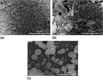
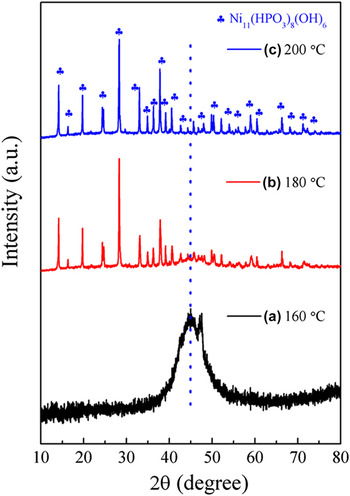
Under keeping the reaction time (16 h) and Ni:P atomic ratio (1:2), the influence of reaction temperature on the morphology and composition of the products was first examined. The results are shown in Figs. 1 and 2. When it was 160 °C, the product was composed of a large amount of regular solid microspheres of a diameter around 2–5 μm [Fig. 1(a)]. However, the XRD pattern of the product synthesized at 160 °C reveals poor crystallinity with a very broad hump at roughly 44.5° in correlation with amorphous Ni–P, Reference Muthuswamy, Savithra and Brock22–Reference Moreau, Ha, Zhang, Hoyden, Muller and Robinson24 suggesting that it is of non-crystalline structure [see Fig. 2(a)]. When it was enhanced up to 180 °C, many particles of irregular thorn-like structures with different sizes as shown in the inset in Fig. 1(b) but few of waxberry-like structures could be observed [Fig. 1(b)], which would be the inchoate formation stage of the waxberry-like microballs, although the XRD pattern of the sample revealed the formation of Ni11(HPO3)8(OH)6 [see Fig. 2(b)]. With increasing temperature because the stability of amorphous Ni–P became weak, it would slowly react with HPO3 2− from the decomposition of NaH2PO2·H2O into Ni11(HPO3)8(OH)6. Reference Liu, Liu, Xu, Qian and Ma25 But the reaction was incomplete at 180 °C, so there still existed lots of amorphous Ni–P microspheres. When it increased up to 200 °C, lots of waxberry-like microballs were obtained [Fig. 1(c)]. At this temperature, the amorphous Ni–P microspheres completely disappeared, which was also confirmed by the XRD pattern in Fig. 2(c). Instead, sharp peaks of Ni11(HPO3)8(OH)6 crystals appeared (Joint Committee on Powder Diffraction Standards [JCPDS] card no. 81-1065), indicating that the amorphous Ni–P microspheres were completely transformed to waxberry-like Ni11(HPO3)8(OH)6 crystalline microballs. These results suggest that 200 °C is suitable for the formation of the present waxberry-like Ni11(HPO3)8(OH)6 crystalline microballs. Thus, in this work, the optimized reaction temperature for the synthesis of waxberry-like Ni11(HPO3)8(OH)6 microballs was determined as 200 °C.
SEM images of the samples prepared at different reaction temperatures: (a) 160, (b) 180, and (c) 200 °C, under keeping the reaction time (16 h) and Ni:P atomic ratio (1:2). The inset in (b) is the magnified image of the rectangle area.

XRD patterns of the samples prepared at different reaction temperatures: (a) 160, (b) 180, and (c) 200 °C, under keeping the reaction time (16 h) and Ni:P atomic ratio (1:2).

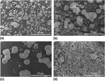
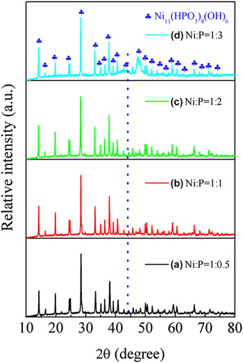
The Ni:P atomic ratio in source materials is another key factor influencing the morphology of the prepared Ni11(HPO3)8(OH)6. As shown in Fig. 3(a), when the molar ratio of Ni:P was 1:0.5, lots of irregular thorn-like structures were agglomerated together, while waxberry-like microballs could not obviously be observed because it lacks P source in the reaction system. When the molar ratio of Ni:P was 1:1, a great deal of waxberry microballs could be observed [Fig. 3(b)], but there were still lots of irregular thorn-like structures agglomerated on the surface of the waxberry microballs. This is probably because the thorn-like structures still needed more P source to react further to form microballs. Furthermore, when the molar ratio of Ni:P was 1:2, a large amount of typical three dimensional waxberry-like microballs composed of numerous highly ordered thorn-like structures can be obtained [Fig. 3(c)]. It is believed that the Ni:P molar ratio of 1:2 is helpful for the formation of waxberry-like microballs. However, when the molar ratio of Ni:P was 1:3, the waxberry-like structures almost disappeared again, while many irregular thorn-like structures and a small amount of microspheres co-existed [Fig. 3(d)], implying that with increasing amount of P source, the growing thorn-like structures might be separated from the waxberry-like microballs. Simultaneously, new amorphous Ni–P microspheres with an average size of 4 μm could be observed, confirming that the excessive P source was prone to form amorphous Ni–P microspheres. In addition, all these observations can be confirmed by their XRD patterns shown in Fig. 4. As seen from Fig. 4, all the sharp XRD peaks can be assigned to those of Ni11(HPO3)8(OH)6 crystals (JCPDS card no. 81-1065). However, a small hump around 44.5° owing to amorphous Ni–P appeared again when the molar ratio of Ni:P was 1:3 in the reaction system. Hence, in this work, the optimized molar ratio of Ni:P was chosen as 1:2.
SEM images of the samples prepared with different Ni:P molar ratios in the source materials: (a) 1:0.5, (b) 1:1, (c) 1:2, (d) 1:3, under keeping the reaction time (16 h) and reaction temperature (200 °C).

XRD patterns of the samples prepared with different Ni:P molar ratios in the source materials: (a) 1:0.5, (b) 1:1, (c) 1:2, (d) 1:3, under keeping the reaction time (16 h) and reaction temperature (200 °C).

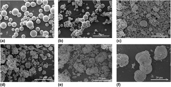
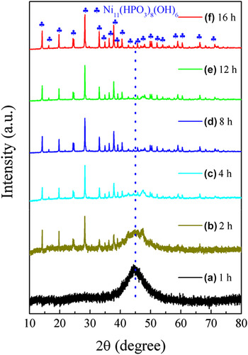
Time-dependent experiments were further carried out to investigate the influence of reaction time on the formation of Ni11(HPO3)8(OH)6 waxberry-like microballs. The results are displayed in Figs. 5 and 6. From the SEM image as shown in Fig. 5(a) together with the XRD pattern in Fig. 6(a), it can be seen that pure amorphous Ni–P microspheres resulted when the dwelling time was no more than 1 h. When it was prolonged for 2 h, irregularly aggregated thorn-like structures appeared [Fig. 5(b)], but amorphous Ni–P was still the main phase [see Fig. 6(b)]. When it was extended up to 4 h, imperfect Ni11(HPO3)8(OH)6 waxberry-like microballs co-existed with amorphous Ni–P microspheres [see Figs. 5(c) and 6(c)], indicating an inchoate formation stage of the waxberry-like microballs. When it increased up to 8 h, Ni–P microspheres almost disappeared and irregular waxberry-like structures could be observed [Fig. 5(d)], which could be confirmed by the XRD pattern of the sample [see Fig. 6(d)], implying that the amorphous Ni–P microspheres were almost transformed into Ni11(HPO3)8(OH)6 microcrystals. When it was prolonged for 12 h, incomplete waxberry-like microballs were still observed [Fig. 5(e)], although its XRD pattern reveals a pure Ni11(HPO3)8(OH)6 phase [Fig. 6(e)]. Finally, when it was further extended to 16 h, perfect 3D waxberry-like Ni11(HPO3)8(OH)6 microballs were obtained [Fig. 5(f)]. And in such case, all the diffraction peaks in the XRD pattern perfectly matched with those of the standard Ni11(HPO3)8(OH)6 crystals [JCPDS card no. 81-1065, see Fig. 6(f)], further confirming the observation from SEM. So, the dwelling time was determined as 16 h.
SEM images of the samples prepared for different dwelling times: (a) 1, (b) 2, (c) 4, (d) 8, (e) 12, and (f) 16 h, under keeping the reaction temperature (200 °C) and Ni:P molar ratio (1:2).

XRD patterns of the samples prepared for different dwelling times: (a) 1, (b) 2, (c) 4, (d) 8, (e) 12, and (f) 16 h, under keeping the reaction temperature (200 °C) and Ni:P molar ratio (1:2).

In summary, the present waxberry-like Ni11(HPO3)8(OH)6 microballs could be successfully prepared under the optimum conditions with a Ni:P molar ratio of 1:2 at 200 °C for 16 h.
B. Composition, structure, and growth mechanism
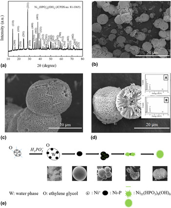
To examine the phase composition of the waxberry-like microballs synthesized under the optimized conditions, a typical XRD pattern is elaborately shown in Fig. 7(a). It can be seen from this figure that all the recorded diffraction peaks can be assigned to those of Ni11(HPO3)8(OH)6 crystallites (JCPDS card no. 81-1065). The peaks are sharp and strong, and no diffraction peaks from other phases can be identified from the pattern. This result reveals that under the optimum conditions, the obtained Ni11(HPO3)8(OH)6 has good crystallinity and high purity.
Typical XRD pattern (a) and SEM results of the waxberry-like microballs synthesized under the optimized conditions with a Ni:P molar ratio of 1:2 at 200 °C for 16 h: (b) low-magnification image; (c) high-magnification image; and (d) EDX measurement on a waxberry-like microball, in which inset A shows the recorded EDX spectrum on the core and inset B is the recorded EDX spectrum on the thorn-like structure. (e) Schematic of the formation process of the present Ni11(HPO3)8(OH)6 waxberry-like microballs.

Figures 7(b) and 7(c) presents typical SEM results of the waxberry-like microballs synthesized under the optimized conditions. The low-magnification SEM image [Fig. 7(b)] demonstrates that the obtained sample consists of many microballs with a diameter of 10–20 μm. The higher magnification SEM image [Fig. 7(c)] reveals that the microballs have a waxberry-like structure. With a close view on the microballs [see Fig. 7(d)], numerous thorn-like structures regularly stacked in high density with one tip pointing toward the center of the microballs, looking like a waxberry. The specific surface area of the sample measured by BET N2 adsorption is 6.8 m2/g, which is a quite small value compared with those of nanoscaled solids, implying weak adsorption ability of the present sample for N2 adsorption and solid structure of the thorn-like structures in the microballs. However, because there are lots of gaps between the thorn-like structures, thus providing large amount of active sites during photocatalysis, it would be beneficial for the improvement of catalytic degradation performance on organic dyes.
To investigate the elemental composition of the as-synthesized waxberry-like microballs, the EDX analysis was carried out. The EDX spectrum acquired from the core of microballs [see inset A in Fig. 7(d)] reveals that the core is composed of O, Ni, and P with an O:Ni:P ratio of 63:11:26, while that from the thorn-like structures [see inset B in Fig. 7(d)] indicates that the flesh of the waxberry consists of O, Ni, and P with an O:Ni:P ratio of 62:14:24. Obviously, both have a composition very close to the stoichiometric ratio of Ni11(HPO3)8(OH)6, which is completely in agreement with the XRD results, further confirming that the as-synthesized sample is of high purity.
In the literature, H2PO2 − ion is a commonly used reducing reagent during the hydrothermal reaction due to its low potential in the aqueous solution. It could react with water into HPO3 2− and PH3 as follows Reference Liu, Liu, Xu, Qian and Ma25 :
 $$3{{\rm{H}}_2}\mathop {{\rm{P}}{{\rm{O}}_2}}\nolimits^ - \to {\rm{P}}{{\rm{H}}_3} + 2{\rm{HP}}\mathop {{{\rm{O}}_{\rm{3}}}}\nolimits^{{\rm{2}} - } + {{\rm{H}}^ + }\quad .$$
In a neutral or acidic solution as in the present experiments, the dismutation process of P is the main reaction. As displayed in Fig. 7(e), during the synthesis of the present product, NiSO4·6H2O and NaH2PO2·H2O were first dissolved into distilled water mixed with ethylene glycol, and thus the Ni2+ ions would be dispersed in the solution evenly to form a stable complex with glycol. With the increment of the reaction temperature, the stability of this complex decreases gradually and slowly releases free Ni ions. Reference Liu, Liu, Xu, Qian and Ma25 Once generated, the active Ni2+ ions reacted quickly with PH3, producing amorphous Ni–P nuclei as expressed by the following equation:
 $${\rm{N}}{{\rm{i}}^{2 + }} + {\rm{P}}{{\rm{H}}_3} \to {\rm{\ Ni-P}} + {{\rm{H}}^ + }\quad ,$$
which would then grow into nanoparticles and further aggregate into bigger microspheres.
The mixed solvent can influence the solubility, reactivity, and diffusion behavior of the HPO3 2− ions and the Ni–P intermediate. With the reaction proceeded, some amorphous Ni–P microspheres aggregated into irregular microspheres of crystalline Ni11(HPO3)8(OH)6 and amorphous Ni–P, further assembling into thorn-like structure due to its reaction with HPO3 2− ions,
 $${\rm{Ni-P}} + {\rm{HP}}\mathop {{{\rm{O}}_{\rm{3}}}}\nolimits^{2 - } + {{\rm{H}}_2}{\rm{O}} \to {\rm{N}}{{\rm{i}}_{11}}{({\rm{HP}}{{\rm{O}}_3})_8}{({\rm{OH}})_6} + {{\rm{H}}^ + }\quad .$$
The surface of these thorn-like structures is rough, so other particles can grow on it, assembling into the final waxberry-like microballs (please also see Figs. 1–6).
C. Photocatalytic properties
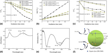
For the photocatalysis tests in the present study, two steps were involved in the decolourization of the organic dyes (MB and RhB): the adsorption and photodegradation of the dye molecules over the catalyst. Figure 8(a) illustrates the decolourizations of 50 mL of 5 mg/L MB and 50 mL of 4 mg/L RhB over 50 mg of the as-synthesized photocatalyst in the dark for 60 min (for the adsorption) and under UV light irradiation for 90 min (for the photodegradation) in comparison with those without catalyst. As is seen from Fig. 8(a), before UV light irradiation, during the adsorption step, the waxberry-like Ni11(HPO3)8(OH)6 microballs presented a certain degree of adsorption effects on MB and RhB. The decolourizations by adsorption on MB and RhB were 5.1% and 7.9%, respectively, which are somewhat small compared with those over many nanoscaled photocatalysts, due to its quite small specific surface area (6.8 m2/g), which means a bad contact between photocatalysts and dye molecules, and relatively small zeta potential (−21.5 mV), which is not of much benefit for the photocatalysts to adsorb the free ions in the solution. During the second, photodegradation step, the decolourized MB and RhB in the presence of the waxberry-like Ni11(HPO3)8(OH)6 microballs under UV light after 90 min increased from 5.1% to 73.9% and from 7.9% to 48.3%, respectively, presenting a significant degradation effect on both organic dyes in the aqueous solution. Owing to the decolourization efficiency of the UV light, the MB and RhB could be decolourized at 33.3 and 32.2% without a catalyst. So, the decolourized MB and RhB by the as-synthesized Ni11(HPO3)8(OH)6 catalyst was 35.5 and 8.2% under UV light. Furthermore, the reaction kinetics of MB and RhB degradation by the present photocatalyst could be evaluated by using the pseudo-first-order reaction to fit the degradation process: ln(C 0/C) = kt, where the rate constant k (min−1) is equal to the slope of the fitted line, C is the constant concentration at time t, C 0 is the initial concentration, and t is the irradiation time. From the fitted lines as shown in Fig. 8(b), the calculated rate constants of MB and RhB degradation are 0.0143 and 0.00661 min−1, respectively, which are all higher than those (0.00407 min−1 for MB degradation and 0.00444 min−1 for RhB degradation) without a catalyst. This results indicate that the as-synthesized waxberry-like Ni11(HPO3)8(OH)6 microballs have a significant photocatalytic activity for the degradation of MB and RhB under UV light irradiation, although it has to admit that the photocatalytic performance of the present Ni11(HPO3)8(OH)6 microballs is quite inferior to those of many of the already reported photocatalysts tested under similar conditions (see in Table S1).
(a) Decolourizations of 50 mL of 5 mg/L MB and 50 mL of 4 mg/L RhB, respectively, over 50 mg of the waxberry-like Ni11(HPO3)8(OH)6 microballs synthesized under the optimized conditions in the dark for 60 min and under UV-light irradiation for 90 min. (b) Their corresponding -ln(C/Co) versus irradiation time (t) plots during the irradiation process. (c) Decolourizations of 50 mL of 5 mg∙L−1 MB and 4 mg∙L−1 RhB, respectively, in the dark and under visible light irradiation over 50 mg of the waxberry-like Ni11(HPO3)8(OH)6 photocatalyst synthesized under the optimized conditions. (d) UV–vis absorption spectrum of the waxberry-like Ni11(HPO3)8(OH)6 microballs synthesized under the optimized conditions. (e) UV–vis reflectance spectrum of the waxberry-like Ni11(HPO3)8(OH)6 microballs synthesized under the optimized conditions. (f) Schematic diagram of the photodegradation mechanism of organic dyes (MB and RhB) over the Ni11(HPO3)8(OH)6 microballs under UV irradiation.

However, as is seen from Fig. 8(c), after visible light irradiation for 100 min, in the photodegradation step, the decolourized MB and RhB over the Ni11(HPO3)8(OH)6 waxberry-like microballs only have a 3.5% and 4.4% increase, respectively, presenting very little decolourization effect on both organic dyes in the aqueous solution. Considering the fact that without any catalyst, the decolourization of MB and RhB under visible light could reach approximately 3.1% and 4.1%, respectively, the decolourized MB and RhB by the Ni11(HPO3)8(OH)6 microball sample were only about 0.4 and 0.3%, revealing that the as-synthesized Ni11(HPO3)8(OH)6 microballs almost have no photocatalytic activity for the degradation of MB and RhB under visible light irradiation.
To tentatively explain the photocatalytic activity of the as-synthesized waxberry-like Ni11(HPO3)8(OH)6 microballs on the degradation of MB and RhB, its UV–vis absorption spectrum was measured. The result is shown in Fig. 8(d). From this figure, it can be seen that the maximum absorption peak of the present microballs is located at 413 nm in the UV range, exhibiting a strong absorption to UV light, which may be why the as-synthesized waxberry-like Ni11(HPO3)8(OH)6 microballs have significant photodegradation activity on MB and RhB under UV light irradiation. However, although two relatively strong absorption peaks at 683 and 766 nm were also observed, the as-synthesized Ni11(HPO3)8(OH)6 microballs almost have no photocatalytic activity for the degradation of MB and RhB under visible light irradiation. In Ref. Reference Zhang, Jianmin, Nishiura and Imamoto26, the strong and broad absorption bands which occurred at 376, 594, and 958 nm were attributed to the typical absorption of d–d transitions of Ni(II) ion. Reference Zhang, Jianmin, Nishiura and Imamoto26 And it was reported that, Reference Ren, Lu, Cai, Shi, Jiang, Chen, Zheng and Liu27,Reference Fukumoto, Yamane, Kase and Yamamoto28 the UV–vis spectrum of [Ni2L(m-OH)](ClO4)2 exhibited a strong peak at 510 nm, a characteristic absorption of a d–d transition of the square-planar four-coordinated Ni(II) center, and the UV–vis spectrum of poly-1-Ni exhibited additional intermediate and small peaks at about 480 and 600 nm, which were assigned to the charge transfer by electronic transition and d–d transition of Ni (2+), respectively. And because of the different splitting width of the d orbital, which is changed by the coordination number and ligand of the materials, the absorption peak of Ni complex would shift more or less. Reference Zhang, Jianmin, Nishiura and Imamoto26–Reference Fukumoto, Yamane, Kase and Yamamoto28 According to the hexagonal structure of Ni11(HPO3)8(OH)6 as is known from the XRD result, the coordination number of Ni11(HPO3)8(OH)6 prepared in this work is 12. Considering all these facts, the two absorption peaks of the present sample might be caused by the absorption of d–d transitions of Ni(II) ion, which is a result of ligand field transition instead of the absorption peaks of Ni11(HPO3)8(OH)6 caused by the charge transfer transition, and will not inspire electron–hole pairs, or the photogenerated electrons and holes might be easily recombined, which cannot degrade the organic dyes. As a consequence, pure waxberry-like Ni11(HPO3)8(OH)6 microballs cannot be used as a visible-driven photocatalyst for the degradation of MB and RhB. In addition, as can be seen from Fig. 8(e), Ni11(HPO3)8(OH)6 microballs have high reflectance around visible light range but low reflectance around 413 nm, further confirming the result of the photocatalytic tests.
To identify the practical energy levels of valence band (VB) and conduct band (CB), the Fermi level (E F) potential of the synthesized Ni11(HPO3)8(OH)6 was determined by electrochemical impedance spectroscopy along with the Mott–Schottky equation, Reference Yeh, Chan, Hsieh and Teng29,Reference Zhu, Yin, Yang, Cui, Wang, Xu, Lin and Huang30
 $${1 \over {{C^2}}} = {2 \over {e\varepsilon {\varepsilon _0}{N_{\rm{A}}}}}\left( {E - {E_{\rm{F}}} - {{kT} \over e}} \right)\quad ,$$
in which the temperature term is generally small and can be neglected. The capacitance values of the space-charge region obtained at various applied potentials are shown in Fig. S1. According to the Mott–Schottky equation, a linear relationship of 1/C 2 versus E can be fitted. The negative slope of the straight line justifies a p-type conductivity of the Ni11(HPO3)8(OH)6 sample, which means that E F is similar to the energy level of VB and the intercept on abscissa was used to calculate the value of E F (0.076 eV versus SCE). According to the conversion relationship of SCE reference electrode with normal hydrogen electrode (NHE), the practical value of E F of the Ni11(HPO3)8(OH)6 sample is 0.32 eV (V versus NHE), which can be regarded as its VB. Furthermore, from the plots of (αhν)2 and (αhν)0.5 against the photon energy for the Ni11(HPO3)8(OH)6 photocatalyst as shown in Figs. S2(a) and S2(b), it was calculated that the apparent energy for its direct band gap was of 2.55 eV and that for the indirect band gap was 2.48 eV. Because of the similar value between the two types of band-gap, the CB can be calculated as −2.23 eV (V versus NHE) approximately.
According to the calculated energy levels of VB and CB, a possible photodegradation mechanism is schematically proposed in Fig. 8(f). During UV irradiation, electron–hole (e−–h+) pairs are generated on Ni11(HPO3)8(OH)6 microballs when they were excited by UV light. These photogenerated e− and h+ would move to the surface of microballs, respectively, as a result of their separation. Then the separated e− and h+ would react with the oxidants or reducers (usually O2 and OH−) adsorbed on the surface of the Ni11(HPO3)8(OH)6 microballs to produce active oxidative radicals (e.g., ·O2 − and ·OH). Subsequently, the active radicals would lead to the degradation of organic dyes (MB and RhB). Significantly, the rough surface of the waxberry-like Ni11(HPO3)8(OH)6 microballs provides a large amount of active sites for the degradation of organic dyes, improving the rate of degradation of organic dyes.
IV. CONCLUSIONS
Waxberry-like Ni11(HPO3)8(OH)6 microballs were successfully synthesized via a hydrothermal route by using NiSO4 as Ni-source and NaH2PO2 as P-source in a mixture solvent of ethylene glycol and water under the optimized conditions with a Ni:P molar ratio of 1:2 at 200 °C for 16 h. A possible formation mechanism for the crystalline waxberry-like Ni11(HPO3)8(OH)6 microballs was proposed. The photocatalytic degradation tests revealed that the as-prepared waxberry-like Ni11(HPO3)8(OH)6 microballs had significant photocatalytic activity on the degradation of MB and RhB under the irradiation of UV light.
Supplementary Material
To view supplementary material for this article, please visit https://doi.org/10.1557/jmr.2017.154.
ACKNOWLEDGMENTS
The authors would like to thank the financial support for this work from the National Natural Science Foundation of China (grant nos. 11274052, 11674035, and 61274015) and the Fund of State Key Laboratory of Information Photonics and Optical Communications (Beijing University of Posts and Telecommunications).