Introduction
Body dysmorphic disorder (BDD) is a psychiatric disorder in which individuals are preoccupied with misperceived defects in their physical appearance, often involving facial features. Like those with BDD, individuals with anorexia nervosa (AN) experience distorted perceptions of appearance, but they mainly stem from the conviction of being overweight despite extreme thinness. AN and BDD share a peak age of onset during adolescence, and have similar diagnostic comorbidities (Phillips & Kaye, Reference Phillips and Kaye2007; Swinbourne & Touyz, Reference Swinbourne and Touyz2007; Kollei et al. Reference Kollei, Brunhoeber, Rauh, de Zwaan and Martin2012). AN and BDD often co-occur – upwards of 32% of patients with BDD report a lifetime eating disorder (Ruffolo et al. Reference Ruffolo, Phillips, Menard, Fay and Weisberg2006), and the converse is true for between 25% and 39% of those with AN (Grant et al. Reference Grant, Kim and Eckert2002, Rabe-Jablonska Jolanta & Sobow Tomasz, Reference Rabe-Jablonska Jolanta and Sobow Tomasz2000). Further, there is over concern with the appearance of specific body parts, such as size of abdomen, hips, and thighs (Grant & Phillips, Reference Grant and Phillips2004). On the other hand, there are important differences; notably in gender distribution, which is far less skewed toward females in BDD (Rief et al. Reference Rief, Buhlmann, Wilhelm, Borkenhagen and Brahler2006; Koran et al. Reference Koran, Abujaoude, Large and Serpe2008; Buhlmann et al. Reference Buhlmann, Glaesmer, Mewes, Fama, Wilhelm, Brahler and Rief2010). BDD is also characterized by more negative self-evaluation, poorer self-worth, and more avoidance of social activities compared to AN (Rosen & Ramirez, Reference Rosen and Ramirez1998; Hrabosky et al. Reference Hrabosky, Cash, Veale, Neziroglu, Soll, Garner, Strachan-Kinser, Bakke, Clauss and Phillips2009; Kollei et al. Reference Kollei, Brunhoeber, Rauh, de Zwaan and Martin2012). These similarities raise the possibility that AN and BDD may experience partially shared abnormalities in visual processing, expressed in pathological extremes of body shape or size perception (Cororve & Gleaves, Reference Cororve and Gleaves2001).
Previous research by the authors suggests abnormalities in the processing of configural and holistic visual information (Feusner et al. Reference Feusner, Townsend, Bystritsky and Bookheimer2007, Reference Feusner, Hembacher, Moller and Moody2011) along with abnormalities in brain network organization and white-matter microstructure in BDD (Arienzo et al. Reference Arienzo, Leow, Brown, Zhan, Gadelkarim, Hovav and Feusner2013; Buchanan et al. Reference Buchanan, Rossell and Maller2013; Feusner et al. Reference Feusner, Arienzo, Li, Zhan, GadElkarim, Thompson and Leow2013). Although several neuroimaging studies in AN suggest abnormal brain activation when visually processing body images (Wagner et al. Reference Wagner, Ruf, Braus and Schmidt2003; Uher et al. Reference Uher, Murphy, Friederich, Dalgleish, Brammer, Giampietro, Phillips, Andrew, Ng, Williams, Campbell and Treasure2005; Sachdev et al. Reference Sachdev, Mondraty, Wen and Gulliford2008) no studies have directly compared visual processing, or patterns of functional connectivity across BDD and AN – an approach that may elucidate transdiagnostic neural phenotypes (Insel & Cuthbert, Reference Insel and Cuthbert2009).
Here we report on functional connectivity within an occipito-temporal visual network specialized for face processing, using a task-functional magnetic resonance imaging (fMRI) experiment. We interrogated this circuit with face stimuli because of the abundant background knowledge of face processing from studies conducted with healthy controls (HC), and because prior studies by our group in BDD allowed for a testable model of an aberrant phenotype shared by BDD and AN (Hrabosky et al. Reference Hrabosky, Cash, Veale, Neziroglu, Soll, Garner, Strachan-Kinser, Bakke, Clauss and Phillips2009).
A model of aberrant visual information processing
Available neurophysiological and MRI data depict circuitry and nodes of information flow governing the processing of visual stimuli at high and low spatial frequency (HSF/LSF, respectively). HSF information conveys the fine details of an image, while LSF information provides the coarse features, including configural and holistic elements; it is the joint processing of these spatial frequency details that form integrated percepts (Bullier, Reference Bullier2001). Consistent with this model, there is evidence of a disassociation in occipital and temporal cortices for HSF and LSF information (Hegde, Reference Hegde2008) and for specialized sub-networks for processing different spatial frequency information (Shipp & Zeki, Reference Shipp and Zeki1995; Shipp, Reference Shipp2001). The well-described face-processing network that includes the occipital face area and right fusiform face area (R FFA; Fox et al. Reference Fox, Iaria and Barton2009) also has specialized sub-networks for HSF and LSF information (Shipp & Zeki, Reference Shipp and Zeki1995; Rotshtein et al. Reference Rotshtein, Vuilleumier, Winston, Driver and Dolan2007; Shipp, Reference Shipp2001).
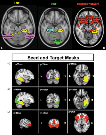
Accordingly, we tested a model proposed by Rotshtein et al. (Reference Rotshtein, Vuilleumier, Winston, Driver and Dolan2007), supported, as well, by multiple previous psychophysical, fMRI and electroencephalographic studies (see Ruiz-Soler & Beltran, Reference Ruiz-Soler and Beltran2006 for a review), which posits convergence of LSF and HSF processing in R FFA from an earlier split in the processing stream. We examine a model of convergence of LSF and HSF processing at the R FFA, wherein LSF information feeds forward bilaterally via an anterior occipital face area to R FFA, and HSF information feeds forward via a posterior occipital face area to left inferior temporal gyrus (ITG), and then to R FFA (Rotshtein et al. Reference Rotshtein, Vuilleumier, Winston, Driver and Dolan2007). This model of face processing, provided a structure for testing our hypotheses regarding visual processing abnormalities in AN and BDD (Fig. 1).
(a) Model of face-processing regions: LSF, low spatial frequency; HSF, high spatial frequency; FFA, fusiform face area; ITG, inferior temporal gyrus. Regions specialized for LSF processing in purple and HSF processing in green and aqua; regions specialized for both LSF and HSF in yellow (adapted from Rotshtein et al. Reference Rotshtein, Vuilleumier, Winston, Driver and Dolan2007). Right-most panel shows regions within the salience network, in red. (b) Seed and target masks for functional connectivity analyses. (a), (b) FFA mask shown in yellow, bilateral anterior occipital face area seeds in purple, right posterior occipital face area seed in green, and left ITG seed in aqua. (c) Salience network mask, in red.

Based on previous fMRI studies in BDD (Feusner et al. Reference Feusner, Townsend, Bystritsky and Bookheimer2007, Reference Feusner, Moody, Townsend, McKinley, Hembacher, Moller and Bookheimer2010, Reference Feusner, Hembacher, Moller and Moody2011), and assuming similar visual processing abnormalities in AN as in BDD, we hypothesized that BDD and AN would have reduced functional connectivity compared to HC within an occipito-temporal face network, specifically between the anterior occipital face area (right and left) and R FFA for LSF stimuli. For HSF stimuli, because previous studies in BDD have not found abnormal activation for HSF images in visual systems, we hypothesized that BDD, AN, and HC would not differ in functional connectivity between the posterior occipital face area and R FFA, nor between left ITG and R FFA. We also predicted that AN and BDD would have greater functional connectivity than HC between the R FFA and regions in the salience network for HSF, but not for LSF, due to greater salience of details of appearance-related features. The salience network is important in processing interoceptive information (Seeley et al. Reference Seeley, Menon, Schatzberg, Keller, Glover, Kenna, Reiss and Greicius2007), and a recent study found reduced salience network activity in AN (McFadden et al. Reference McFadden, Tregellas, Shott and Frank2014). In additional exploratory analyses, we investigated whole-brain connectivity using R FFA as a seed to probe for other aberrant neural circuitry.
Participants and method
Participants
We recruited 60 adults, aged 20–48 years, in three groups: BDD (n = 20), weight-restored AN (n = 20), and HC (n = 20). All participants gave written informed consent for the UCLA Institutional Review Board-approved protocol.
Clinical evaluations were performed for BDD participants by J.D.F. and for AN participants by C.B. or M.S. We screened participants for primary or co-morbid diagnoses with the Mini-International Neuropsychiatric Interview (MINI v. 6.0; Sheehan et al. Reference Sheehan, Lecrubier, Sheehan, Amorim, Janavs, Weiller, Hergueta, Baker and Dunbar1998). We assessed for severity of psychiatric symptoms using the Hamilton Anxiety Rating Scale (HAMA; Hamilton, Reference Hamilton1959), Brown Assessment of Beliefs Scale (BABS; Eisen et al. Reference Eisen, Phillips, Baer, Beer, Atala and Rasmussen1998), and the Montgomery–Åsberg Depression Rating Scale (MADRS; Montgomery & Asberg, Reference Montgomery and Asberg1979). In addition, BDD participants received the BDD version of the Yale–Brown Obsessive–Compulsive Scale (BDD-YBOCS; Phillips et al. Reference Phillips, Hollander, Rasmussen, Aronowitz, DeCaria and Goodman1997), and AN participants received a modified version of the Eating Disorder Evaluation Edition 16.0D (EDE; Fairburn et al. Reference Fairburn, Cooper and O'Connor2008). All of our AN participants were restricting type and were weight-restored.
All participants had normal/corrected visual acuity, as tested with a Snellen eye chart, and all were right-handed, per the Edinburgh Handedness Inventory (Oldfield, Reference Oldfield1971). One BDD and one AN participant was taking psychiatric medications and the rest were unmedicated.
AN and BDD inclusion/exclusion criteria
BDD individuals were eligible who met DSM-IV criteria using the BDD Diagnostic Module (Phillips et al. Reference Phillips, Atala and Pope1995) and had a score of ⩾20 on the BDD-YBOCS. Any screened BDD individual positive for a lifetime diagnosis of AN or bulimia nervosa was excluded. Individuals with AN were eligible if they met DSM-IV criteria (excluding the requirement of amenorrhea) and had a body mass index (BMI) of ⩾18.5. We chose to study only weight-restored AN individuals to avoid confounds of starvation on brain activity. Six of the 20 AN participants and three of the 20 BDD participants were in treatment at the time of the study. Individuals with co-morbid AN and BDD were excluded, as were those with concurrent Axis I disorders besides major depression, dysthymia, panic disorder, social phobia, generalized anxiety disorder. These diagnoses were allowed given their frequency in BDD and AN, and our wish to recruit a representative clinical sample. Individuals were excluded if they had current substance use disorders.
HC exclusion criteria
HC participants could not meet any criteria for Axis I disorders, including substance use disorders.
Ethical standards
The authors assert that all procedures contributing to this work comply with the ethical standards of the relevant national and institutional committees on human experimentation and with the Helsinki Declaration of 1975, as revised in 2008.
Faces: stimuli and task
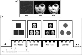
Visual stimuli included neutral-expression faces, spatial frequency-filtered to include only HSF and LSF as previously described (Iidaka et al. Reference Iidaka, Yamashita, Kashikura and Yonekura2004). The forced-choice, two-sample task consisted of a target face and two selection faces that appeared simultaneously on the screen for 4 s (Fig. 2). Participants pressed the right or left button to choose the selection face that was identical to the target face, and after the participant responded, the fixation cross appeared during the inter-stimulus interval of 0.5 s. We told participants to respond as quickly as possible. The control task consisted of matching ovals and circles. (Normal spatial frequency face images were also shown but were not part of this analysis.) A total of 48 sets of face stimuli were presented in alternating blocks of HSF, LSF, NSF, or control, with four sets of images per block. The order of stimuli was counterbalanced across participants using a Latin squares design.
(a) Example face stimuli. HSF, High spatial frequency; LSF, low spatial frequency; NSF, normal spatial frequency. (b) Functional magnetic resonance imaging experimental design: matching task in blocks of HSF, LSF, and NSF faces, alternating with control task of circles and ovals.

MRI acquisition protocol
Scanning was performed on a 3-T Siemens TRIO at the Ahmanson-Lovelace Brain Mapping Center at University of California, Los Angeles. We collected blood oxygenation level dependent (BOLD) functional echo-planar images (EPI) using: repetition time (TR) 2.5 s; echo time (TE) 25 ms; flip angle, 80°; voxel dimensions, 3 × 3 × 3 mm; 0.75 mm gap; field-of-view, 192 mm; matrix, 64 × 64; 133 measurements; 32 slices. Data collected during the first three TRs were discarded for T1 equilibration. A matched-bandwidth T2 structural scan, co-planar to the EPIs, was acquired: TR of 5 s; TE, 34 ms; flip angle, 90°; 32 slices; voxel dimensions, 1.5 × 1.5 × 3 mm, field-of-view, 192 mm; and matrix, 128 × 128. We obtained a high-resolution MPRAGE T1-weighted image to provide detailed brain anatomy with: TR 1.9 s, TE 2.26 ms, and voxel dimensions 1 × 1 × 1 mm.
MRI data processing
MRI data were processed using FEAT (FMRI Expert Analysis Tool) version 6.0, part of FSL (FMRIB's Software Library; http://www.fmrib.ox.ac.uk/fsl). Image processing steps included motion correction, skull-stripping, spatial smoothing, normalization, and temporal filtering. Registration to standard space was carried out using a three-step transformation to register each functional image to a co-planar structural image, then to MPRAGE, and finally to Montreal Neurological Institute (MNI) standard brain space. We visually inspected all images for proper registration as well as relative and absolute motion to identify motion spikes. There was not a significant effect of motion among groups as measured by DVARS (F 57,2 = 1.41, p = 0.25) where DVARS is D: temporal Derivative of time courses; VARS: root mean square VARiance over voxels, a measure of the change rate of signal across the brain. Additional details of motion calculation and exclusions are given in the Supplementary material.
Face-processing model: seeds and targets
The model of face processing which underlies our analysis is diagrammed in Fig. 1a . We tested LSF and HSF streams separately: functional connectivity between left and right anterior occipital face areas and R FFA for LSF faces; and between right posterior occipital face area and R FFA, and left ITG and R FFA, for HSF faces. Functional seeds for each group were derived from data during face processing, using the leave-one-subject-out (LOSO) procedure to reduce bias due to non-independence (Esterman et al. Reference Esterman, Tamber-Rosenau, Chiu and Yantis2010). We created separate group LOSO masks of right and left anterior occipital face area, right posterior occipital face area, left ITG, and right R FFA, to functionally localize face visual processing areas (Fig. 1b ). The salience network mask was derived from the Harvard Oxford Atlas, comprised of orbitofrontal, anterior cingulate, and insula masks thresholded at 50% (Seeley et al. Reference Seeley, Menon, Schatzberg, Keller, Glover, Kenna, Reiss and Greicius2007). (See Supplementary method for LOSO analysis.)
Statistical analyses
We used one-way ANOVAs and χ 2 tests to assess differences among groups for our demographic, clinical, and task performance measures.
We calculated functional connectivity using psychophysiological interaction (PPI) analyses (Friston et al. Reference Friston, Buechel, Fink, Morris, Rolls and Dolan1997). PPI is a hypothesis-driven method of analyzing functional connectivity of brain regions during a specific task, based upon correlation of the time-series between ‘seed’ and ‘target’ brain regions. It is used to test for interactions between a ‘psychological’ state (in this experiment, matching human faces) and a ‘physiological’ state (coupling of activation between seed and target time series).
A first-level PPI analysis of faces stimuli contrasted to control task was obtained for all participants for each set of LSF and HSF stimuli. Then first-level PPI results, specific for each functional seed and spatial frequency, were carried up to group-level analyses specific for each target region. First-level contrast Z statistic images were entered in between-group analyses using each subject as a random factor. All Z statistic images were thresholded using clusters determined by Z > 2.0, and a corrected-cluster significance threshold of p= 0.05 (Worsley, Reference Worsley, Jezzard, Matthews and Smith2001), with the exception of two exploratory analyses that used Z > 1.7, as indicated, and a cluster-corrected significance threshold of p = 0.05).
For LSF processing, we evaluated connectivity between the left and right anterior occipital face area and R FFA. For HSF processing, we evaluated connectivity between right posterior occipital face area and R FFA, and between left ITG and R FFA. For salience network connectivity, we evaluated connectivity between R FFA and a salience-network target mask. For exploratory analyses we tested connectivity between R FFA and all other voxels in the brain. We performed one-way between-group ANOVAs for each seed-target pair hypothesis, followed by t tests for AN v. HC and BDD v. HC to test group specificity. As post-hoc exploratory analyses, we also compared AN v. BDD for the aforementioned connectivity.
Results
Demographics and behavioral results
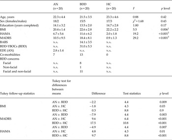
Results of one-way ANOVAs revealed that BDD, AN, and HC groups did not differ in mean age or years of education. A χ 2 test showed no differences in sex between groups. Post-hoc Tukey tests were performed to determine the direction and significance of the between-group comparisons. All demographics and behavioral comparisons are summarized in Table 1. There were significant differences between groups for BMI, HAMA, and MADRS: BMI was significantly lower in AN compared to BDD and HC, HAMA was significantly lower in HC compared to AN and BDD, and in AN compared to BDD, and MADRS was significantly lower for HC than for AN and BDD, and for AN compared to BDD. ANOVA results: F 57,2 = 5.5, p = 0.006; F 57,2 = 19.2, p < 0.001; F 57,2 = 29.2, p < 0.001. Tukey tests: BMI (AN < BDD, t 18 = 4.4, p = 0.009; AN < HC, t 18 = 3.7, p = 0.03; BDD > HC, t 18 = 0.68, p = 0.88); HAMA (AN < BDD, t 18 = 4.4, p = 0.007; AN > HC, t 18 = 4.3, p = 0.01; BDD > HC, t 18 = 8.76, p < 0.001); MADRS (AN < BDD, t 18 = 4.9, p = 0.003; AN > HC, t 18 = 5.9, p < 0.001; BDD > HC, t 18 = 10.8, p < 0.001).
Demographics and psychometrics

AN, Anorexia nervosa; BDD, body dysmorphic disorder; HC, healthy controls; BMI, body mass index; HAMA, Hamilton Anxiety Rating Scale; MADRS, Montgomery–Asberg Depression Rating Scale; BABS, Brown Assessment of Beliefs Scale; BDD-YBOCS, BDD version of the Yale-Brown Obsessive Compulsive Scale; EDE, Eating Disorder Evaluation; n.a., not applicable.
Co-morbidities and types of appearance concerns in BDD (facial and non-facial) are also listed in Table 1. A complete list of co-morbidities can be found in Supplementary Table S4 (see also Supplementary method). There were no significant group differences in response times or accuracy rates during the matching tasks (see Supplementary Figs S3 and S4).
PPI analysis results
R FFA connectivity with occipital-temporal face network
LSF connectivity with R FFA
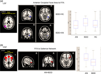
The ANOVA results demonstrated significant differences between groups for LSF connectivity. Contrary to our hypotheses, comparisons between groups found greater connectivity between left anterior occipital face area and R FFA for BDD v. HC (Fig. 3a , left). Also contrary to our hypotheses, there were no significant differences in connectivity between right anterior occipital face area and the R FFA for either AN v. HC, or BDD v. HC. There was greater connectivity between left anterior occipital face area and R FFA for BDD v. AN (Fig. 3a , right). This cluster was examined as a region of interest to explicate group differences, with the t statistic group averages shown in the box plots. (See Supplementary method: ‘Method for examining direction of connectivity differences’ for details of the method.)
(a) Regions within fusiform face area (FFA) target (blue) with significantly greater connectivity from the left anterior occipital face area seed for low spatial frequency (LSF) faces, for body dysmorphic disorder (BDD) compared to healthy controls (HC), left side, and BDD compared to anorexia nervosa (AN), right side, (Z > 2.0, P = 0.05, corrected). Box plots indicate the direction of the connectivity, showing the average t statistic for each group for the cluster. (b) Regions within the salience network target (red) with significantly greater connectivity from the FFA seed for high spatial frequency (HSF) faces, for AN compared to BDD (Z > 2.0, p = 0.05, corrected).

In addition, we reran all of our significant between-group results with sex as a covariate, and found that the results were virtually identical (see Supplementary Fig. S7).
HSF connectivity with R FFA. Consistent with our hypotheses, for HSF connectivity the ANOVA revealed no significant differences between groups for connectivity between right posterior occipital face area and R FFA or for left ITG and R FFA.
R FFA connectivity with the salience network
LSF connectivity with the salience network
Consistent with our hypotheses, for LSF stimuli the ANOVA revealed no significant differences between groups for connectivity between R FFA and the salience network.
HSF connectivity with the salience network
For HSF stimuli, the ANOVA resulted in significant differences between groups. Contrary to our hypotheses, follow-up comparisons found no differences for AN v. HC or BDD v. HC for connectivity between R FFA and the salience network; however, there was greater connectivity for AN v. BDD in salience network regions, including right insula and right orbitofrontal cortex (Fig. 3b ).
R FFA connectivity to whole brain – exploratory
LSF connectivity – exploratory
BDD < HC and AN < HC
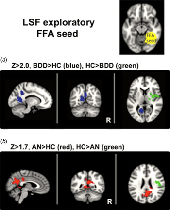
There was lower LSF connectivity for BDD v. HC between R FFA and right insula, putamen, thalamus, and central opercular cortex (Fig. 4a ). For AN < HC, there were no significant differences at Z > 2.0, but reducing the statistical threshold (Z > 1.7, p< 0.05, corrected) revealed lower connectivity for AN between R FFA and right insula and central opercular cortex (Fig. 4b ).
Exploratory results for connectivity between the fusiform face area (FFA) seed and the rest of the brain for low spatial frequency (LSF) faces. (a) Body dysmorphic disorder (BDD) > healthy controls (HC) in blue; HC > BDD in green; Z > 2.0, p = 0.05, corrected. (b) anorexia nervosa (AN) > HC in red; HC > AN in green; Z > 1.7, p = 0.05, corrected.

BDD > HC and AN > HC
There was greater LSF connectivity for BDD v. HC between R FFA and right and left precuneus, right posterior cingulate cortex (PCC), and left lingual gyrus (Fig. 4a ). For AN > HC, there were no significant differences at Z > 2.0, but reducing the statistical threshold (Z > 1.7, P < 0.05, corrected) revealed greater connectivity for AN between R FFA and right and left precuneus, left PCC, and right thalamus (Fig. 4b ). Additional details of LSF significant results can be found in Supplementary Table S1 and Fig. S1.
HSF connectivity–exploratory
HSF significant results for all groups are shown in Supplementary Fig. S2.
Post-hoc regression analyses
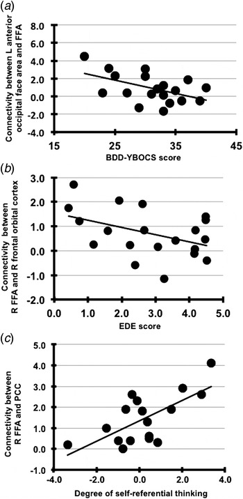
To follow up on the significant between-groups results, we performed post-hoc regression analyses on functional connectivity and the main symptom measure for each of the AN and BDD groups, using Bonferroni-adjusted alpha levels as a correction for multiple comparisons. The relationship between LSF connectivity between the left anterior occipital face area and the R FFA and BDD-YBOCS scores (see Fig. 5a ), showed that higher connectivity strength showed a trend for association with lower BDD-YBOCS scores (R 2 = 0.26, F 1,19 = 5.8, P = 0.027), not surviving Bonferroni correction (α = 0.05/3 = 0.017). For AN, we examined the relationship between degree of HSF connectivity between the R FFA and the orbitofrontal cortex (in the salience network), and clinical symptom severity (EDE) scores for AN (see Fig. 5b ). Higher connectivity strength showed a trend for association with lower scores (R 2 = 0.19, F 1,19 = 4.14, P = 0.057), although not surviving Bonferroni correction (α = 0.05/5 = 0.01).
(a) Low spatial frequency (LSF) functional connectivity between left (L) anterior occipital face area and fusiform face area (FFA) trend for association with body dysmorphic disorder–Yale-Brown Obsessive Compulsive Scale (BDD-YBOCS) score for BDD. (b) High spatial frequency functional connectivity between right (R) FFA and R frontal orbital cortex trend for association with Eating Disorder Evaluation (EDE). (c) LSF functional connectivity between R FFA and posterior cingulate cortex (PCC) is associated with degree of self-referential thinking for anorexia nervosa. Note: values for self-referential thinking scores are demeaned.

To understand how abnormal functional connectivity patterns in face-processing systems may relate to subjective experience in AN and BDD participants when viewing facial images, we performed additional regression analyses between the degree of functional connectivity and scores on post-scan questionnaires that quantified the degree to which each face viewed in the scanner triggered thoughts about their own appearance (see Supplementary Table S3 and Supplementary method for linear regressions). For AN, we found that greater connectivity for LSF faces between R FFA and PCC was significantly associated with higher self-referential thinking scores (R 2 = 0.46, F 1,16 = 12.7, p = 0.003) (Fig. 5c ), passing Bonferroni correction (α = 0.05/3 = 0.017). For BDD, there was no significant relationship between connectivity and the subjective experience scores.
Discussion
To our knowledge, our results provide the first evidence of aberrant functional connectivity in BDD within an occipito-temporal face-processing network, specifically for faces that contain configural and holistic elements. This pattern was not observed in AN, although there was a remarkably similar pattern of functional connectivity for these image types in AN and BDD between the R FFA and higher-order regions including insula, central opercular cortex, and PCC/precuneus. For high detail images, BDD and AN showed differences from each other and HC in functional connectivity patterns between R FFA and higher-order regions, but not in occipito-temporal networks. This profile suggests BDD and AN have similar LSF processing-stream abnormalities in higher-order systems; however, LSF connectivity within occipito-temporal fac- processing networks is affected in BDD, but not AN.
Although we predicted lower connectivity for LSF stimuli for both AN and BDD groups, we found greater connectivity between right anterior occipital face area and R FFA, in the BDD group only. We had predicted decreased connectivity based on the relative hypoactivity for LSF faces observed in a previous study examining patterns of regional fMRI activation (Feusner et al. Reference Feusner, Moody, Townsend, McKinley, Hembacher, Moller and Bookheimer2010). However, abnormalities in degree of regional activation do not always indicate a corresponding alteration in functional connectivity. Moreover, the regions found to be hypoactive in the previous study were earlier in the visual processing stream (V1 and V2) than those examined in the current study. It is possible therefore that increased connectivity may reflect compensatory mechanisms for abnormal processing in earlier visual regions (Bolte et al. Reference Bolte, Hubl, Feineis-Matthews, Prvulovic, Dierks and Poustka2006). This is supported by the observation that those with higher connectivity strength had lower symptom severity (BDD-YBOCS scores).
While the phenomenology of AN suggests that individuals may experience perceptual distortions, this may not be reflected in abnormalities in these occipito-temporal visual networks for faces. Instead, this may be a function of disturbances in higher-order processing of consciously perceived images. Alternatively, the absence of significant abnormalities in occipito-temporal visual network could be a reflection of the clinical observations (J. D. Feusner et al. unpublished observations) that faces do not seem to be as much a focus of attention in AN as other appearance features, such as abdomen, thighs, and hips. By contrast, facial features, including facial skin, are common appearance concerns in BDD (Phillips, Reference Phillips2005), which is consistent with the pattern of specific concerns found in our sample (see Table 1).
Another possibility is that aberrant connectivity in AN is limited to the acutely malnourished state, remitting when BMI is normalized. Consistent with this idea Kingston et al. (Reference Kingston, Szmukler, Andrewes, Tress and Desmond1996) showed that underweight AN improved their performance on the Rey Complex Figure Task (RCFT) after gaining 10% of body weight (although the RCFT task taps into both visuo-spatial and executive functioning systems; Shin et al. Reference Shin, Park, Park, Seol and Kwon2006). Moreover, a recent effective connectivity fMRI study in underweight individuals with AN found abnormal functional organization within a body-processing visual network for body visual stimuli (Suchan et al. Reference Suchan, Bauser, Busch, Schulte, Gronemeyer, Herpertz and Vocks2013). Future within-subject study designs, comparing face and non-face stimuli in AN scanned prospectively, will help discern these possibilities.
R FFA connectivity to salience network is greater for high detail images in AN than BDD
Contrary to our hypotheses, we did not find greater connectivity for HSF stimuli for either AN or BDD compared to HC. Instead, we found greater HSF connectivity for AN relative to BDD between R FFA and right fronto-insular regions. This could be related to aberrant insular and orbitofrontal activity (Frank et al. Reference Frank, Reynolds, Shott, Jappe, Yang, Tregellas and O'Reilly2012) and/or insular connectivity (Kullmann et al. Reference Kullmann, Giel, Teufel, Thiel, Zipfel and Preissl2014) that previous studies have found in AN, and could reflect abnormal integration of high-detail face information in the context of general body awareness. Why this was observed for AN only in contrast to BDD is unclear. The observation that individuals with higher connectivity between R FFA and the orbitofrontal cortex had lower severity of eating disorder symptoms could be an indication of a compensatory process. As hypothesized, compared to HC, neither the BDD nor the AN group had greater connectivity for LSF images between R FFA and the salience network.
R FFA has similar connectivity to higher-order regions in AN and BDD
Our results in AN and BDD showed similar, abnormally strong, connectivity between R FFA and precuneus and adjacent PCC for LSF stimuli, although the magnitude of the differences from HC was greater for BDD than AN (as reflected in results significant only at a lower Z threshold for AN). Connectivity in response to face stimuli has been observed between R FFA and precuneus in HC (Davies-Thompson & Andrews, Reference Davies-Thompson and Andrews2012). Moreover, the precuneus and PCC are implicated in memory, visual imagery (Cavanna & Trimble, Reference Cavanna and Trimble2006), retrieving episodic memories associated with faces (Gobbini & Haxby, Reference Gobbini and Haxby2007), and, particularly as network hubs in the default mode network (DMN) (Gusnard et al. Reference Gusnard, Akbudak, Shulman and Raichle2001; Raichle et al. Reference Raichle, MacLeod, Snyder, Powers, Gusnard and Shulman2001), self-referential thinking (Berman et al. Reference Berman, Peltier, Nee, Kross, Deldin and Jonides2011). Our results are consistent with a recent study in AN that found increased activation in DMN regions associated with self-referential thought (Cowdrey et al. Reference Cowdrey, Filippini, Park, Smith and McCabe2014). One interpretation of our results is that enhanced connectivity between face-processing regions and precuneus/PCC in AN and BDD could reflect similar tendencies across disorders to engage in self-referential thinking when viewing images of others, particularly since individuals with both disorders spend an inordinate amount of time thinking about their appearance and comparing their appearance to others. Facial stimuli could also trigger thoughts of self or thoughts of others’ perspectives of themselves (Cavanna & Trimble, Reference Cavanna and Trimble2006), or they could initiate thoughts of others as suggested by a recent AN study examining perspective-taking related to self-identity that found greater activation in precuneus for thoughts of friends and their appearance (McAdams & Krawczyk, Reference McAdams and Krawczyk2014). These interpretations are speculative; however, we have evidence that specific thought patterns may be related to connectivity between these regions in the current study, as the post-hoc regression analysis revealed that the degree to which the facial stimuli triggered self-referential thinking is associated with activity in precuneous and PCC for AN participants. It is unclear at present why this relationship was not observed in BDD participants.
Regions of decreased LSF connectivity for both BDD and AN groups compared to HC were found in insula and in central opercular cortex. These results are in line with recent studies that report aberrant insula and salience network activity in women with AN (Kim et al. Reference Kim, Ku, Lee, Lee and Jung2012; Kullmann et al. Reference Kullmann, Giel, Teufel, Thiel, Zipfel and Preissl2014; McFadden et al. Reference McFadden, Tregellas, Shott and Frank2014). The insula is important for recognition and response to salient stimuli and for processing interoceptive information. The decrease in connectivity between the R FFA and the insula could disrupt interoceptive functions and result in misinterpretation of interoceptive information, contributing to disturbances in integration of configural and holistic information about face with general body awareness, ultimately exacerbating AN and BDD symptoms.
Clinical significance
The findings have clinical relevance. Distortions of perception may contribute to poor insight in BDD, limiting engagement in treatment (Eisen et al. Reference Eisen, Phillips, Coles and Rasmussen2004), and unremitted perceptual disturbance may foreshadow increased risk of relapse in AN (Keel et al. Reference Keel, Dorer, Franko, Jackson and Herzog2005). Aberrant processing in occipito-temporal face networks of configural and holistic visual elements in BDD could be associated with a reduced ability to contextualize details, which, in turn, could maintain perceptual distortions expressed clinically as a conviction of flaws and imperfections.
The results also have implications for understanding phenotypic overlaps in AN and BDD. Given evidence for similar abnormal functional connectivity for face processing in higher-order integrative and emotional systems, this abnormality may explain heightened self-referential thinking across body image disorders. In contrast, the existence of distinct phenotypes of abnormal connectivity patterns within visual networks may shed light on syndrome-specific clinical-behavioral variations. In this regard, the combined results for LSF and HSF point to abnormalities that could inform the development of innovative perceptual retraining treatments. For BDD, therapies using tasks that mediate configural and holistic elements of a stimulus by engaging the dorsal visual stream (Nan et al. Reference Nan, Wan, Lou, Vai and Rosa2013), may hold benefit by normalizing LSF connectivity if it compensates for deficient activation in the visual cortical areas that normally feed forward LSF information to the dorsal stream. Likewise, for AN, there may be utility in perceptual retraining that involves face and body awareness/integration (Vocks et al. Reference Vocks, Busch, Schulte, Gronermeyer, Herpertz and Suchan2010). For both AN and BDD patients, empirical study of psychotherapy techniques that reduce self-referential thinking, such as those used in existing psychotherapies – Acceptance and Commitment Therapy (Hayes, Reference Hayes2005) being one recent example, mindfulness therapy being another, has at least theoretical justification.
Limitations
There are several limitations. We did not have functional localizer data available to optimally identify individual visual seed regions; instead we created functional seeds using the LOSO method which reduces statistical non-independence bias (Esterman et al. Reference Esterman, Tamber-Rosenau, Chiu and Yantis2010). Results from PPI analyses do not specify directionality, preventing conclusions about direction of information flow. We chose face stimuli due to prior knowledge of visual processing with respect to spatial frequencies in HC and in BDD; however, face stimuli may be more salient and symptom-relevant in BDD compared to AN. A comparison of BDD subgroups with face v. non-face concerns could not be explored because only one BDD patient had only non-face concerns. Future connectivity studies should examine responses to body stimuli in body processing networks. Co-morbidities differed between AN and BDD groups, which could have affected results. We did not have data on the length of weight restoration for all AN participants (for the three participants for whom we have data, the length was 5 months, 2 years, and 3 years). This information is difficult to obtain because typically part of the treatment for those with AN is to not weigh themselves, and often their therapists or dieticians do not tell them their weight.
Conclusions
We found similar abnormal functional connectivity for face processing in higher-order systems in BDD and AN, but distinct abnormal connectivity patterns within occipito-temporal visual networks. The observed abnormalities in functional connectivity for configural and holistic visual information within face-processing networks in individuals with BDD may contribute to distorted perception due to formation of an atypical visual template. For individuals with AN, the absence of similar abnormalities in connectivity in earlier face-processing networks may be due to a lesser degree of emotional relevance of faces, or perhaps to attenuation of aberrant connectivity with normalization of weight. Findings have implications for understanding relationships between these disorders and the pathophysiology underlying perceptual distortions.
Supplementary material
For supplementary material accompanying this paper visit http://dx.doi.org/10.1017/S0033291715001397.
Acknowledgements
We thank all the individuals who participated in this study, Nathan Hutcheson and Sarah Madsen for helpful comments on the manuscript, and Giulia Salgari for statistical analysis of ratings scores. Dr Strober received support from the Resnick endowed Chair in Eating Disorders. Funding sources are: Dr Feusner (R01MH093535 and 5K23MH079212) and Dr Bohon (2T32MH073517).
Declaration of Interest
None.