Isolation of the subclavian artery is a rare vascular anomaly, with the subclavian artery separated from the aortic arch and arising from a ductus or ligamentum arteriosus connected to the same-sided pulmonary artery.Reference Edwards1 This anomaly can cause a congenital pulmonary or subclavian steal with perfusion of the arm or lung via the vertebrobasilar system. Isolation of the subclavian artery is seen in association with DiGeorge syndrome and with conotruncal cardiac defects.Reference Momma2–Reference Jones, Garabedian and Grifka4
We report a case of an isolated right subclavian artery arising from a right-sided ductus arteriosus and supplied retrograde via the right vertebral artery, creating a congenital pulmonary and subclavian steal phenomenon. To our knowledge, only three prior cases of isolation of the right subclavian artery without associated congenital cardiac disease have been described.Reference Brill, Peyster, Keller and Galtman5–Reference Garti and Aygen7
Case report
The patient was first evaluated at 8 months of age for a murmur. She was a fraternal twin and was significantly smaller than her brother, plotting at the 5th percentile for both height and weight. Blood pressures were equal in all four extremities. A grade II/VI continuous murmur was heard along the left sternal border with radiation to the left infraclavicular region and to the back. The transthoracic echocardiography showed a moderate-sized patent ductus arteriosus with continuous left-to-right shunt and associated left atrial dilation. The decision was made to proceed with percutaneous occlusion to eliminate the left-to-right shunt and to minimise long-term endocarditis risk.
Cardiac catheterisation showed a mixed venous saturation of 64%, with a step up to 86% in the right pulmonary artery but no step up into the left pulmonary artery. Distal pulmonary artery pressures were normal with a 24-millimetre mercury gradient from the left and a 16-millimetre mercury gradient from the right to the main pulmonary artery. No additional gradients were present.
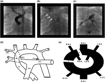
An initial angiogram showed a trivial left-sided patent ductus arteriosus. The aortic arch was leftward with a normal origin of the right carotid, left carotid, and left subclavian arteries. There was no right subclavian artery arising from the aorta (Figs 1a and 2). There was late, retrograde filling of the right vertebral artery with subsequent filling of the right subclavian artery and right pulmonary artery via a large right-sided patent ductus arteriosus (Figs 1b and 2). The angiography confirmed the diagnosis of an isolated right subclavian artery and also identified discrete stenosis of both branch pulmonary arteries (Figs 1c and 2). The procedure was aborted to obtain further imaging of the brachiocephalic and cerebral vasculature in anticipation of likely surgical intervention.
(a) Aortogram shows a left-sided aortic arch with the right carotid artery arising as the first head vessel. There is no right-sided subclavian artery seen. (b) Late phase of a subsequent carotid artery injection shows faint retrograde fill of the right vertebral artery (arrows) with fill of the right pulmonary artery using a right-sided ductus arteriosus. The right subclavian is not well seen as contrast preferentially fills the lower resistance pulmonary circulation. (c) Contrast injection in the main pulmonary artery shows discrete right pulmonary artery narrowing at the insertion of the right-sided ductus arteriosus. (d) Schematic of the isolated right subclavian artery and associated major arteries Ao represents aorta; LPA and RPA represent left and right pulmonary artery; MPA represents main pulmonary artery; PDA represents patent ductus arteriosus; RSA and LSA represent right and left subclavian artery; RCC and LCC represent right and left common carotid; VA represents vertebral artery. (e) Edward's hypothetical aortic arch showing the embryology of an isolated right subclavian artery. Asterisk (*) represents involution of the right dorsal aorta and right fourth arch. RSA and LSA represent right and left subclavian artery; RCC and LCC represent right and left common carotid; RDA and LDA represent right and left ductus arteriosus; ASAo and DSAo represent ascending and descending aorta.

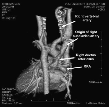
Three-dimensional reconstruction of computed tomography angiography from a posterior view shows the right-sided ductus arteriosus arising from the right pulmonary artery (RPA) and supplying the isolated right subclavian artery.

Subsequent computed tomography angiography confirmed the diagnosis of an isolated right subclavian artery connected to a right vertebral artery with an additional connection to the right pulmonary artery via a right-sided patent ductus arteriosus. The anatomy resulted in a steal phenomenon from the cerebral circulation to the right subclavian artery and ultimately into the right pulmonary artery. The patient subsequently underwent successful bilateral ductal ligation, implantation of her right subclavian artery into the ascending aorta, and right pulmonary arterioplasty. There were no intra-operative or post-operative complications. She was discharged home on post-operative day 3. At 1-year follow-up, blood pressures were equal in both upper extremities and her right radial pulse was easily palpable. Subsequent chromosomal microarray confirmed a diagnosis of DiGeorge syndrome with a microdeletion of chromosome 22q11.
Discussion
In summary, we present a patient with DiGeorge syndrome, bilateral patent ductus arteriosus, and associated isolation of the right subclavian artery. The patient presented with a murmur but did not demonstrate abnormal right extremity blood pressure. The anomalous subclavian artery was diagnosed at the time of catheterisation.
An isolated subclavian artery is the rarest described arch anomaly.Reference Stewart, Kincaid and Titus8 The isolated subclavian artery is usually connected to the pulmonary artery by an ipsilateral ligamentum or patent ductus arteriosus. There is no vascular ring as the ductus is ipsilateral to the isolated subclavian artery and is not attached to the aorta. Development requires dual ipsilateral breaks in Edwards's hypothetical aortic arch.Reference Edwards1 In our case, involution of the right dorsal aorta and right fourth arch resulted in migration of the right seventh intersegmental (subclavian) artery to the right sixth (ductal) arch. Most described cases of isolated subclavian artery involve isolation of the left subclavian artery associated with a right aortic arch or with conotruncal defects including tetralogy of Fallot, double-outlet right ventricle, interrupted aortic arch, and D-transposition of the great arteries.Reference Momma2–Reference Jones, Garabedian and Grifka4 Isolation of the right subclavian artery is extremely rare, particularly in the absence of other intracardiac defects.Reference Brill, Peyster, Keller and Galtman5–Reference Garti and Aygen7, Reference Nath9
A pulmonary steal was present in our patient with the right lung perfused via retrograde flow from the vertebrobasilar system, stealing from the circle of Willis. NathReference Nath9 described a similar patient presenting at 18 months of age with secondary pulmonary hypertension. This patient underwent surgical repair but died at the age of 3 years, with the autopsy showing grade IV pulmonary vascular disease. Thus, the primary aim of intervention is to limit pulmonary overcirculation.
Prior reports have documented successful resolution of pulmonary overcirculation with percutaneous ductal occlusion.Reference Jones, Garabedian and Grifka4 However, this approach is associated with a residual subclavian steal phenomenon from the vertebral artery. The long-term implications of a subclavian steal are not well known. Symptoms are well described in the adult population, and include cerebral insufficiency with focal deficits, as well as dizziness, headache, and syncope. Symptoms related to underperfusion of the arm are also common and include paresthesias, weakness, and fatigue with activity.Reference Heidrich and Bayer10 In an interesting case report, Brill et alReference Brill, Peyster, Keller and Galtman5 attributed cerebral underdevelopment to embryonic subclavian steal in a patient with an isolated right subclavian artery. For these reasons, we elected to proceed with complete surgical repair so as to normalise the patient's circulation with resolution of both the pulmonary and subclavian steal phenomena.
In conclusion, this case represents the fourth description of an isolated right-sided subclavian artery in a patient with otherwise normal intracardiac anatomy. This anomaly has not been previously shown with computed tomography angiography and three-dimensional reconstruction as the most recent previous description was published in 1987. Circulation was normalised with re-implantation of the subclavian artery into the aorta and ductal ligation. The patient continues to do well at 1-year follow-up.