The incidence of isolated patent ductus arteriosus in healthy individuals after the neonatal period is 0.5–1%.Reference Steinberger, Moller, Berry and Sinaiko1 Patent ductus arteriosus in full-term infants is abnormal and related to significant structural abnormalities.Reference Nora2 Usually it is silent, displaying no symptoms, and is of no prognostic significance. Symptomatic ductus accounts for approximately 5–10% of all types of congenital cardiac diseases.Reference Schneider and Moore3 However, the indications for closing the ductus are unclear, even if they are reasoned in term infants as the prevention of heart failure, pulmonary hypertension, and bacterial endocarditis.Reference Morgan, Gray, Miller and Oldershaw4–Reference Espino-Vela, Cardenas and Cruz6 In Sweden, with a population of around nine million people, two out of nearly three million deaths during the period 1960–1993 were attributed to patent ductus arteriosus and infective endarteritis. In both cases, the patent ductus arteriosus was haemodynamically significant. Thus, prevention of endarteritis is not an indication by its own for routine closure of patent ductus arteriosus.Reference Thilén and Alström-Olsson7 A quite widely accepted indication for closing open ductus in full-term infants is the existence of a continuous heart murmur associated with ductal flow documented by echocardiography, even if the existence of continuous flow murmur has been suggested to be due to ductal jet direction and not the amount of flow.Reference Bennhagen and Benson8 In some centres, the ductus is closed with a device always after finding patent ductus arteriosus in an ultrasound study. In infants born at a gestational age of less then 29 weeks, the incidence of patent ductus arteriosus is 40–55% because of incomplete maturation of the ductal tissue.Reference Reller, Rice and McDonald9 Without question, a significant number of premature babies with respiratory difficulties are aggravated by ductus and need treatment. The indication for treatment in pre-term infants is to prevent pulmonary overperfusion and systemic underperfusion.Reference Jim, Chiu and Chen10, Reference Shimada, Kasai, Konishi and Fujiwara11
A pulmonary-to-systemic flow ratio over 1.5 is used as an indication for closing a ventricular septal defect. Using the same indication for closing an open duct as that used in other shunt lesions would be logical. We previously documented a significant correlation in lambs between the percentage of colour pixels in a pulmonary artery longitudinal cross-section, representing ductal flow, and the pulmonary-to-systemic flow ratio measured with electromagnetic flow transducers (curvilinear r2 = 0.84).Reference Harling, Jansson, Gudmundsson and Pesonen12 When the shunt was 1.4:1 or more, eight out of nine lambs had 50% or more of the region of interest covered with colour pixels – sensitivity 80%, specificity 100%. A quantitative colour Doppler examination would be an easy and harmless non-invasive method to quantify the shunt and useful in the treatment decision. In the present study, we aimed to clarify whether the method can be used to determine a clinically significant ductal shunt in children.
Materials and methods
The regional Ethics Board in Lund, Sweden, reviewed the study and found no ethical impediment.
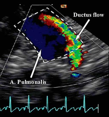
We retrospectively evaluated the echocardiographic studies of 51 infants and children scheduled for device closure of patent ductus arteriosus between 1998 and 2007. The quality of the echocardiograms performed by four different observers during the years was not always ideal. The images included in the study had to contain the pulmonary artery including the valves and ductal opening close to the level of the origins of the pulmonary artery branches visualised from a high left parasternal longitudinal short-axis view close to the plane of the pulmonary artery branch origins from the main pulmonary artery. This compelled us to limit the study series to 20 good-quality recordings best fulfilling the criteria above (Fig 1). The median age of the 20 children included in the study was 2 years (range 0.6–15 years) and the median weight was 12 kilograms (range 7–48 kilograms).
Region of interest lined out in the pulmonary arterial longitudinal cross-section.

An Acuson Sequoia™ C256 echocardiography system (Acuson Mountain View, CA, USA) equipped with a 5- or 7-megahertz transducer was used to detect the pulmonary artery and ductal flow. The echocardiographic studies were performed just before device closure of patent ductus arteriosus, with the patient in general anaesthesia and pulmonary-to-systemic flow ratio measured under the heart catheterisation. The calculation of pulmonary-to-systemic flow ratio was based on blood saturations in the superior cava, left pulmonary artery branch, and the aorta.
Closure of the duct was performed with the Cook detachable coils or the Amplatzer Duct Occluder. The colour pixel analyses were made from cine loops including the main stem of the pulmonary artery in longitudinal sections. The region of interest was outlined to include the ductal orifice and the main pulmonary artery from the pulmonary valve to the pulmonary bifurcation (Fig 1). By a computer analysis, colour pixels were calculated in this area. Electrocardiogram was present in the images that were used to find a late diastolic frame closest to the QRS complex. A frame in late diastole was selected to obtain pure ductal flow without a right ventricular contribution of flow to the pulmonary artery. A pulmonary-to-systemic flow ratio between 1:1 and 2.3:1 was found in the 20 children who fulfilled the image criteria.
Colour Doppler scanning
Images were obtained by four different observers, which generated a variation in echocardiographic settings as colour scale maximum of 55–130 centimetres per second, colour Doppler frequency of 2.5–5 megahertz, colour Doppler gain of 43–61 units given by the ultrasound machine, and frame rate of 18–75 frames per second. The off-line analysis was made on exported bitmap image files in Digital Imaging and Communications in Medicine format from cine loops saved on magneto-optical disk.
Computer analysis of colour Doppler images
An image frame in diastole closest to the beginning of the R-peak in the Electrocardiogram was selected. The analysis was made in a custom-designed program written in matrix laboratory (MATLAB®, The MathWorks, Natick, MA, USA). Colour pixels in the colour Doppler image represented the direction of flow – red towards and blue away from the transducer – or turbulence – for example, a large variance of the flow velocity estimate, represented by green. Anatomical information was shown in grayscale where no flow is detected. In the bitmap format, the hue in a pixel was represented by three numbers corresponding to the base colours, that is red, green, and blue, which make up the colour in question. If the pixel showed grayscale information only, the three numbers were equal. The images were read into matrix laboratory program, separating the image into three matrices, each containing one of the base colours.
We first calculated the number of pixels that indicated flow towards or away from the transducer and their mean velocity within the pulmonary artery in longitudinal sections or turbulence. The number of pixels inside the pulmonary artery in longitudinal sections with no colour, or non-colour pixels, was also registered. The ratio between colour and non-colour pixels in the pulmonary artery in longitudinal sections was calculated. The number of pixels of each colour were added together and divided by the area of the pulmonary artery in longitudinal sections to calculate the total percentage of colour pixels in the pulmonary artery in longitudinal sections. We calculated the total sum of green pixels, separated out the other two base colours, and calculated the percentage of total green pixels. The relative volume of the jet was calculated by multiplying the size of the ductus, that is, ductus lumen calculated as pi × radius2, with the mean velocity of the pixels divided by body surface area. To determine the intra-observer variation, the area was measured twice at 1-day intervals. In addition, the inter-observer variation was measured.
Statistical analyses
The percentages of pixels in the area of the pulmonary artery in longitudinal sections covered by colour and non-colour were correlated with the pulmonary-to-systemic flow ratio and the diameter of the ductus measured from the angiograms using simple linear, as well as curvilinear, regression analysis. The positive predictive value was plotted to test the prediction power of the model and find the threshold for the best sensitivity and specificity for classifying a clinically significant patent ductus arteriosus. The statistical package for the Social Sciences, version 15.0 for Windows (SPSS Inc., Chicago, IL, USA), was used. Reliability analysis was used to test the significance of intra- and inter-observer variability. A p-value less than 0.05 was considered significant.
Results
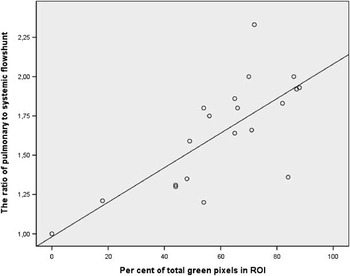
The intraclass correlation of repeated measurements of the percentage of total green pixels between the same observer was 0.99 (Table 1), and was 0.86 between two observers (Table 2). The ductal diameter correlated with the ratio between colour and non-colour pixels (r = 0.68, p < 0.05). The linear correlation between pulmonary-to-systemic flow ratio and the ductal diameter measured from the angiograms was borderline significant (r = 0.59, r2 = 0.34, p < 0.05). We did not observe better results even when the area of the ductus lumen was calculated and divided by the body surface area (r = 0.5, r2 = 0.25, p < 0.01). The ratio between colour and non-colour pixels correlated with Qp/Qs (r = 0.59, r2 = 0.33, p < 0.05), but the total sum of colour pixels to pulmonary-to-systemic flow ratio had a stronger correlation (r = 0.69, r2 = 0.48, p < 0.002). The estimated jet volume showed a similar correlation as the sum of colour pixels (r = 0.67, r2 = 0.45, p < 0.002). The sum of the percentage of green colour pixels in the pulmonary artery in longitudinal sections had the best correlation with Qp/Qs (r = 0.73, r2 = 0.54, p < 0.001, Fig 2). In the multiple regression analysis, pulmonary-to-systemic flow ratio was explained by the sum of green pixels, ductus diameter, body surface area, the difference in diastolic blood pressure between the pulmonary artery and the aorta, and weight (r = 0.82, r2 = 0.67, p < 0.03). In a stepwise regression analysis, only green pixels remained as a significant explanatory variable.
Intraclass correlation between two analyses made by the same examiner A (20 children).

ICC = intraclass correlation; Md = median; SD = standard deviation
Intraclass correlation between examiner A and B (20 children).

ICC = intraclass correlation; Md = median; SD = standard deviation
Percentage of total green pixels in the pulmonary artery longitudinal cross-section correlated with the ratio of pulmonary to systemic flow between 1:1 and 2.23:1 (r = 0.73, r2 = 0.54, p < 0.001).

When the shunt was 1.5:1 or more, 12 out of 13 infants had 50% or more of the area in the region of interest covered with green pixels, which gave a sensitivity of 92%, specificity of 71%, and a positive predictive value of 85%.
Discussion
Computer analyses were made from saved images from children who had undergone device closure of patent ductus arteriosus in our centre between 1998 and 2007. We found 51 recordings with Electrocardiogram performed under anaesthesia right before the placement of the device. In 20 records, the anatomical landmarks – the ductal orifice and the main pulmonary artery from the pulmonary valve to the pulmonary bifurcation – were observed. Even though these 20 children were a heterogeneous group with a varied age range (0.6–15 years) and weight (7–48 kilograms), and the settings regarding ultrasound examination differed, the computer-based analysis of colour pixels representing ductal flow showed a good correlation to the shunt, between 1:1 and 2.3:1.
The sensitivity for a clinically significant shunt was already high in this small series with acceptable specificity, which suggests that the method can be used to find shunts with a pulmonary-to-systemic flow ratio over 1.5. The method could be used to provide an objective indication for ductus arteriosus closure. Our previous experimental study on newborn lambs showed a good correlation between colour Doppler measurements and the pulmonary-to-systemic flow ratio measurement according to Fick's principle. The percentage of colour pixels in the pulmonary artery in longitudinal sections had a very high correlation with the measured pulmonary-to-systemic flow ratio values (curvilinear r2 = 0.84).Reference Harling, Jansson, Gudmundsson and Pesonen12 The increased rate of false-positive samples in the present study as compared with those in the experimental study might result from less optimal image quality.
The aim of the present study was to test the applicability of the computer-based analysis of colour pixels to find a significant ductal shunt in children. Computer-based quantitative analyses like ours, where each pixel is matched with a colour bar displayed in the image (colour value) and percentage of pixels in the region of interest (colour pixel density), are validated in adults by Delorme et al.Reference Delorme, Weisser, Zuna, Fein, Lorenz and van Kaick13 In the present study, we further developed our previously published methodReference Harling, Jansson, Gudmundsson and Pesonen12 by separating the total sum of green colour pixels, representing high velocity or turbulence, from the red and blue pixels. Turbulence was slightly more strongly correlated with pulmonary-to-systemic flow ratio than with the total sum of colour pixel percentages. Turbulent flow appeared as green pixels because the variance in blood cell velocity is high. With increasing flow velocity, the variance of the blood cell velocity also increases. Ductal flow appears usually as a high-velocity jet to the pulmonary artery with a turbulence, expressed as green pixels in the colour flow recording. Therefore, it is understandable that green pixels in the pulmonary artery in longitudinal sections correlate well with pulmonary-to-systemic flow ratio.
If indicated, the treatment of choice in term children is intervention by device occlusion under interventional heart catheterisation. This procedure can be performed safely in infants who have a body weight of 6 kilograms or more,Reference Moore, Schneider and Dimeglio14, Reference Pass, Hijazi, Hsu, Lewis and Hellenbrand15 even though it has been done in young infants with a lower body weight.Reference Roberts, Adwani, Archer and Wilson16 In most centres, ducts are closed with a device if the shunt does not look too small upon colour evaluation and a continuous murmur is heard, whereas ductus arteriosus is closed in some centres with a device even if only a trace of ductal shunting is observed. This predisposes the patient to possible complications of cardiac catheterisation and intervention. Even if the risks are small, they are apparent. Moreover, unnecessary procedures get expensive for the community. We suggest that the criterion for an intervention should be based on the shunt size (pulmonary-to-systemic flow ratio) over 1.5 estimated by echocardiography. This colour Doppler evaluation might be good for decision making of device closure of ductus arteriosus and for complementary use in very premature infants under neonatal intensive care.
The standard of care in many centres is to perform routine echocardiography to detect and evaluate ductus arteriosus within the first 72 hours of life in infants with extremely low gestational age. Knowing the shunt size is important for giving the infant the best treatment at the right moment so as to prevent serious complications of the disease or treatment. The treatment for closing the duct might have serious side effects, and thus the indications should be clear. Some cyclo-oxygenase inhibitors used for ductal closure in pre-term infants have a disadvantage of not only constricting the duct, but also other arteries.Reference Lundell, Sonesson and Sundell17, Reference Van Bel, Guit, Schipper, van de Bor and Baan18 Surgical ligation of ductus arteriosus in infants with small anatomic structures, and often sick lungs, also includes a high risk.Reference Chiang, Lin, Lien and Chou19
At present, the clinical significance of a patent ductus arteriosus is evaluated from ductal diameter, left heart dimensions, the colour Doppler information about the amount of ductal flow and symptoms. The investigator should not even consider the Qp/Qs. However, probably a change of attitude is needed. In particular, in pre-term infants a harmful shunt can be detected before enlargements in left heart dimensions and symptoms are observed, and shunts without haemodynamic importance are also found, thus reducing the amount of unnecessary treatments.
Despite the group of infants being heterogeneous, and the documentation made by four different examiners not being homogenous, the sensitivity was high (92%) and specificity 71%. We used two different transducers with different settings regarding colour scale maximum, colour Doppler frequency, and gain, as well as the frame rate. Measuring colour pixels using the same settings for the ultrasound machine should make the method more accurate. The anatomic landmarks necessary to mark out the region-of-interest were not always optimal. We believe that a prospective study with better images could increase the specificity of the method. Studies on premature babies are required for better evaluating the value of this method in this target group. The concept might be useful in other settings, with different types of shunts.
Limitations of the study
We used recordings made in everyday clinical practice. However, to obtain reliable data, the same transducer with the same colour scale, frequency, and gain should be used. The variability of the recordings is increased if the method is not standardised. Images with unclear anatomic landmarks decrease the sensitivity and specificity of the method. Images for the analysis were obtained from bitmap images, which suffered from image quality degradation. Optimally, vendor-specific analysis software, which operates on image data acquired before the transfer to the Digital Imaging and Communications in Medicine system, would be more optimal. The pulmonary-to-systemic flow ratio was calculated from the saturation of blood in the vena cava, the left branch of the pulmonary artery, and the aorta. It is also uncertain whether using oxygen saturation in the left pulmonary artery adequately describes the amount of the shunt flow. This uncertainty probably increases the variability of measured ductal shunting and decreases the correlation coefficient.
Conclusion
A computer-based quantitative analysis of ductal flow by colour Doppler could be a useful non-invasive tool to determine a haemodynamically important ductal shunting in children. Even if echocardiographic settings differed, good correlation was observed, indicating this method to be practical. However, the significance of the method must further be validated in prospective studies.
Acknowledgements
The study was supported by grants from University Hospital of Skåne and Region Halland, Halmstad, Sweden. The authors thank research nurse Annika Maxedius for excellent assistance in collecting data.