Introduction
Oncogenic osteomalacia is a rare syndrome characterised by the presence of osteomalacia secondary to a mesenchymal tumour which produces renal phosphate wasting and altered vitamin D metabolism. Most cases of oncogenic osteomalacia are caused by what is currently referred to as ‘phosphaturic mesenchymal tumour mixed connective tissue variant’.Reference Weidner and Santa Cruz1 The hallmark of these tumours is overexpression of fibroblast growth factor 23,Reference Koriyama, Nishimoto, Kodama, Nakazaki, Kurona and Yoshida2 a polypeptide hormone of the fibroblast growth factor family which suppresses phosphate reabsorption and 1,25-dihydroxyvitamin D3 synthesis. This eventually results in osteomalacia due to phosphaturia, hypophosphataemia, inappropriately low serum 1,25-dihydroxyvitamin D3 concentration and increased alkaline phosphatase concentration. The consequence of this is demineralisation and subsequent weakening or fractures of bones.
The causative tumour in oncogenic osteomalacia is usually benign and inconspicuous, and a long interval may thus ensue between symptom presentation and tumour identification. These patients are often misdiagnosed as having nutritional osteomalacia and are treated for prolonged periods without any success. In one study, the period between onset of symptoms and excision of the causative tumour was five years.Reference Weidner, Bar, Weiss and Strottmann3 Once the tumour is identified and resected, the clinical symptoms and biochemical abnormalities typically disappear. The majority of tumours associated with oncogenic osteomalacia occur in the bone and soft tissue of the upper and lower extremities.Reference Koriyama, Nishimoto, Kodama, Nakazaki, Kurona and Yoshida2, Reference Folpe, Fanburg-Smith, Billings, Bisceglia, Bertoni and Cho4 Less than one-third of these tumours involve head and neck sites.Reference Beech, Rokade, Gittoes and Johnson5 Oncogenic osteomalacia caused by tumours of the sphenoethmoidal region is extremely rare. Only six reports of an association with such tumours have been published.Reference Beech, Rokade, Gittoes and Johnson5–Reference Clunie, Fox and Stamp10
The present report highlights the need for early recognition of the possibility of a sinonasal neoplastic aetiology for osteomalacia in the elderly. We also emphasise the manner in which detailed head and neck evaluation should be performed in order to detect the causative lesion.
Case report
A 55-year-old man presented complaining of progressive, diffuse muscle and joint pains with difficulty in standing and walking for two years. He had become accustomed to mobilising with a wheelchair. He did not have any ear, nose or throat complaints. He was referred from the Department of Internal Medicine to the Endocrinology department for evaluation of suspected osteomalacia.
Investigation for causes of osteomalacia revealed that the patient had hypophosphataemia, phosphaturia, decreased serum 1,25-dihydroxyvitamin D3 concentration and elevated parathyroid hormone concentration. The maximum phosphate transportation rate in the renal proximal tubules (calculated as phosphate tubule maximum divided by glomerular filtration rate) was also low. The serum calcium concentration was normal.
After ruling out causes for primary and secondary phosphaturia, the possibility of a tumour causing the same was considered. Although the patient was asymptomatic, an ENT evaluation was sought to exclude sinonasal involvement. Routine history-taking and ENT examination (including rigid nasal endoscopy and X-ray of the paranasal sinuses) failed to reveal any ENT abnormalities. Clinical examination and radiology of the skeletal system, abdomen and lungs did not reveal any masses. The patient was thus diagnosed with nutritional osteomalacia, and was treated with oral vitamin D and calcium supplements.
The patient was reviewed after three months and found to have no improvement in his symptoms and biochemical abnormalities. Suspicion of an underlying neoplastic aetiology was heightened by the persistence of these clinical and biochemical abnormalities. Thus, the patient was referred to the ENT department to specifically and conclusively exclude an occult tumour of the paranasal sinuses causing oncogenic osteomalacia.
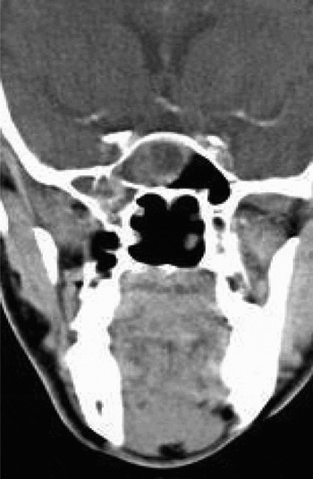
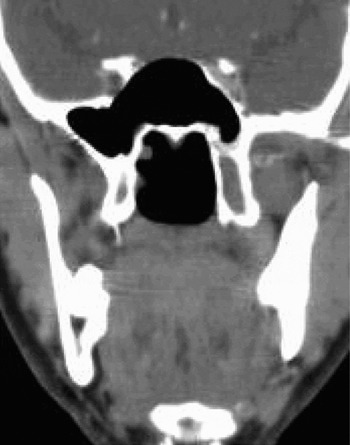
Once again, clinical examination was normal. However, a contrast-enhanced computed tomography (CT) scan of the paranasal sinuses demonstrated a moderately enhancing mass with central hypodense areas in the right sphenoethmoid region (Figure 1).
Pre-operative, coronal computed tomography scan showing a moderately enhancing mass with central hypodense area in the right sphenoid sinus.

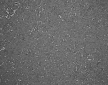
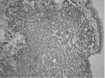
The patient underwent endoscopic excision of the mass under hypotensive anaesthesia. Intra-operatively, a mucosa-covered, friable, vascular mass was found involving the right posterior ethmoid and sphenoid sinuses. The histopathological diagnosis was a sinonasal-type haemangiopericytoma with positive immunohistochemical staining for smooth muscle actin and vimentin (Figures 2 and 3) and negative staining for cluster of differentiation 34 (CD34) glycoprotein.
Photomicrograph showing whorls and short fascicles of bland spindle cells with eosinophilic cytoplasm (H&E; ×200).

Photomicrograph showing tumour cells with positive cytoplasmic staining for smooth muscle actin (×400).

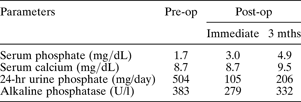
In the immediate post-operative period, as well as a week after surgery, there was substantial improvement in the patient's serum phosphate and 24-hour urinary phosphate levels (Table I).
Pre- and post-operative changes in biochemical parameters
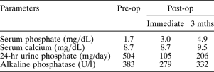
Pre-op = pre-operative; post-op = post-operative; mths = months; hr = hour
When the patient returned for follow up three months later, he had no bone pains and no longer required a wheelchair. Repeat testing of serum phosphate and 24-hour urinary phosphate levels showed further improvement (Table I).
At six-month follow up, repeated contrast-enhanced CT scanning of the paranasal sinuses showed no evidence of any residual lesion in the right sphenoid and posterior ethmoid sinuses (Figure 4). Based on the clinical features, histopathological diagnosis and post-operative course of events, a diagnosis of phosphaturic mesenchymal tumour or haemangiopericytoma causing oncogenic osteomalacia was confirmed.
Post-operative, coronal computed tomography scan of the paranasal sinuses, showing no residual mass in the sphenoid sinus.

At most recent follow up, three years after diagnosis, the patient was ambulant with no bone pains and normal serum and urinary phosphate levels.
Discussion
Oncogenic osteomalacia is a rare paraneoplastic syndrome which chiefly involves the extremities.Reference Koriyama, Nishimoto, Kodama, Nakazaki, Kurona and Yoshida2, Reference Folpe, Fanburg-Smith, Billings, Bisceglia, Bertoni and Cho4 Other sites of involvement include the maxillary sinus,Reference Koriyama, Nishimoto, Kodama, Nakazaki, Kurona and Yoshida2, Reference Weidner, Bar, Weiss and Strottmann3, Reference Weiss, Bar, Weidner, Wener and Lee11 buccal vestibule,Reference Ahn, Kim, Cha, Kim, Yim and Kim12 temporal bone,Reference Kaylie, Jackson and Gardner13 lung,Reference Schapira, Ben Izhak, Nachtigal, Burstein, Shalom and Shagrawi14 subcutaneous tissue,Reference Oka, Kamo, Sasaki, Kaji, Nishizawa and Imanishi15 metacarpophalangeal jointReference Jacob, Finny, Thomas, Thomas, John and Oncogenic16 and frontal lobe.Reference Sandhu and Martuza8
Our patient presented to the endocrinology department with complaints of generalised body aches and bone pain. Nutritional osteomalacia was the initial diagnosis at presentation, because of the initially negative ENT investigation. A second ENT consultation was sought in view of persistent phosphaturia. The occult nature of the tumour was in accordance with other reported cases in which nasal complaints were absent but sinonasal tumours eventually discovered (Table II).Reference Beech, Rokade, Gittoes and Johnson5–Reference Clunie, Fox and Stamp10
Previous reports of tumours of the sphenoethmoidal region associated with oncogenic osteomalacia

Y = year; M = male; F = female; MRI = magnetic resonance imaging; CT = computed tomography; post-op RT = post-operative radiotherapy; Ca = carcinoma
Given the vague symptomatology in patients with this condition, it is imperative in the first instance to perform at least a contrast-enhanced CT scan of the paranasal sinuses. Routine examination by anterior rhinoscopy and rigid nasal endoscopy alone could result in the tumour being missed. In previously reported patients with a focus of tumour in the ethmoid sinus, the neoplasm was detected solely by CT scanning or other radiological tests. Thus, both the endocrinologist and the otolaryngologist must have a high index of suspicion, and should proceed with CT or magnetic resonance imagingReference Nelson, Mason, Robinson, Hogan, Martin and Ahlström17 even if clinical evaluation is negative. Other reported imaging modalities which may be of use are 111In-pentetreotide scanning,Reference Ungari, Rocchi, Rinna, Agrillo, Lattanzi and Pagnoni7, Reference Casari, Rossi, Varenna, Gasparini, Parafioriti and Failoni18 octreotide scanningReference Harish, Jurriaans, Jan, Sur and Colterjohn19 and positron emission tomography.Reference Chua, O'Connor, Wong and Ganatra20
Oncogenic osteomalacia is associated with a number of benign bone and soft tissue tumours, such as haemangiopericytoma and giant cell tumour.Reference Harish, Jurriaans, Jan, Sur and Colterjohn19 Weidner and Santa Cruz,Reference Weidner and Santa Cruz1 in their landmark study, reviewed previously reported cases of oncogenic osteomalacia and found that most of the tumours associated with this condition had certain histological features in common. They coined the term phosphaturic mesenchymal tumour mixed connective tissue variant to describe these unique lesions. These tumours are characterised by a distinctive admixture of spindle cells, osteoclast-like giant cells and prominent blood vessels which range from capillary-sized channels to hyalinised, thick-walled and haemangiopericytoma-like vessels. The other distinctive feature of this tumour is its matrix, which ranges from myxoid to myxochondroid with foci of calcification and metaplastic bone.
However, the typical features of phosphaturic mesenchymal tumour mixed connective tissue variant are not seen in sinonasal tumours causing oncogenic osteomalacia. In Folpe and colleagues'Reference Folpe, Fanburg-Smith, Billings, Bisceglia, Bertoni and Cho4 meta-analysis of a series of 32 cases of oncogenic osteomalacia, there were two sinonasal tumours, both of which lacked the typical features of phosphaturic mesenchymal tumour mixed connective tissue variant; these two tumours resembled sinonasal-type haemangiopericytoma, with small, myoid-appearing, spindle-shaped cells arranged around blood vessels, as seen in our case. Both these tumours lacked matrix production and osteoclast-type giant cells. Furthermore, these tumours' immunohistochemical profile was similar to that of our case, viz, positive for smooth muscle actin and negative for cluster of differentiation 34 glycoprotein.
Sinonasal-type haemangiopericytomas represent less than 1 per cent of all sinonasal tumours. They are composed of short fascicles of uniform, oval- to spindle-shaped cells arranged around vessels which often show perivascular hyalinisation. Sinonasal-type haemangiopericytomas are invariably positive for vimentin, smooth muscle actin and factor XIIIa.Reference Wenig21
• Oncogenic osteomalacia is a rare syndrome characterised by the presence of hypophosphataemic, phosphaturic osteomalacia secondary to a benign mesenchymal tumour
• As the tumour is often occult and usually benign, its detection poses a diagnostic challenge
• Radiological investigations, particularly contrast-enhanced computed tomography scans of the sinuses, should be undertaken despite negative clinical findings
• Surgical excision is the mainstay of treatment
• The possibility of a benign sinonasal tumour causing phosphaturic, hypophosphataemic oncogenic osteomalacia should be considered by the treating physician, and should prompt early referral to the otolaryngologist
Sinonasal haemangiopericytoma causing oncogenic osteomalacia is best treated by wide surgical excision. Long term follow up is required to detect any recurrence, and revision surgery may be needed to achieve complete tumour excision.Reference Fuentealba, Pinto, Ballesteros, Pacheco, Boettiger and Soto6 Post-operative radiotherapy has been recommended in patients with aggressive tumours, although there is limited published evidence for this. One patient who had a sinonasal tumour with intracranial extension received post-operative radiotherapy because of the presence of dural and brain parenchymal invasion.Reference Sandhu and Martuza8
Biochemical markers are employed both for diagnosis and for monitoring of tumour activity. In patients with suspected oncogenic osteomalacia, measurement of serum phosphate, as part of routine biochemical investigation, is of utmost importance to distinguish this condition from other cases of osteomalacia.
Conclusion
A diagnosis of hypophosphataemic, phosphaturic osteomalacia is arrived at by clinical and biochemical evaluation, and warrants a complete search for causes of oncogenic osteomalacia. A detailed ENT evaluation should be performed, which should include (at the very least) a contrast-enhanced CT scan of the paranasal sinuses to exclude an occult tumour. Histological characterisation of the tumour is useful in determining whether it is a phosphaturic mesenchymal tumour mixed connective tissue variant or a sinonasal haemangiopericytoma. Wide surgical excision is the treatment of choice. Following excision of the causative tumour, clinical and biochemical improvement is both immediate and dramatic.