Introduction
The human filarial parasite Onchocerca volvulus causes onchocerciasis, also known as river blindness. Onchocerciasis is characterized by chronic skin and eye lesions. It is transmitted by a blackfly vector from the genus Simulium. In 1995, the World Health Organization (WHO) estimated that 17 million people were infected with O. volvulus, of whom 270,000 were blind and another 500,000 were visually impaired (WHO, 1995). In addition to visual impairment, onchocerciasis is associated with serious pathological changes of the skin. The disease is endemic in many tropical regions of Africa, Asia, Central and South America. Different surveys indicate that this disease is dramatically underestimated, also by WHO calculations: 37 million people are now thought to be infected, with 90 million at risk in Africa and more than 400,000 infected in Central and South America (Basanez et al., Reference Basanez, Pion, Churcher, Breitling, Little and Boussinesq2006).
According to the Cameroonian public health ministry in 2004, about 28% of the population is affected by the disease, with more than 1 million suffering from skin alteration due to onchocerciasis. Furthermore, about 110,000 km2 of land in Cameroon has been left uncultivated owing to the disease, with economical consequences. In Ngaoundere and in the Adamawa region, the prevalence of O. volvulus and Onchocerca ochengi are estimated to be around 30% and 65%, respectively (Wahl et al., Reference Wahl, Enyong, Ngosso, Schibel, Moyou, Tubbesing, Ekale and Renz1998). Both parasites are transmitted to humans and cattle by the same vector (Simulium) (Wahl et al., Reference Wahl, Enyong, Ngosso, Schibel, Moyou, Tubbesing, Ekale and Renz1998).
Adult worms (macrofilariae) live under the skin, where they form nodules. The excision of nodules, which is the most widespread form of treatment to eliminate macrofilariae, is associated with the therapeutic administration of ivermectin, being the only existing drug against this parasite (WHO, 1995). However, it kills only the microfilariae (WHO, 1995; Borsboom et al., Reference Borsboom, Boatin, Nagelkerke, Agoua, Akpoboua, Alley, Bissan, Renz, Yameogo, Remme and Habbema2003) and continuous use of ivermectin is necessary until the adult worm dies. The increasing reliance of onchocerciasis control on a single drug risks the potential emergence of ivermectin resistance, particularly in the setting of ongoing parasite transmission. Osei-Tweneboana et al. (Reference Osei-Tweneboana, Eng, Boakye, Gyapong and Prichard2007) have reported that ivermectin resistance is developing in O. volvulus in some communities in Ghana, and is manifesting itself as a more rapid return to high microfilarial counts after treatment.
Wolbachia-targeted chemotherapy is an interesting novel approach; however, the length of treatment is still a stumbling block (Pfarr & Hoerauf, Reference Pfarr and Hoerauf2006; Nfon et al., Reference Nfon, Makepeace, Njongmeta, Tanya and Trees2007); as there is yet no effective drug that can kill the macrofilariae. Due to damage caused by onchocerciasis, there is a great need to search for new filaricidal drugs from other sources, such as botanicals that are strongly macro- and microfilaricidal.
Since there is no laboratory host for O. volvulus, model organisms are often needed. Accordingly, the bovine filarial parasite O. ochengi has been used in some promising antifilarial drug studies (Gilbert et al., Reference Gilbert, Nfon, Makepeace, Njongmeta, Hastings, Pfarr, Renz, Tanya and Trees2005). In addition, Caenorhabditis elegans serves as a suitable model organism for research into nematode parasites (Burglin et al., Reference Burglin, Lobos and Blaxter1998). This 1 mm long, free-living bacteriphagous nematode can be cultivated easily on agar plates. Under optimal conditions, it has a reproductive cycle of 3 days. After four juvenile stages, immature worms grow to adults (Wixon et al., Reference Wixon, Blaxter, Hope, Barstead and Kim2000). The lifetime of C. elegans is estimated to be about 3 weeks. The simple morphology, rapid development, easy cultivation and accessibility to genetics and molecular biology make C. elegans a powerful analytical tool for investigations of biological effects of toxic substances and for identification of suitable pharmacological targets (Kampkotter et al., Reference Kampkotter, Gombitang-Nkwonkam, Zurawski, Timpel, Chovolou, Watjen and Kahl2007).
In Africa and other developing countries, most people depend on traditional herbal medicine for health needs, and these herbal remedies are usually safer than active compounds isolated from plants (Fabricant & Farnsworth, Reference Fabricant and Farnsworth2001). Plant-derived anthelmintic products have the advantage of being more biodegradable and environmentally friendly (Hammond et al., Reference Hammond, Fielding and Bishop1997).
We have selected the following medical plants from Cameroon and Ghana based on information obtained from herdsmen and pastoralists. These plants are used regularly against human and livestock parasites. Leaves of Ficus exasperata Vahl (Moraceae) are traditionally used as an ingredient in a vermifuge preparation in Liberia. An aqueous decoction of leaves has been shown to possess anti-ulcer activity (Akah et al., Reference Akah, Orisakwe, Gamaniel and Shittu1998). A poultice of leaves of Annona senegalensis Pers (Annonaceae) is used for the treatment of worm infestation and diarrhoea (Burkill, Reference Burkill1997; Fall et al., Reference Fall, Sambou, Seck, Wele, Ndoye, Gleye and Laurens2008). A bark decoction of Khaya senegalensis (Desr.) A. Juss (Meliaceae) is used for the treatment of fever, menstrual disorders, venereal diseases, worm infections, dysentery and stomach complaints (Burkill, Reference Burkill1997). Leaf and bark decoctions of Anogeissus leiocarpus (DC.) Guill & Perr (Combretaceae) are used for the treatment of yellow fever, jaundice and as a vermifuge in folklore medicine (Burkill, Reference Burkill1997). Euphorbia hirta L. (Euphorbiaceae) is used for the treatment of asthma, syphilis, dysentery, wounds and skin diseases in traditional medicine (Burkill, Reference Burkill1997). Furthermore, this plant has been shown to have anti-amoebic properties (Tona et al., Reference Tona, Cimanga, Kambu, Totte, Pieters and Vlietinck2009). Leaves and roots of Parquetina nigrescens (Afzel.) Bullock (Asclepiadaceae) are applied as poultice for the treatment of wounds, stomach complains, worm infections, boils and carbuncles (Burkill, Reference Burkill1997; Agyare et al., Reference Agyare, Asase, Niehues, Lechtenberg, Deters and Hensel2009).
In the current study we prepared aqueous and ethanolic extracts of the above-mentioned medicinal plants and investigated their nematocidal potential on adults and larvae of the free-living C. elegans as well as adults of the filarial parasite O. ochengi.
Materials and methods
Plant materials and chemicals
Leaves of Ficus exasperata Vahl and Parquetina nigrescens (Afzel.) Bullock were collected in July, 2007 from the Bosomtwi-Atwima-Kwanwoma area, Ghana and identified by Dr Alex Asase, Department of Botany, University of Ghana. Voucher specimens have been deposited at the Ghana Herbarium, University of Ghana, Ghana. Leaves of Anogeissus leiocarpus (DC.) Guill & Perr, Euphorbia hirta L., Annona senegalensis Pers and Khaya senegalensis (Desr.) A. Juss (and stem bark) were collected in February, 2009 in Ngaoundere area, Cameroon and identified by Mr Froumsia Moksia, Department of Life Science, University of Maroua, Cameroon. Voucher specimens have been deposited at the National Herbarium in Yaounde, Cameroon. Unless stated otherwise, all chemicals were purchased from Sigma (Deisenhofen, Germany).
Preparation of plant extracts
Plant species were dried at room temperature, weighed, ground finely and sieved on a 0.5 mm mesh screen. Ten grams of the powdered material were extracted with 100 ml distilled water at 90°C for 10 min or with 100 ml of ethanol (60% v/v) at 70°C for 30 min, centrifuged (3500 g, 10 min) and filtered over filter papers No. 413 (VWR International, Darmstadt, Germany). The clear filtrate was concentrated by a rotary evaporator at a temperature not exceeding 40°C under reduced pressure, lyophilized and the resulting powder was stored at 4°C.
For further investigation the dried plant extracts were dissolved in ethanol 50% (v/v), diluted in 1% dimethylsulphoxide (DMSO) to a final concentration of 100 mg/ml, centrifuged and aliquots taken to determine their activity on C. elegans.
Monoxenic and axenic culture of C. elegans
Caenorhabditis elegans wild type (N2 Bristol) was grown at 20°C under standard monoxenic conditions on NGM-agar (Nematode Growth Medium: 2.5 g peptone from casein, 3 g NaCl, 17 g agar, 0.5% cholesterin, 1 mm CaCl2, 1 mm MgSO4, 25 mm KH2PO4/K2HPO4 in 1 litre of water) in Petri dishes containing the food bacterium Escherichia coli OP50. To synchronize worm cultures or to initiate axenic worm cultures, the alkaline bleaching method was applied (Lenaerts et al., Reference Lenaerts, Walker, Hoorebeke, Gems and Vanfleteren2008). Briefly, monoxenic worm cultures were harvested in M9 buffer (3 g KH2PO4, 6 g Na2HPO4, 5 g NaCl, 0.25 g MgSO4.7H2O, in 1 litre of water) and subsequently incubated for 7 min in freshly prepared chlorox solution consisting of 5.5 ml water, 4 ml commercial chlorox bleach (8 degrees) and 0.5 ml 10N NaOH. While worms and bacteria were lysed, worm eggs resisted the harsh treatment. Following three washing steps in M9 buffer, isolated eggs were used to initiate synchronous monoxenic or axenic cultures. Liquid axenic medium consisted of 3% (w/v) dry yeast extract (Becton-Dickinson, Franklin Lake, New Jersey, USA), 3% (w/v) soy peptone supplemented with 0.5 mg/ml bovine haemoglobin (50 mg/ml haemoglobin stock solution in 0.1N KOH autoclaved at 121°C for 10 min). Penicillin/streptomycin solution (10,000 U/10,000 μg/ml) was added to the complete medium (Lenaerts et al., Reference Lenaerts, Walker, Hoorebeke, Gems and Vanfleteren2008).
Sampling and in vitro screening assay of O. ochengi
Nodules of O. ochengi were removed from the skin of cattle at the Ngaoundere's slaughterhouse and brought to the laboratory for dissection. After dissection of nodules, O. ochengi were isolated and washed three times in sterile PBS (phosphate-buffered saline). Worms were subsequently transferred to RPMI-1640 medium supplemented with l-glutamine (Sigma, Deisenhofen, Germany) and washed twice. Following the protocol of Borsboom et al. (Reference Borsboom, Boatin, Nagelkerke, Agoua, Akpoboua, Alley, Bissan, Renz, Yameogo, Remme and Habbema2003), adult worms (six individuals per well) were incubated with different concentrations of plant extracts (0, 0.125, 0.25, 0.5, 1 and 2 mg/ml) in RPMI supplemented with penicillin/streptomycin (10,000 U/10,000 μg/ml). Assays were incubated at 37°C and mortality was determined after 6 h, 12 h and 24 h.
In vitro screening assay of C. elegans
Synchronized C. elegans (approx. 15 worms: young adults or L4 larvae) from liquid axenic medium were transferred into fresh M9 medium supplemented with 2% glucose, 0.5% cholesterin, 25 mm potassium phosphate, pH 6.0, 1 mm CaCl2, 1 mm MgSO4 penicillin/streptomycin (10,000 U/10,000 μg/ml). Subsequently, 500 μl of aliquots of supplemented M9 medium were transferred to 24-well sterile plates and increasing concentrations of plant extracts (0, 0.25, 0.5, 0.75, 1, 2, 4, 6 and 8 mg/ml) were added. Assays were incubated at 20°C and mortality was determined after 24 h, 48 h and 72 h.
In addition, the effect of plant extracts (1 mg/ml) was tested on C. elegans grown monoaxenically in the presence of E. coli OP50 on NGM-agar. After chlorox treatment, isolated eggs were introduced on NGM-agar plates to initiate synchronous culture. After egg-hatching, L1 were transferred on to NGM-agar plates (six worms per plate) supplemented with 1 mg/ml plant extracts. Worms were cultured under standard conditions and analysed after 3 days and 7 days. Control plates did not contain plant extracts. NGM-plates were cooled for 30 min to paralyse worms for photography.
Worm mortality and LC50 determinations
The numbers of dead and living worms were counted under a binocular microscope. Worms that were completely straight or curved and immotile after shaking were considered to be dead. The percentage mortality was expressed as the percentage of dead worms in relation to the number of living, swimming worms. Each plant extract, together with the respective control groups, was tested in three independent duplicate determinations. LC50 values (lethal concentration of the extract required to kill 50% of helminths) of different extracts were calculated using GraphPad (GraphPad Software Inc., La Jolla, California, USA). Results are presented as mean values ± standard error (SEM). Negative controls were treated with 0.02% DMSO and 0.05% ethanol and ivermectin (0–2 mg/ml) was used as the positive control.
Phytochemical screening
Preliminary phytochemical screening for the presence of flavonoids, alkaloids, carbohydrates and saponins was conducted on dried leaves from P. nigrescens, F. exasperata, A. leiocarpus, A. senegalensis and E. hirta, as well as on dried leaves and stem bark of K. senegalensis (Harborne, Reference Harborne1998). The quantity of tannins was determined according to the method of Glasl (Reference Glasl1983) using pyrogallol (purity 99.5%, high-performance liquid chromatography) as reference compound.
Results and discussion
In the present work, aqueous and ethanolic extracts of selected plants used in traditional medicine in Cameroon and Ghana were tested for their anthelmintic activity. Two models were employed: the free-living nematode C. elegans and the bovine filarial nematode O. ochengi. Caenorhabditis elegans is usually cultured monoxenically in the presence of the food bacterium E. coli OP50. However, to avoid possible unwanted biological side-effects, like degradation, detoxification or activation of bioactive ingredients caused by co-cultured E. coli, plant extracts were first tested against axenically maintained C. elegans. According to their inhibition efficacy, extracts were rated as exhibiting high (LC50 < 2.5 mg/ml), moderate (LC50 between 2.5 and 4 mg/ml) or no anthelmintic activity (LC50 > 4 mg/ml) (figs 1–3).
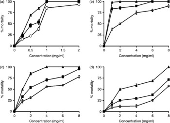
Plant extracts with high activity against C. elegans. Mortality rates of axenically cultured C. elegans after (○) 24 h, (■) 48 h and (▲) 72 h exposure to increasing concentrations (0–8 mg/ml) of crude ethanolic extracts from (a) A. leiocarpus bark, (b) K. senegalensis bark, (c) K. senegalensis leaves, (d) E. hirta leaves. Extracts affected the survival of C. elegans in a time- and concentration-dependent manner. Data are means ± SEM from three independent duplicate experiments.

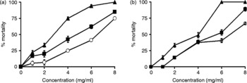
Plant extracts with moderate activity against C. elegans. Caenorhabditis elegans cultured under axenic conditions were exposed to increasing concentrations (0–8 mg/ml) of crude aqueous extracts from (a) A. senegalensis and (b) P. nigrescens leaves. Mortality rates were determined after (○) 24 h, (■) 48 h and (▲) 72 h. Data are means ± SEM from three independent duplicate experiments.

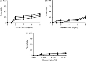
Plant extracts showing no activity against C. elegans. Axenically maintained C. elegans were incubated in the presence of increasing concentrations (0–8 mg/ml) of (a) crude alcoholic extracts from F. exasperata leaves and (b) aqueous extracts from E. hirta leaves. Mortality rates were determined after (○) 24 h, (■) 48 h and (▲) 72 h. Controls (c) with DMSO and ethanol are included. Data are means ± SEM from three independent duplicate experiments.

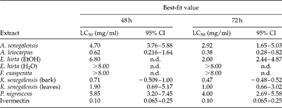
Ethanolic extracts of the bark of A. leiocarpus and K. senegalensis as well as the leaves of K. senegalensis and E. hirta displayed the highest anthelmintic activity, with LC50 values of 0.38 mg/ml, 0.47 mg/ml, 1.0 mg/ml and 2.0 mg/ml after 72 h, respectively (fig. 1, table 1), while aqueous extracts of leaves from P. nigrescens and A. senegalensis showed moderate anthelmintic activity (fig. 2). This result is in accordance with observations of Igweh & Onabanjo (Reference Igweh and Onabanjo1989) and Alawa et al. (Reference Alawa, Adamu, Gefu, Ajanusi, Abdu, Chiezey, Alawa and Bowman2003), demonstrating potent effects of A. senegalensis against protozoans and gastro-intestinal nematodes. Ethanolic extracts of F. exasperata leaves and aqueous extracts of E. hirta leaves hardly affected the survival of C. elegans (fig. 3).
LC50 values of plant extracts tested against axenically cultured C. elegans. Worms were incubated with increasing concentrations of DMSO–ethanol-dissolved plant extracts for 48 h and 72 h. Confidence intervals (CI) are also shown; n.d., not determined; H2O, aqueous extract; EtOH, ethanolic extract.
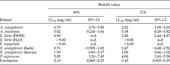
Generally, active extracts inhibited the survival of young or adult worms in a time- and concentration-dependent manner. Highly effective extracts (the bark of A. leiocarpus and K. senegalensis) killed worms at 1 mg/ml after 48 h and 72 h, respectively. Respective controls indicated that neither ethanol nor DMSO concentrations used in these tests had any effect on the worms (fig. 3c).
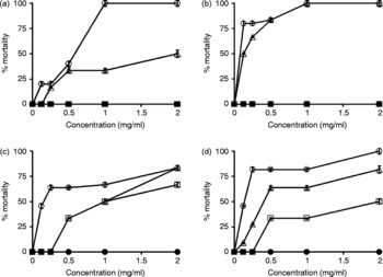
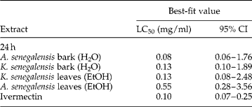
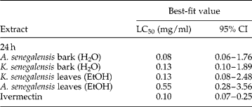
Similar results were obtained with the leaves and bark of A. senegalensis, and leaves and bark of K. senegalensis tested against O. ochengi (fig. 4 a, b, c and d). Aqueous extracts from the bark of A. senegalensis and K. senegalensis as well as ethanolic extracts from the leaves of K. senegalensis and A. senegalensis displayed high anthelmintic activity with LC50 values of 0.08 mg/ml, 0.13 mg/ml, 0.13 mg/ml and 0.55 mg/ml, respectively, after 24 h (table 2). Similar to the data obtained for C. elegans, plant extracts inhibited the survival of O. ochengi adults in a time- and concentration-dependent manner. The ethanolic extract from leaves of A. senegalensis as well as aqueous extracts of the bark of A. senegalensis and K. senegalensis killed O. ochengi almost completely at 1 mg/ml after 24 h (fig. 4 a, b and d). According to the calculated LC50 values, these plant extracts show an efficacy similar to or in the case of the aqueous bark extract of A. senegalensis (LC50 = 0.08 mg/ml) even higher than that of the positive control ivermectin (LC50 = 0.1 mg/ml) (table 2).
Plant extracts with activity against O. ochengi. Mortality rates of O. ochengi after (●) 0 h, (□) 6 h, (△) 12 h and (○) 24 h exposure to increasing concentrations (0–2 mg/ml) of (a) crude alcoholic extract from A. senegalensis leaves, (b) crude aqueous extract from A. senegalensis bark, (c) crude alcoholic extract from K. senegalensis leaves and (d) crude aqueous extract from K. senegalensis bark. Extracts affected the survival of O. ochengi in a time- and concentration-dependent manner. Data are mean ± SEM from three independent duplicate experiments.

LC50 values for plant extracts tested against cultured O. ochengi. Worms were incubated with increasing concentrations of plant extracts that were dissolved in DMSO–ethanol for 24 h. Confidence intervals (CI) are also shown. H2O, aqueous extract; EtOH, ethanolic extract.
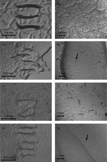
We next examined the biological effect of plant extract candidates with high anthelmintic activity (A. leiocarpus, A. senegalensis and the bark of K. senegalensis) on monoxenically cultured C. elegans at a concentration of 1 mg/ml. Starting with eggs, control worms developed to the adult stage within 3 days, having a body length of approximately 1 mm. On day 7 they had laid eggs and were about 1.5 mm long (fig. 5a). In contrast, worms that were maintained in the presence of plant extracts were found to be smaller than 0.7 mm on day 3 (fig. 5b1, c1 and d1). Worms exposed to A. senegalensis aqueous leaf extract showed a drastically delayed L1 to adult development (fig. 5c2) and worms treated with ethanolic extracts of A. leiocarpus and K. senegalensis did not reach the adult stage even after 7 days incubation and were unable to lay eggs (fig. 5b2 and d2). Therefore, in addition to their anthelmintic activity under axenic conditions, plant extracts strongly affected the egg-to-adult development and survival of C. elegans under standard culture conditions, indicating that co-cultured E. coli do not significantly alter the bioactivity of the used plant extracts. After a 7-day incubation period, only 7% of worms were alive on plates containing A. leiocarpus and K. senegalensis (fig. 5b2 and d2).
The effect of selected plant extracts on monoxenically cultured C. elegans. (a) Control worms reached the adult stage within 3 days (a1) having a body length of approx. 1 mm. On day 7 (a2) they were about 1.5 mm long. Worms cultured on NGM-agar plates supplemented with 1 mg/ml of (b) A. leiocarpus, (c) A. senegalensis and (d) K. senegalensis (bark) were smaller than 0.7 mm on day 3 (b1, c1 and d1). After a 7-day incubation period they were dead or did not reach the adult stage (b2 and d2). In c2, worms became adult; however, they developed more slowly than control worms. Arrows indicate worms.

These results strongly indicate that plant extracts derived from A. leiocarpus and K. senegalensis bark contain promising bioactive compounds that might be useful in the control of helminth infections by interrupting the worms' life cycle and preventing their growth. Since the motility of worms is also affected, it may be possible that these plant extracts also act on worm muscles and paralyse them.
Remarkably, none of these selected plants have been previously tested against C. elegans and Onchocerca spp. Our data reinforces the existing knowledge and the regular use of plants by herdsmen and pastoralists for the treatment of worm infections. Consistent with this, recent studies carried out with some of the plants used in the present study have also demonstrated efficacy against other parasitic nematodes (Musongong et al., Reference Musongong, Nukenine, Ngassoum, Gangue, Messine, Fokunang, Zalom, Njongmeta and Tanya2004; Monglo et al., Reference Monglo, Njongmeta, Musongong, Ngassoum and Nukenine2006; Fall et al., Reference Fall, Sambou, Seck, Wele, Ndoye, Gleye and Laurens2008; Adenola et al., Reference Adenola, Fagbemi and Idowu2009). Anogeissus leiocarpus and K. senegalensis were found to have anthelmintic activity against different animal gastrointestinal worms (Monglo et al., Reference Monglo, Njongmeta, Musongong, Ngassoum and Nukenine2006; Adenola et al., Reference Adenola, Fagbemi and Idowu2009) and crude extracts of A. leiocarpus, A. senegalensis and Coriandrum sativum demonstrate anthelminthic properties against eggs and infective larvae of Haemonchus contortus (Monglo et al., Reference Monglo, Njongmeta, Musongong, Ngassoum and Nukenine2006; Eguale et al., Reference Eguale, Tilahun, Debella, Feleke and Makonnen2007; Adenola et al., Reference Adenola, Fagbemi and Idowu2009). Musongong et al. (Reference Musongong, Nukenine, Ngassoum, Gangue, Messine, Fokunang, Zalom, Njongmeta and Tanya2004) also reported a strong toxic effect of A. senegalensis against infective larvae of Strongyloides papillosus.
Extracts from other medicinal plants such as Piliostigma thonningii, Ocimum gratissimum and Nauclea latifolia have been shown to affect Onchocerca worms and to inhibit the glutathione S-transferases (Njoku et al., Reference Njoku, Okeyode and Asuzu1996; Fakae et al., Reference Fakae, Campbell, Barrett, Scott, Teesdale-Spittle, Liebau and Brophy2000).
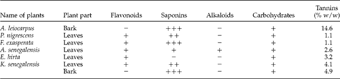
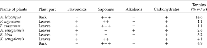
Preliminary phytochemical screening of our plant extracts revealed the presence of flavonoids, alkaloids, saponins, carbohydrates and tannins (table 3). Plant extracts of A. leiocarpus, K. senegalensis and A. senegalensis exhibited high anthelmintic activity. Currently, we are investigating the bioactive compounds responsible for this nematocidal activity using bioactivity-guided fractionation and characterization of the active compounds.
Phytochemical screening of selected plants. The phytochemical screening revealed the presence or absence of flavonoids, alkaloids, saponins, carbohydrates and tannins in the bark and leaves of plants. +, present; −, absent; % w/w, % weight/weight, relative to pyrogallol.
This study revealed that crude extracts of A. leiocarpus, A. senegalensis and K. senegalensis exhibited strong in vitro anthelmintic activity against the model organism C. elegans and the filarial parasite O. ochengi, affecting the egg to adult development and the survival of young and adult worms. In future studies, we will isolate active compounds and investigate their mode of action. Additionally we are looking into safety aspects to determine the feasibility of developing effective and safe drug preparations for the treatment of onchocerciasis and other helminth infections.
Acknowledgements
This research was supported by the fellowship of the Alexander von Humboldt Foundation (AVH) to D.N. and the German Academic Exchange Service (DAAD) to C.A. Further support was provided by the Deutsche Forschungsgemeinschaft (DFG) grant LI 793/5-1 to the Cameroonian-German Cooperation Project (CGCP), http://www.cameroon.uni-muenster.de.