Introduction
The inter-relationships between sleep, cognition and major depression (MD) are complex in the older population. Not only is the ageing process associated with changes to sleep timing, maintenance and architecture (Reference Ohayon and Vecchierini1,Reference Kupfer, Reynolds, Ulrich, Shaw and Coble2), but it is also associated with cognitive impairment, neurodegenerative processes and differential aetiological paths to depression (Reference Hickie and Scott3–Reference Reynolds, Kupfer and Taska5).
Sleep disturbance in older people may be a prognostic indicator for cognitive decline. This has been shown in healthy older samples (Reference Cricco, Simonsick and Foley6) as well as in those with neurodegenerative disease (Reference Mortimer, Ebbitt, Jun and Finch7) wherein the presence of this feature is associated with neuropsychiatric symptoms, functional decline, disability, poor quality of life, carer burden and the need for institutionalisation (Reference Naismith, Lewis, Rogers, Kerkhof and Van Dongen8). Sleep disturbance is also common in patients with depression and is part of the core diagnostic criteria (Reference Reynolds, Kupfer and Taska5,Reference Naismith, Lewis, Rogers, Kerkhof and Van Dongen8). It often persists despite symptom resolution and large, community-based studies of older people suggest that this feature is a risk factor for depression onset and recurrence (Reference Cho, Lavretsky, Olmstead, Levin, Oxman and Irwin9). These findings collectively suggest that sleep disturbance may be aetiologically linked to neurodegenerative and neuropsychiatric disorders in older people and may even be a prodromal, prognostic or mediating feature (Reference Naismith, Norrie, Lewis, Rogers, Scott and Hickie10,Reference Naismith, Rogers, Hickie, Mackenzie, Norrie and Lewis11).
Despite the apparent commonalities, few studies have examined concurrently the inter-relationships between affective and sleep disturbance and cognitive impairment. This is despite the fact that both depression (Reference Duman and Monteggia12,Reference Naismith, Hickie and Turner13) and sleep disturbance (Reference Stickgold14) have been shown in independent studies to relate to a range of neuropsychological deficits and to modulate levels of neurotrophic factors relevant for neurogenesis. Preliminary studies have shown that in older patients with MD, sleep–wake disturbance is correlated with memory impairment and to later ages of depression onset (Reference Naismith, Norrie, Lewis, Rogers, Scott and Hickie10), features which are particularly associated with cerebrovascular disease, treatment resistance and progression to dementia (Reference Steffens, Otey and Alexopoulos15). However, no known studies have examined these associations in those meeting criteria for mild cognitive impairment (MCI), an ‘at risk' group for conversion to dementia longitudinally.
MCI is a syndrome defined as cognitive decline (i.e. significant changes in memory and thinking functions) but with preservation of daily functioning (Reference Petersen, Doody and Kurz16). There is a conversion rate to dementia (particularly Alzheimer's disease) of around 50% within 5 years (Reference Gauthier, Reisberg and Zaudig17). Traditional MCI criteria focussed on memory impairment as being a key feature [amnestic MCI (aMCI)] although definitions have recently been extended to include a more heterogeneous group without predominant memory change [non-amnestic MCI (naMCI)] (Reference Petersen, Doody and Kurz16–Reference Petersen and Morris18).
Recent reviews suggest that between 14 and 59% of patients with MCI have sleep disturbance as assessed by clinician-ratings and semi-structured interviews (Reference Beaulieu-Bonneau and Hudon4). However, prior studies have not specifically examined the relationship between depressive symptoms and sleep complaints in these MCI samples. Thus, it has been noted that further studies are required in this area (Reference Beaulieu-Bonneau and Hudon4). Although a preliminary analysis utilising actigraphy indicated that the relationship between neuropsychological dysfunction and sleep disturbance in MCI was not accounted for by depressive symptoms (Reference Naismith, Rogers, Hickie, Mackenzie, Norrie and Lewis11), no known studies have specifically sought to examine the impact of current and remitted MD on the range of sleep complaints reported. Delineation of these inter-relationships will help to elucidate the extent to which sleep disturbance in MCI is reflective of the ‘state' phenomena of depressive symptom severity and/or to underlying and persistent neurobiological change within the sleep–wake system. This knowledge, in turn, may help to guide early management approaches specifically targeting the sleep–wake and mood systems. In this study, we therefore aimed to evaluate the impact of current and remitted depression on self-reported sleep disturbance in patients with MCI.
Methods
Sample
Seventy-seven adults over the age of 50 years and who met criteria for MCI were recruited from outpatient neurology and psychiatry clinics at the Brain & Mind Research Institute, Sydney, Australia. Seventeen healthy control participants were recruited from the community. Exclusion criteria included: dementia diagnosis or mini-mental status examination (MMSE) score <24 (Reference DePaulo and Folstein19); neurological disease (e.g. Parkinson's disease, epilepsy); psychosis; bipolar I or bipolar II disorder; prior stroke or head injury (with loss of consciousness ≤30 min) and inadequate English for neuropsychological assessment. This study was approved by the University of Sydney Human Research Ethics Committee and all participants gave written informed consent.
Measurements
Clinical ratings. A Geriatric Psychiatrist conducted a semi-structured interview to determine medical history, medication details and clinical history. The structured clinical interview for DSM-IV (SCID-II) (Reference First, Gibbon, Spitzer, Williams and Benjamin20) was employed to document current and lifetime MD history, and current symptom severity was rated using the 17-item Hamilton Depression Rating Scale (HDRS) (Reference Hamilton21). Participants were classified into (a) CON, healthy controls, (b) DEP-C, mild cognitive impairment and meeting criteria for current major depression, (c) DEP-R, mild cognitive impairment and remitted major depression and (d) MCI, those meeting criteria for MCI but with no depression history.
Clinical diagnosis of MCI was obtained using Peterson's criteria of decline of at least 1.5 standard deviations on standardised neuropsychological tests, relative to age adjusted normative data (Reference Petersen and Morris18). As per criteria, each participant was required to have subjective and objective cognitive decline, but with preservation of function (as reflected by a Global Deterioration Scale score of ‘3'(Reference Reisberg, Ferris and de Leon22). Both aMCI and naMCI subtypes were included. Classification of aMCI was consensus rated (SN, KD, LN, ZT) using rigorous criteria that required the participant to manifest deficits on tests of delayed memory recall, not merely encoding (Reference Dubois, Feldman and Jacova23). As previously described, a full neuropsychological assessment was conducted by a Clinical Neuropsychologist or trained research psychologist to assist with determination of MCI diagnosis and exclusion of patients with suspected dementia (Reference Naismith, Rogers, Hickie, Mackenzie, Norrie and Lewis11). For descriptive purposes, the results of some neuropsychological tests are reported herein. Premorbid intellectual ability was estimated using the Wechsler Test of Adult Reading (24). Encoding of verbal material was assessed using the Wechsler Memory Scale-III Logical Memory subtest (Logical Memory-encoding, maximum = 75) (Reference Wechsler25). Visuomotor speed and executive functioning were assessed using Parts A and B of the Trailmaking Test respectively (Trailmaking A and Trailmaking B, seconds) (Reference Spreen and Strauss26).
Sleep quality assessment. Participants completed the Pittsburgh Sleep Quality Index (PSQI) (Reference Buysse, Reynolds, Monk, Berman and Kupfer27) to determine an overall subjective measure of sleep disturbance over the previous month. The PSQI comprises 17 items with most questions rated on a 4-point Likert scale. It provides a global score (maximum = 21) based on seven components (maximum sub-scale score = 3) including sleep quality, latency, duration, habitual sleep efficiency (i.e. time in bed - time spent asleep), sleep disturbance, use of sleeping medication and daytime dysfunction due to poor sleep quality. A global score greater than 5 is a commonly used cut-off score for significant sleep disturbance.
Statistical analysis
All analyses were conducted using SPSS Version 16 (SPSS Inc., Chicago, IL, USA). To examine between-groups data, One-Way Analysis of Variance was used with post hoc Scheffé tests and in the event of unequal variances non-parametric group comparisons were employed. Categorical data was analysed using Chi-square while continuous data was analysed using Pearson Product Moment correlations. All analyses were two-tailed and employed an alpha level of 0.05.
Results
Of the 77 patients, 29 had aMCI and 48 had naMCI. Four participants were taking benzodiazepines regularly to assist with sleep and three took benzodiazepines occasionally. Thirty-one patients were taking antidepressants (n = 26, newer generation serotonergic and noradrenergic agents; n = 3, tricylic antidepressants; n = 2, tetracyclics). Four patients took mood stabilisers including (n = 2, lithium; n = 1, sodium valproate; n = 1, lamotrigine). One patient used antipsychotic medication (quetiapine).
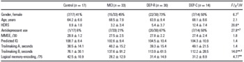
Table 1 displays demographic data for the samples. There were no significant differences between the groups in terms of age, sex, years of education or MMSE score, though as expected, those in the MCI groups tended to have poorer performance on tests of memory and executive functioning than CON, and the DEP-C group had higher HDRS scores. For the whole sample, there was no association between PSQI global scores and performance on tests of gross cognitive abilities (MMSE; r = 0.15, ns), visuomotor speed (Trailmaking A; r = −0.07, ns), memory-encoding (Logical Memory-encoding; r = 0.08, ns), or mental flexibility (Trailmaking B; r = −0.07, ns).
Demographics (mean, sd) for controls, patients with MCI and patients with a past (DEP-R) and current (DEP-C) history of major depression

† Chi-square.
‡ Kruskal–Wallis non-parametric analyses.
* p < 0.001.
** p < 0.01.
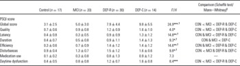
Table 2 displays the results of the between-group comparisons and shows that there was a significant group difference on the PSQI global score as well as component scales of sleep quality, latency, duration, efficiency, sleep disturbance and daytime dysfunction. The groups did not differ in their use of medication to aid sleep. Post hoc tests specifically showed that overall sleep disturbance, as measured by the PSQI global score, was greatest in the DEP-R and DEP-C groups and differed significantly in comparison to CON and MCI groups. Those with DEP-C had global PSQI scores almost three times as great as CON and twice as great as MCI. The MCI group as a whole reported global PSQI scores at precisely the cut-score of 5 for sleep disturbance. Component scale analysis showed that DEP-C and DEP-R had significantly poorer sleep quality, efficiency and greater daytime dysfunction and sleep disturbances than the CON and MCI groups. Latency to sleep onset was also longer: those with DEP-C self-reported a sleep latency of 52 min, compared to 29 min for DEP-R, 18 min for CON and 16 min for the MCI group. To ensure that the MCI group analyses were not influenced by having a heterogeneous sample of aMCI and naMCI, these analyses were repeated with the exclusion of those with aMCI and there were no appreciable differences in the results.
PSQI global and component scores for controls, patients with MCI and patients with a past (DEP-R) and current (DEP-C) history of major depression

Global score has a maximum of 21. Component scores have a maximum of 3.
† Kruskal–Wallis non-parametric analyses.
‡ Multiple comparisons utilise Scheffé tests and Mann–Whitney U tests for normal and non-parametric data respectively.
* p < 0.05.
** p < 0.01.
*** p < 0.001.
When sleep disturbance was further examined according to the commonly used cut-score of 5 on the PSQI, there was, as expected, a significant overall group effect (X 2 = 23.6, df = 3, p < 0.001). Specifically, only 12% (2/17) of CON showed sleep disturbance in comparison to 30% (10/33) of those with MCI, 63% (19/30) of DEP-R and 86% (12/14) of DEP-C. Again, these data suggest that those patients who are currently depressed show considerable sleep disturbance, whilst those who are remitted continue to show disturbance at a rate higher than MCI patients with no depression history.
Correlations between sleep disturbance and depression severity
To examine the relationship between depressive symptom severity and sleep disturbance, Pearson correlations for the entire patient sample were calculated with the sleep items extracted from the total HDRS score. The resultant analyses showed that ‘state' depressive symptoms correlated with global PSQI score (r = 0.52, p < 0.001) and the component scores of sleep quality (r = 0.35, p < 0.01), latency (r = 0.40, p < 0.001), duration (r = 0.39, p < 0.001), efficiency (r = 0.38, p < 0.001), disturbance (r = 0.23, p < 0.05), medication use (r = 0.30, p < 0.05) and daytime dysfunction (r = 0.44, p < 0.001).
Discussion
This study has shown for the first time that self-reported sleep disturbances in patients with MCI are not limited to those with current MD, but are also persistent in those who are in remission. Both of the depression groups showed more pronounced overall sleep disturbance than those with MCI and controls and this was manifest pervasively across the domains of sleep quality, latency, efficiency, sleep disturbance and daytime dysfunction. The pertinence of depressive history is further illustrated when sleep disturbance is considered according to commonly used PSQI cut-offs (>5) for sleep quality impairment. Using this classification, almost 90% of the patients with current MD manifested sleep disturbance, as well as over two-thirds of those who were in remission. Importantly, the rates of sleep disturbance in those with remitted or current depression were almost double and treble of those with MCI but no mood disturbance respectively.
The observation that depression and sleep disturbance are closely tied supports an abundance of clinical and research data in this field. Indeed, this patient group has also been shown to have altered sleep architecture when undergoing polysomnographic assessment, including decreased latency to rapid eye movement (REM) (Reference Reynolds, Kupfer and Taska5) as well as reduced amounts of slow wave sleep (Reference Kupfer, Reynolds, Ulrich, Shaw and Coble2). Previously, we have shown that moderate to severe depressive symptoms are associated with reduced neuropsychological functioning in older people with depression (Reference Naismith, Norrie, Lewis, Rogers, Scott and Hickie10). Here, we extend these findings by showing that even in those who meet formal criteria for MCI, sleep disturbance is prominent and persistent for those with a depression history, suggesting that such features may reflect underlying neurobiological changes in the sleep–wake system. As large community studies have indicated that sleep disturbance persists with symptom resolution (Reference Cho, Lavretsky, Olmstead, Levin, Oxman and Irwin9), and is predictive of cognitive decline (Reference Cricco, Simonsick and Foley6), this feature may be a marker for poor prognostic course, including relapse (Reference Thase, Fasiczka, Berman, Simons and Reynolds28) and/or progression to dementia (Reference Naismith, Norrie, Lewis, Rogers, Scott and Hickie10,Reference Naismith, Rogers, Hickie, Mackenzie, Norrie and Lewis11).
When examined collectively, depression severity accounted for around 27% of the variance in global PSQI scores. Although this is a significant contribution, there remains a large amount unaccounted for. In this regard, it is also worth noting that the remitted group had only minimal levels of depressive symptomatology. Thus, the pronounced sleep disturbance in the remitted group was not merely a result of residual ‘state' effects of depressive symptoms. In this older sample, other contributors to these inter-relationships include illness-specific factors such as chronicity, disease duration, depressive subtype and medication history as well as cerebrovascular disease, which is particularly implicated in the pathophysiology of late-onset depression (Reference Hickie and Scott3,Reference Naismith, Hickie and Turner13,Reference Hickie, Scott and Naismith29). Thus, the underlying neurobiological changes in late-life depression may selectively impact the sleep–wake system via involvement of various neurotransmitter systems, inflammatory, immunological and glial cell changes, alterations in the level of neurotrophins or via the location and burden of vascular lesions (Reference Duman and Monteggia12). By contrast, for those with MCI alone, the cognitive disturbance may largely reflect incipient Alzheimer's pathology (Reference Gauthier, Reisberg and Zaudig17). That is, the location of neuritic plaques and tangles may be paramount as well as the amount of acetylcholine depletion, since this is critical to REM sleep (Reference Hobson, Datta, Calvo and Quattrochi30) and possibly even to memory consolidation during slow wave sleep (Reference Gais and Born31). Notably, in this sample, however, a large proportion met criteria for naMCI (using rigorous criteria where aMCI required impairment on measures of delayed recall, not simply poor learning) and since the underlying aetiology for this subtype requires further research, the nature and distribution of pathology relevant to the sleep–wake system cannot be inferred. Further studies incorporating neuroimaging and longitudinal follow-up would be useful here as well as those incorporating more objective measures of the circadian system. In this regard, data has shown changes in altered sleep–wake timing, levels of melatonin and changes in the suprachiasmatic nucleus in those with Alzheimer's disease. Thus, circadian disturbance may prove to be a useful biomarker of those who will ultimately progress to more severe forms of dementia (Reference Naismith, Lewis, Rogers, Kerkhof and Van Dongen8).
These findings have clinical implications for the diagnosis and management of MCI. For example, MCI with sleep disturbance and mood change could warrant a trial of pharmacological and/or non-pharmacological interventions targeting the sleep and circadian systems. Thus, adequate delineation of clinical features at this stage, may, in turn, offer opportunities for secondary prevention or neuroprotective therapies (Reference Naismith, Glozier, Burke, Carter, Scott and Hickie32). Although the mechanism by which sleep is critical for cognition is unknown, emerging data from animal and neuroimaging studies indicates that sleep is critical for hippocampal neurogenesis and memory consolidation (Reference Naismith, Lewis, Rogers, Kerkhof and Van Dongen8,Reference Mazzoni, Gori and Formicola33). Further, newer antidepressant agents concurrently targeting both serotonergic and melatonergic system (e.g. Agomelatine) may offer synergistic benefits in the management of sleep, circadian disruption and mood (Reference Arendt and Rajaratnam34), although have yet to demonstrate efficacy in older people with cognitive impairment.
Limitations and future research
This study included a heterogenous clinical sample of patients with MCI as well as those whom have a history of depression. In this study, a number of patients were taking psychotropic and other forms of medication. Thus, the impact of medication on sleep–wake functioning cannot be ruled out as a confounding factor. Since naMCI is not yet well characterised, those with MCI in this sample may not be representative of the broader MCI population. Indeed, those with late-life depression and MCI but with preferential memory impairment may be more likely to progress to Alzheimer's disease (Reference Steffens, McQuoid and Potter35) thus highlighting the limitation of cross-sectional data. Future studies in MCI would also be enhanced by additional self-report and objective measures including actigraphy and polysomnography to provide further delineation of the nature and degree of sleep disturbance.
A key finding of this study is that effects of both current and remitted depression are evident on the sleep–wake system in MCI regardless of the aetiology of cognitive decline. Longitudinal analyses will ultimately elucidate the dynamic multi-system interplay between those circuits mediating mood, cognitive and sleep–wake systems and should determine the predictive utility of sleep–wake disturbance as a biomarker for ongoing decline.
Acknowledgements
The authors would like to thank Phoebe Carter, Jennifer Mackenzie and Loren Mowszowski for their assistance with neuropsychological assessments. We would also like to thank the patients who participated in this research. Professor Naismith is supported by NHMRC Clinical Research Fellowship No. 402864. Professor Hickie, Professor Rogers, Dr Norrie and Dr Diamond are supported by an NHMRC Australian Fellowship awarded to Professor Hickie. Dr Terpening is supported by NHMRC Project Grant 632689. Dr Lewis is supported by a University of Sydney Rolf Edgar Lake Postdoctoral Fellowship. The authors declare that they have no conflict of interests.