Introduction
The order Spiriferinida (Ivanova, Reference Ivanova1972) may be regarded as a peculiar stock amidst rhynchonelliform brachiopods. It groups punctate, strophic, ventribiconvex spire bearers, commonly with well-developed ventral interareas, which lingered since mid-Paleozoic times and, together with some Athyridida, were the last to survive into the Early Jurassic. Members of the order Spiriferinida are profusely and widely recorded throughout the western Tethys in the Early Jurassic and remarkably in the Subbetic area (Jiménez de Cisneros, Reference Jiménez de Cisneros1903, Reference Jiménez de Cisneros1921a, Reference Jiménez de Cisnerosb, Reference Jiménez de Cisneros1923, Reference Jiménez de Cisneros1935; Iñesta, Reference Iñesta1988, Reference Iñesta1993; Baeza-Carratalá, Reference Baeza-Carratalá2008, Reference Baeza-Carratalá2013; Baeza-Carratalá et al., Reference Baeza-Carratalá, Giannetti, Tent-Manclús and García Joral2014).
Despite their conspicuous occurrences, Mesozoic spiriferinides have been arranged only in a very few long-established genera (Manceñido, Reference Manceñido2004; Carter and Gourvennec, Reference Carter and Gourvennec2006), likely due to the problematical erection of new taxa within this group. Taxonomy of the Early Jurassic spiriferinids is far from being solved due to the controversial selection of the type species for the genus Spiriferina, depending on the author (Comas-Rengifo et al., Reference Comas-Rengifo, García Joral and Goy2006). Some previous authors (e.g., Pitrat, Reference Pitrat1965; Rousselle, Reference Rousselle1977) considered Spirifer walcotti Sowerby, 1822 as the type species of Spiriferina, thereby including most of the ribbed species within genus Spiriferina. Acceptance of this view led to a search for a new denomination for the smooth forms; hence, Rousselle (Reference Rousselle1977) established the genus Liospiriferina, with Terebratulites rostratus Schlotheim, Reference Schlotheim1822 as type species, encompassing within its broad scope the representatives of Cisnerospira analyzed herein.
Conversely, some other authors (e.g., Dagys, Reference Dagys1974; Manceñido, Reference Manceñido1981, Reference Manceñido2004; Carter et al., Reference Carter, Johnson, Gourvennec and Hou1994) considered Terebratulites rostratus as the type species of Spiriferina (see Manceñido, Reference Manceñido1981 for a discussion of this matter), relegating the genus Liospiriferina as an objective synonym. However, most recently, Carter (Reference Carter2006), in the latest revision of this group for the ‘Treatise,’ reestablished S. walcotti as type species of the genus Spiriferina, thus reinstating the genus Liospiriferina and adopting practically the same arrangement formerly set up by Rousselle (Reference Rousselle1977). Proper settling of this compounded nomenclatural issue would require intervention of the International Commission on Zoological Nomenclature (ICZN) with its plenary powers.
Manceñido (Reference Manceñido2004), inspired by the diagrams for the systematic grouping of the Spiriferina species in the Subbetic area depicted by Jiménez de Cisneros (Reference Jiménez de Cisneros1921a), enhanced the systematics of the group by discriminating the genus Cisnerospira, which comprises spiriferinides with highly developed, subpyramidal, ventral valves that show a biostratigraphic distribution restricted to the Early Jurassic, with their last occurrence in the early Toarcian, when they became extinct with the remaining spire bearers worldwide, round about the early Toarcian oceanic anoxic event (ETOAE; Ager, Reference Ager1987; Vörös, Reference Vörös2002; Comas-Rengifo et al., Reference Comas-Rengifo, García Joral and Goy2006; García Joral et al., Reference García Joral, Gómez and Goy2011; Baeza-Carratalá et al., Reference Baeza-Carratalá, García Joral, Giannetti and Tent-Manclús2015).
Manceñido (Reference Manceñido2004) proposed Spiriferina adscendens Deslongchamps, Reference Deslongchamps1858 as the type species of Cisnerospira in a statement pointing out the key characters that differentiate the genus. He also suggested Spiriferina angulata Oppel, 1861 and Spiriferina acuta Stur in Geyer, Reference Geyer1889 might belong to the new taxon. The validity of the generic name was implicitly recognized in the ‘revised Treatise,’ as attested by its inclusion, with relevant data, among other supplemental additions in the last volume (Williams et al., Reference Williams2007, p. xlix). Subsequent authors (e.g., Comas-Rengifo et al., Reference Comas-Rengifo, García Joral and Goy2006, Reference Comas-Rengifo, Duarte, García Joral and Goy2013, Reference Comas-Rengifo, Duarte, Félix, García Joral, Goy and Rocha2015; Vörös and Dulai, Reference Vörös and Dulai2007; Baeza-Carratalá, Reference Baeza-Carratalá2008, Reference Baeza-Carratalá2013; Vörös, Reference Vörös2009; Mandl et al., Reference Mandl, Dulai, Schlögl, Siblík, Szabó, Szente and Vörös2010; Baeza-Carratalá et al., Reference Baeza-Carratalá, García Joral and Tent-Manclús2011, Reference Baeza-Carratalá, Giannetti, Tent-Manclús and García Joral2014; Vörös and Kandemir, Reference Vörös and Kandemir2011; Baeza-Carratalá and García Joral, Reference Baeza-Carratalá and García Joral2012) have referred to Cisnerospira those species originally included by Manceñido (Reference Manceñido2004) plus several others from western Tethyan basins, thus expanding the usage of this genus-group nominal taxon.
The present work describes in detail the genus Cisnerospira Manceñido, Reference Manceñido2004, resorting mostly to taxa recorded in the Subbetic Zone, reassessing and broadening its initial diagnosis as well as formally establishing its suprageneric position, unspecified hitherto. The diagnostic criteria, combining external features with internal structure, are accurately ascertained. In addition, the possible inclusion of several species into the genus Cisnerospira is proposed and discussed in the light of new data from the Subbetic records emphasizing scope of variability. By further comparing with nearby basins, their regional (Subbetic) and global (western Tethys) biostratigraphical range and paleobiogeographical significance are worked out.
Therefore, an outcome of this work is contributing to solve the complex systematics of the Spiriferina s.l. group, where the external ribbing pattern acquired a special relevance as diagnostic criterion, in the absence of more reliable generic criteria, so far. Finally, a morphofunctional analysis involving most significant features has been undertaken in discussing presumable epibenthonic ways of life and adaptive strategies for the genus Cisnerospira.
Geographical and geological setting
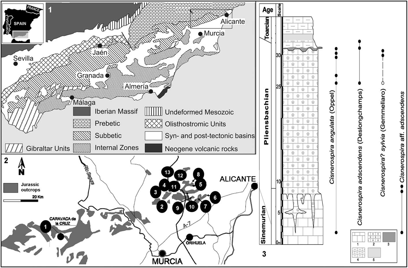
The studied specimens come from 13 Early Jurassic calcareous outcrops (Fig. 1) located in Alicante and Murcia provinces (SE Spain), forming part of the External Zones of the Betic Cordillera within the easternmost Subbetic domain (Fig. 1). In the Early Jurassic, this region was situated in the South Iberian Paleomargin (westernmost Tethys Ocean) and was characterized by the predominance of shallow carbonate platforms and then by pelagic seamount facies during most of the Jurassic and Early Cretaceous (Vera et al., Reference Vera, Martín-Algarra, Sánchez-Gómez, Fornós and Gelabert2004).
(1) Geological sketch showing the studied area within the context of the Betic Cordillera (modified after Vera et al., Reference Vera, Martín-Algarra, Sánchez-Gómez, Fornós and Gelabert2004). (2) Map showing the locations of the localities where Cisnerospira was recorded among the Jurassic outcrops from Alicante and Murcia provinces (Spain): 1. Rincón de Egea; 2. Sierra de La Espada; 3. Sierra de Quibas; 4. Rabillo de Quibas; 5. Sierra Pelada; 6. Sierra de Orts; 7. Tarabillo section; 8. Cerro de La Cruz; 9. Sierra de Los Frailes; 10. Sierra de Algayat; 11. Moleta de Togores; 12. Collado de La Campana; 13. Cruz de La Algueña. (3) Synthetic Lower Jurassic lithostratigraphical section showing the distribution of Cisnerospira species in the eastern Subbetic. Lithologic keys: 1: massive white limestone; 2: red crinoidal grainstone; 3: hardground with grey and red crinoidal limestone; 4: marly limestones; 5: marls.
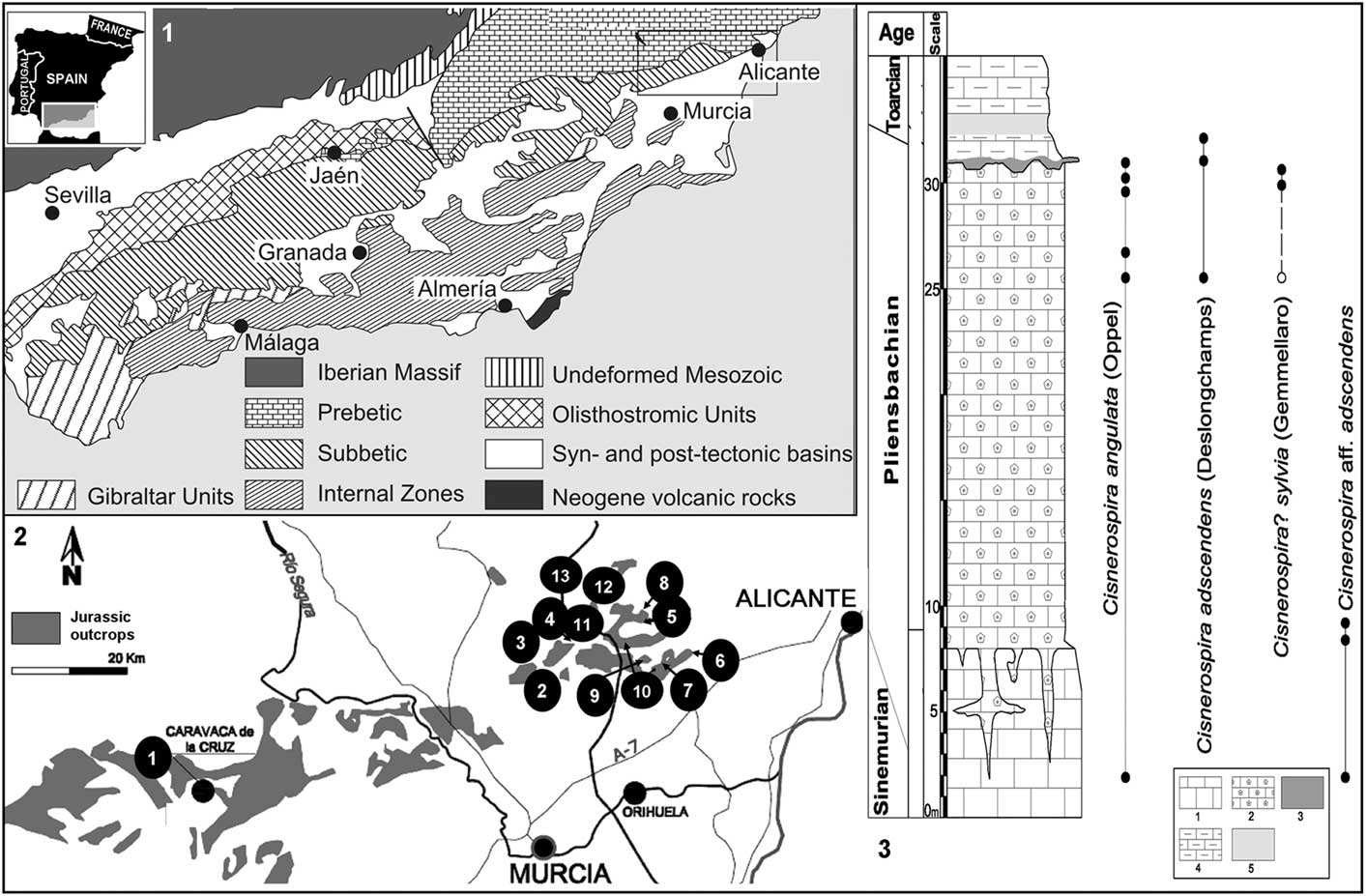
In the easternmost Subbetic area, the genus Cisnerospira is recorded in massive micritic and pseudo-oolithic whitish wackestone beds with lateral and upward transition to oolithic grainstone/packstone with intraclasts and peloids that belong to the intermediate member of the Gavilán Formation (Van Veen Reference Van Veen1969), which is assigned to the Sinemurian-Pliensbachian transition spanning the Echioceras raricostatum–Gemmellaroceras aenigmaticum zones (Baeza-Carratalá and García Joral, Reference Baeza-Carratalá and García Joral2012; Baeza-Carratalá, Reference Baeza-Carratalá2013). Simultaneously or immediately successive, occurrences of Cisnerospira are also found in dark pelmicritic limestone beds with chert and abundant crinoids (Fig. 1.3).
Overlying both previous deposits, Cisnerospira is also recorded in the upper member of the Gavilán Formation (Pliensbachian–earliest Toarcian), consisting of red crinoidal grainstone beds with abundant glauconite and occasionally calcarenite levels interspersed. These sediments are often found infilling extensional fractures. In the upper part, the red crinoidal grainstone beds show irregular tops with condensed pavements interpreted as hardgrounds with ammonoids, belemnites, and brachiopods overlain by lenticular deposits of glauconitic sandy limestone with a dark greyish matrix or limestone beds with wackestone texture also containing Cisnerospira. Finally, in the latest Pliensbachian–early Toarcian, the onset of the marly sedimentation in the Subbetic basin typified by the Zegrí Formation (Molina, Reference Molina1987) bears the last occurrences of Cisnerospira in yellowish/greenish marl and marly limestone beds with calcarenites and sandy marlstone sporadically interbedded (Fig. 1.3).
Materials and methods
A total number of 170 specimens were collected and studied from 13 localities (Fig. 1.2) where the Lower Jurassic deposits from the easternmost Subbetic area crop out and were summarized in a synthetic stratigraphical section (Fig. 1.3), displaying the complete record for the genus Cisnerospira. The taxonomic identifications were complemented by material held in the Jiménez de Cisneros historical collection (JdC collection hereafter) after a systematic revision (Baeza-Carratalá, Reference Baeza-Carratalá2008). The symbols used in the synonymic/chresonymic lists follow conventions summarized in Matthews (Reference Matthews1973).
The ammonite zonal standard scheme used is according to Cariou and Hantzpergue (Reference Cariou and Hantzpergue1997) for the Early Jurassic of the Mediterranean domain. Chronostratigraphical data mainly from Braga (Reference Braga1983), Iñesta (Reference Iñesta1988), and Caracuel et al. (Reference Caracuel, Fernández-López, Tent-Manclús and Yébenes2004) were also used.
For the taxonomical analysis, the main biometric parameters and indices were measured whenever possible. The internal structure was studied using the conventional method of preparing oriented transverse serial sections and taking acetate peels. The distance between serial sections was 0.1 mm. High-resolution microphotographs of acetate peels were taken under an optical microscope (Nikon CFI60 E600POL).
Repositories and institutional abbreviations
All specimens examined as well as the obtained peels are deposited at Departamento de Ciencias de la Tierra y del Medio Ambiente of the University of Alicante (DCTMA) and in the JdC collection held at the Paleontological Museum of Murcia (Murcia, Spain).
Biostratigraphical record of the genus Cisnerospira
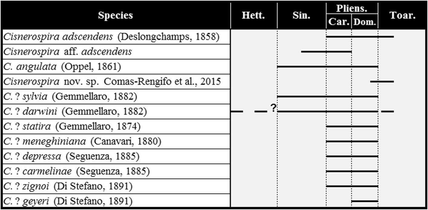
The global biostratigraphical distribution of the representatives of Cisnerospira is restricted to the Early Jurassic, showing their last occurrence in the early Toarcian (Dactylioceras polymorphum Zone), when they became extinct worldwide with the remaining spire bearers, i.e., the early Toarcian mass extinction event (Vörös, Reference Vörös2002; Comas-Rengifo et al., Reference Comas-Rengifo, García Joral and Goy2006; Baeza-Carratalá et al., Reference Baeza-Carratalá, García Joral, Giannetti and Tent-Manclús2015). Figure 2 summarizes the stratigraphical range of the taxa assigned or tentatively adscribed to Cisnerospira in the western Tethys Ocean as a whole.
Biostratigraphical distribution range of Cisnerospira species (and taxa tentatively assigned to this genus) in the western Tethys. Sources cited in the text.
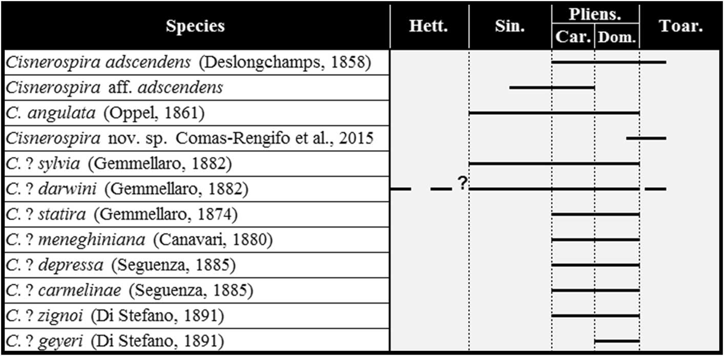
The earliest record of the genus seems to be C.? darwini (pending the analysis of its internal structure), which has a wide distribution from the Hettangian? to the late Pliensbachian–early Toarcian. The Hettangian records of this species have some uncertainty. Thus, Vörös (Reference Vörös1997) first recorded C.? cf. darwini from the Hettangian of the Bakony Mountains but finally assigned Gemmellaro’s species without reservation to the Sinemurian–Pliensbachian (Vörös and Dulai, Reference Vörös and Dulai2007; Vörös, Reference Vörös2009). Moreover, this species is widely recorded in several Tethyan basins throughout the Early Jurassic (e.g., Di Stefano, Reference Di Stefano1891; De Gregorio, Reference De Gregorio1930; Dulai, Reference Dulai2003; Vörös and Dulai, Reference Vörös and Dulai2007; Vörös, Reference Vörös2009; Mandl et al., Reference Mandl, Dulai, Schlögl, Siblík, Szabó, Szente and Vörös2010), including C.? darwini graeca in Western Greece (Renz, Reference Renz1932; Manceñido, Reference Manceñido1978, Reference Manceñido1993) and even a record from the Moluccas Islands in eastern Seram (Wanner and Knipscheer, Reference Wanner and Knipscheer1951; Manceñido, Reference Manceñido1978).
C.? sylvia and C. angulata range from the Sinemurian to the late Pliensbachian. The first species is recorded in the Sinemurian from the Transdanubian Range (Vörös, Reference Vörös1993; Dulai, Reference Dulai2002, Reference Dulai2003; Vörös and Dulai, Reference Vörös and Dulai2007) as well as in the Sinemurian of the Northern Calcareous Alps (Mandl et al., Reference Mandl, Dulai, Schlögl, Siblík, Szabó, Szente and Vörös2010). Jiménez de Cisneros (Reference Jiménez de Cisneros1926) assigned this species in Spain to the ‘middle Lias,’ and Baeza-Carratalá (Reference Baeza-Carratalá2008, Reference Baeza-Carratalá2013) recorded this taxon in the Pliensbachian from the Subbetic area. On the other hand, C. angulata has been widely recorded in the western Tethys. It is assigned to the Sinemurian in Hungary (Vörös, Reference Vörös1993; Dulai, Reference Dulai2002, Reference Dulai2003; Vörös and Dulai, Reference Vörös and Dulai2007) and Austria (Vörös et al., Reference Vörös, Szabó, Dulai, Szente, Ebli and Lobitzer2003; Mandl et al., Reference Mandl, Dulai, Schlögl, Siblík, Szabó, Szente and Vörös2010); the Carixian from Pyrenees (Alméras and Fauré, Reference Alméras and Fauré2000); the latest Sinemurian–early Carixian from Western Algeria (Elmi et al., Reference Elmi, Alméras, Benhamou, Mekahli and Marok2003); and the early Pliensbachian from the Caucasus (Ruban, Reference Ruban2004). Comas-Rengifo et al. (Reference Comas-Rengifo, García Joral and Goy2006) recorded C. angulata in the North Iberian margin from the E. raricostatum–latest Davoei zones (late Sinemurian–Pliensbachian). In the Betic Cordillera, Jiménez de Cisneros (Reference Jiménez de Cisneros1921a, Reference Jiménez de Cisneros1923, Reference Jiménez de Cisneros1935) assigned this species to the ‘middle Lias,’ and Iñesta (Reference Iñesta1988, Reference Iñesta1993) and Baeza-Carratalá (Reference Baeza-Carratalá2008, Reference Baeza-Carratalá2013) assigned it to the Pliensbachian, finally supporting in this work a distribution range from the late Sinemurian to the late Pliensbachian in the Subbetic basin.
In the Sinemurian–Pliensbachian transition, the species C. aff. adscendens is restricted in the Subbetic area to the E. raricostatum-G. aenigmaticum zones (Baeza-Carratalá and García Joral, Reference Baeza-Carratalá and García Joral2012; Baeza-Carratalá, Reference Baeza-Carratalá2013). Some other species tentatively attributed to Cisnerospira, such as statira (Gemmellaro, Reference Gemmellaro1874), meneghiniana (Canavari, Reference Canavari1880), depressa (Seguenza, Reference Seguenza1885) carmelinae (Seguenza, Reference Seguenza1885), and zignoi (Di Stefano, Reference Di Stefano1891) are recorded throughout the Pliensbachian.
Finally, the last representatives of the genus are recorded in the late Pliensbachian–early Toarcian transition, with the type species, C. adscendens, reported by Deslongchamps (Reference Deslongchamps1858, p. 166) from the ‘middle Liassic’ (Pliensbachian) and from the ‘Couche à Leptaena’ (early Toarcian, D. polymorphum Zone) but noticed that they may be reelaborated (‘remaniés’) in these latter deposits. It was not possible for us to sort out such presumptive reelaborated condition in the present type area. This taxon was also assigned to the ‘middle Liassic’ by Jiménez de Cisneros (Reference Jiménez de Cisneros1935), and Alméras (Reference Alméras1964) adscribed it to the Carixian–Domerian. British occurrences of this species were assigned to the late Pliensbachian by Thomas (Reference Thomas1978) and Ager (Reference Ager1987), while Baeza-Carratalá (Reference Baeza-Carratalá2008, Reference Baeza-Carratalá2013) recorded C. adscendens in the Pliensbachian–earliest Toarcian from the Subbetic area. The latest record probably corresponds to the subpyramidal forms assigned to Cisnerospira n. sp. from the D. Polymorphum Zone in the Lusitanian Basin (Comas-Rengifo et al., Reference Comas-Rengifo, Duarte, Félix, García Joral, Goy and Rocha2015), yet a more accurate systematic allocation is required for these forms.
The aforementioned biochronological and spatial data suggest a possible migration trend of the genus Cisnerospira taking into account that the early records of C. angulata are restricted to the most intra-Tethyan areas of the Alpine region, and later Cisnerospira is recorded in the surrounding Tethyan basins, culminating with the occurrences of C. adscendens in the westernmost European areas. A comparable phenomenon, involving other brachiopod taxa, has already been noticed by Ager (e.g., Reference Ager1960, Reference Ager1967, Reference Ager1993) and recently by Baeza-Carratalá et al. (Reference Baeza-Carratalá, García Joral, Giannetti and Tent-Manclús2015).
Systematic paleontology
Phylum Brachiopoda Duméril, Reference Duméril1805
Subphylum Rhynchonelliformea Williams et al., Reference Williams, Carlson, Brunton, Holmer and Popov1996
Class Rhynchonellata Williams et al., Reference Williams, Carlson, Brunton, Holmer and Popov1996
Order Spiriferinida Ivanova, Reference Ivanova1972
Suborder Spiriferinidina Ivanova, Reference Ivanova1972
Superfamily Spiriferinoidea Davidson, Reference Davidson1884
Family Spiriferinidae Davidson, Reference Davidson1884
Subfamily Paralaballinae Carter, Carter in Carter et al., Reference Carter, Johnson, Gourvennec and Hou1994
Genus Cisnerospira Manceñido, Reference Manceñido2004 .
Type species
Spiriferina adscendens Deslongchamps, Reference Deslongchamps1858
Diagnosis
Smooth, medium-sized spiriferinid with very large ventral valve, subpyramidal in outlook, deeper than the weakly convex (operculiform) dorsal one. Microornamentation consists of very fine and dense spinules with quincuncial arrangement. Dental plates are placed close to each other. Hinge teeth inserted in broad and shallow sockets. High ventral median septum. Cardinal process is a ctenophoridium with densely striated myophore; short hinge plates nearly fused to the socket ridges and crural bases developed very close (even attached) to the dorsal valve. Dorsal median septum and septalium absent. Spiralium oriented ventrally up to posteriorly.
Etymology
As a tribute to Jiménez de Cisneros, paleontologist who formerly endeavored to split systematically the Spiriferina group in the Subbetic area.
Occurrence
Cisnerospira is widely distributed in the Sinemurian–early Toarcian of the westernmost Tethys margin, showing more profuse records in the Mediterranean bioprovince but also occurring in some Subboreal basins of the NW European one, such as the Iberian Range, Pyrenées, Portugal, Normandy, or the Dorset basins, and occasionally reaching eastward up to Indonesia. The palaeogeographical distribution and the biostratigraphical range of Paralaballinae are thus extended, ranging from the Late Triassic of Tibet, with Paralaballa as the earliest representative, to the Early Jurassic (earliest Toarcian) of the western Tethys, with Cisnerospira as the second and last known genus of this subfamily.
Remarks
The genus Cisnerospira Manceñido, Reference Manceñido2004 is systematically allocated into the subfamily Paralaballinae Carter, Carter in Carter et al., Reference Carter, Johnson, Gourvennec and Hou1994, consisting up to now of the single genus Paralaballa Sun, Reference Sun1981 from the Late Triassic of Tibet (Carter et al., Reference Carter, Johnson, Gourvennec and Hou1994; Carter, Reference Carter2006). The relationship between Cisnerospira and Paralaballa is apparent in both external and internal features shared by them. The external similarity is especially based on the cyrtiniform shape, with smooth shells, subpyramidal ventral valves, and the presence of median dorsal fold and ventral sulcus. The internal structures show a remarkable likeness mainly concerning the articulation, with well-developed dental plates and ventral median septum; absence of spondylium, septalium, and dorsal median septum; and with development of crural bases very close or even fused to the dorsal valve by short dorsal adminicula. Consequently, the type species of Paralaballa, P. zangbeiensis (Sun, Reference Sun1981, 7, 32; text-fig. 14; Carter, Reference Carter2006, p. 1934, fig. 1289, 1 a–g), shows the architecture of the hinge area and the early stages of the cardinalia comparable to those present in the genus Cisnerospira.
The analysis of the particular spiralium architecture of this genus deserves special attention. Between-subfamily analysis reveals that the singular orientation of the spiralia in Cisnerospira, with the apices pointing into the internal cavity of the ventral beak (Ager, Reference Ager1987, text-fig. 2), markedly differs from the orientation shown by the representatives of other closely related subfamilies, such as Spiriferininae Davidson, Reference Davidson1884; e.g., Triadispira Dagys, Reference Dagys1961; Spiriferina d’Orbigny, Reference d’Orbigny1847; Calyptoria Cooper, Reference Cooper1989; Cingolospiriferina Pozza, Reference Pozza1992; or Liospiriferina Rousselle, Reference Rousselle1977 (formerly Cisnerospira species had been arranged within this latter genus). In this sense, in addition to the differences in the spiralium and the external shape, the genus Liospiriferina possesses a dorsal median septum and septalium and distinctive structure of the cardinalia.
Within-subfamily analysis reveals that the earliest representative of Paralaballinae shows spiralia with laterally directed apices but substantially occupying the ventral valve cavity. This ventral development is more clearly manifested in Early Jurassic representatives, from the Sinemurian onward; thus, although the analysis of the internal structure in Cisnerospira angulata (distributed throughout the Early Jurassic) is not conclusive about the orientation of the apices, the ventral development of the spiralia seems to be increased noticeably, as will be discussed in the following when describing C. angulata. Finally, the late Pliensbachian–early Toarcian representatives of Cisnerospira (C. adscendens) clearly show apices oriented toward the ventral beak (Thomas Reference Thomas1978, text-fig. 14; also in Ager, Reference Ager1987, text-fig. 2). Hence, an overall trend implicit in this interpretation suggests that the spiralia with posteriorly pointing apices would represent a derived condition fully attained in younger members of the subfamily.
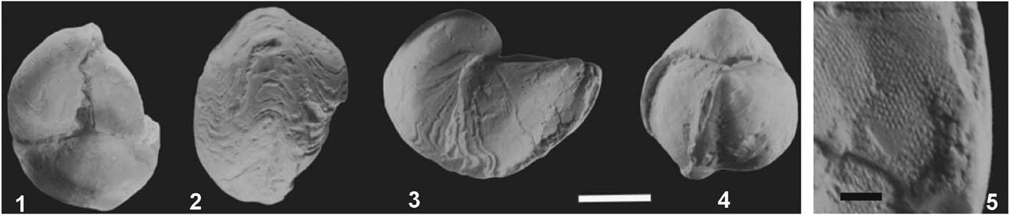
On the basis of the aforementioned diagnostic criteria, besides the type species Cisnerospira adscendens (Deslongchamps, Reference Deslongchamps1858), of which a plaster cast is figured herein (Fig. 3), other taxa that have been assigned to Cisnerospira are C. aff. adscendens (herein), C. angulata (Oppel, 1861), and Cisnerospira n. sp. Comas-Rengifo et al. (Reference Comas-Rengifo, Duarte, Félix, García Joral, Goy and Rocha2015). Pending further studies to ascertain their detailed taxonomic status, in the absence of data about their internal structure, and due to their high resemblance in the external shape, the following nominal species are provisionally referred to Cisnerospira as well: C.? sylvia (Gemmellaro, Reference Gemmellaro1882), C.? darwini (Gemmellaro, Reference Gemmellaro1882), C.? statira (Gemmellaro, Reference Gemmellaro1874), C.? meneghiniana (Canavari, Reference Canavari1880), C.? depressa (Seguenza, Reference Seguenza1885), C.? carmelinae (Seguenza, Reference Seguenza1885), C.? geyeri (Di Stefano, Reference Di Stefano1891), and C.? zignoi (Di Stefano, Reference Di Stefano1891).
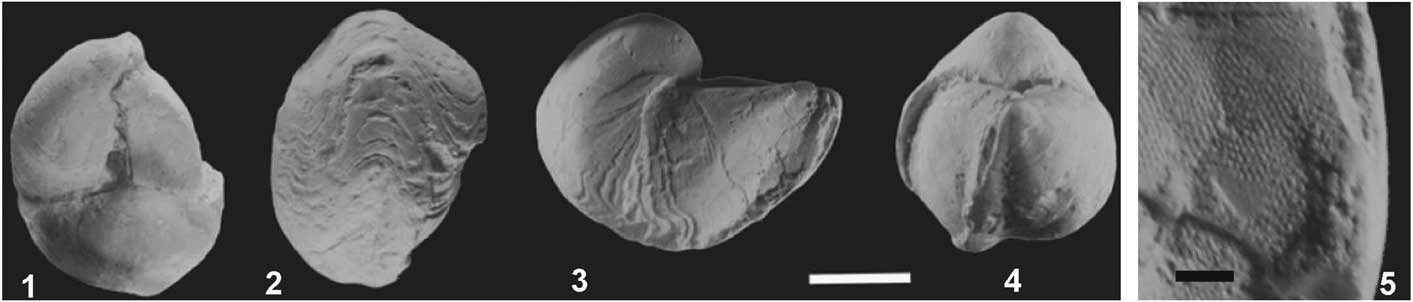
Cisnerospira adscendens (Deslongchamps, Reference Deslongchamps1858). Plaster cast of hypotype specimen (Natl. Mus. Wales 59.410.643.A) studied by Thomas (Reference Thomas1978, pl. 2, fig. 3c, e, f, text-fig. 14; see also Ager, Reference Ager1987, text-fig. 2) from Thorncombe Beacon, Dorset, England, ‘Junction Bed’ (Inter-paltus layer), late Pliensbachian (Spinatum Zone). (1) Dorsal view; (2) anterior view; (3) lateral view; (4) posterior view; (5) detailed view showing the arrangement of the microornamentation consisting of very fine and dense spinules. (1–4) Scale bar = 1 cm; (5) scale bar = 1 mm.
On the other hand, taxa with a similar subpyramidal shape and unribbed median fold and sulcus, but awaiting to identify accurate data of their internal architecture, are provisionally excluded from Cisnerospira due to the presence of faint radial costation on the anterior flanks (instead of the weak striae that are occasionally present in some specimens of Cisnerospira). This is the situation of the nominal species Spiriferina guillieroni Haas, Reference Haas1887, systematically arranged in Callospiriferina Rousselle, Reference Rousselle1977 by Comas-Rengifo et al. (Reference Comas-Rengifo, García Joral and Goy2006), whereas S. gibba Seguenza, Reference Seguenza1885, was assigned to Callospiriferina? by Baeza-Carratalá (Reference Baeza-Carratalá2008) and to Cisnerospira by Vörös and Kandemir (Reference Vörös and Kandemir2011). In addition, the smooth Spiriferina acuta (Stur in Geyer, Reference Geyer1889) is cautiously excluded from Cisnerospira for having wider than high interareas and a rather incurved beak, suggesting a closer resemblance to representatives of Liospiriferina (Vörös, 2015, personal communication).
Cisnerospira adscendens (Deslongchamps, Reference Deslongchamps1858)
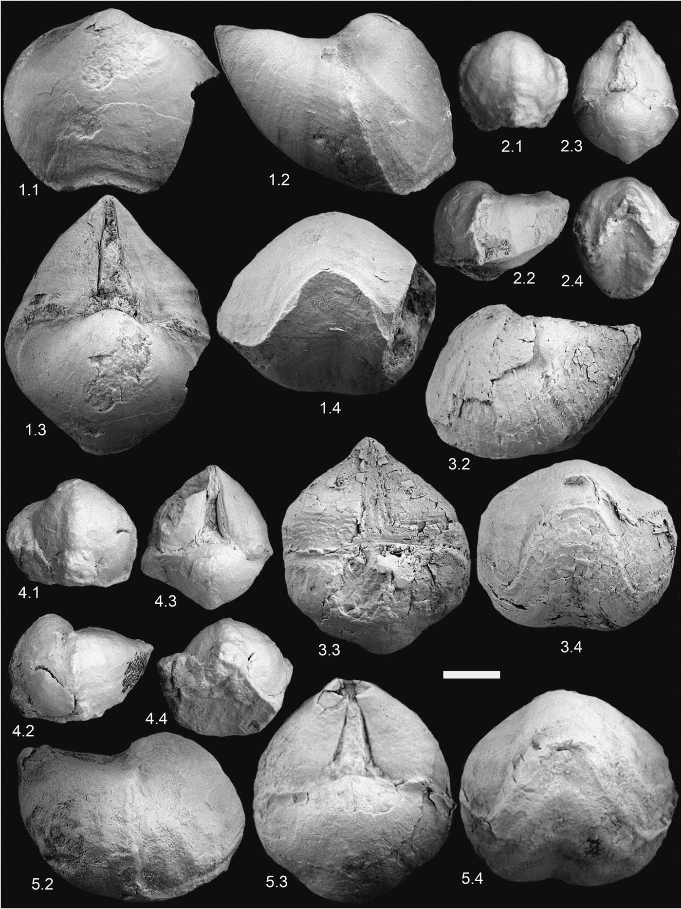
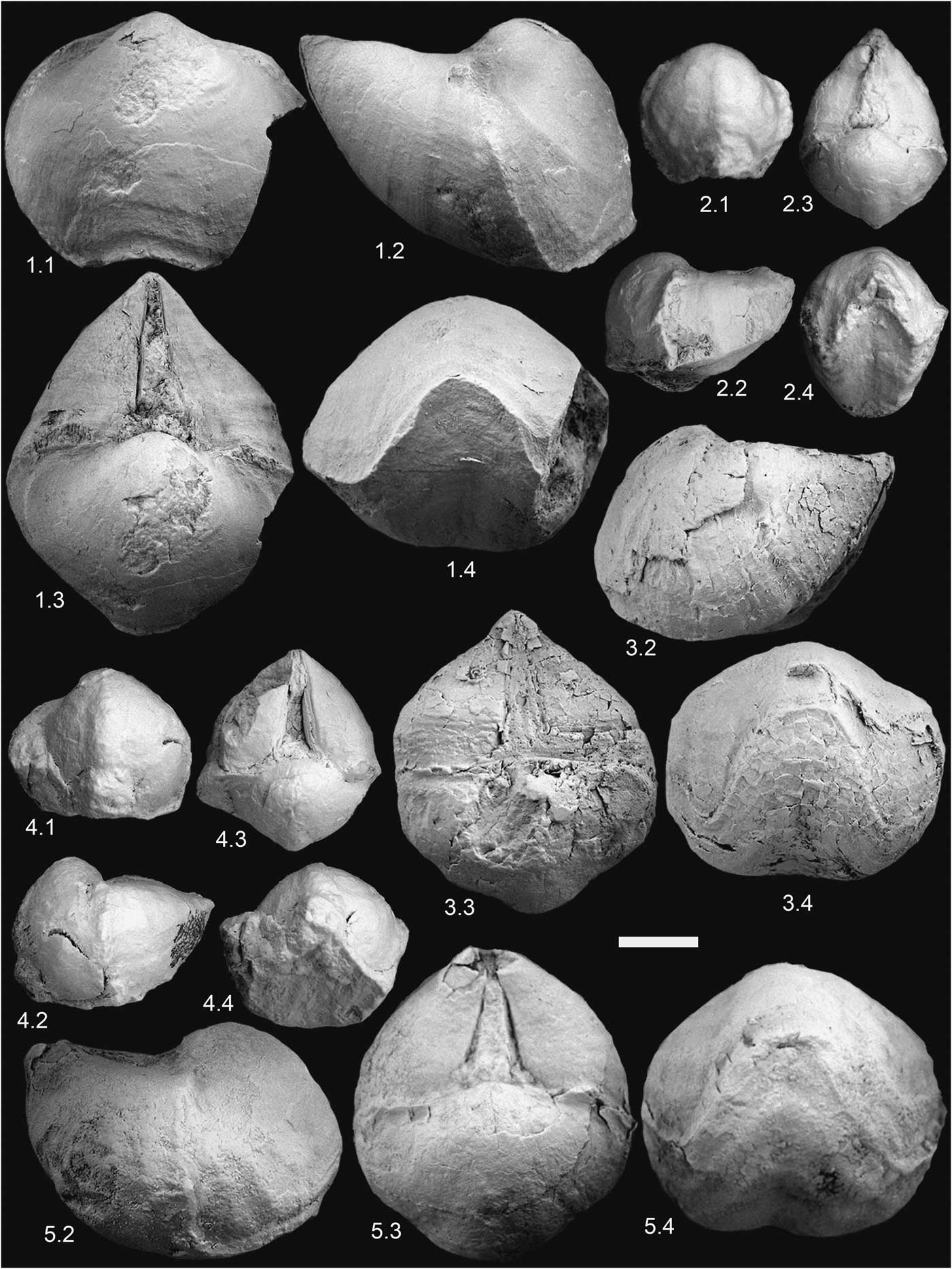
Cisnerospira adscendens (Deslongchamps) from the easternmost Subbetic area. Views of each specimen are ordered consecutively in (1) dorsal, (2) lateral, (3) posterior, and (4) anterior views. All specimens were coated with magnesium oxide. (1) Specimen SHF.ad.1 from the Tarabillo section; (2) specimen O.IX.23.2 from JdC collection; (3) specimen SHN.ad.1 from Sierra de Orts; (4) Specimen O.IX.23.1 from JdC collection; (5) specimen SHN.ad.2 from Sierra de Orts, sectioned in Figure 5.1. Scale bar = 1 cm.
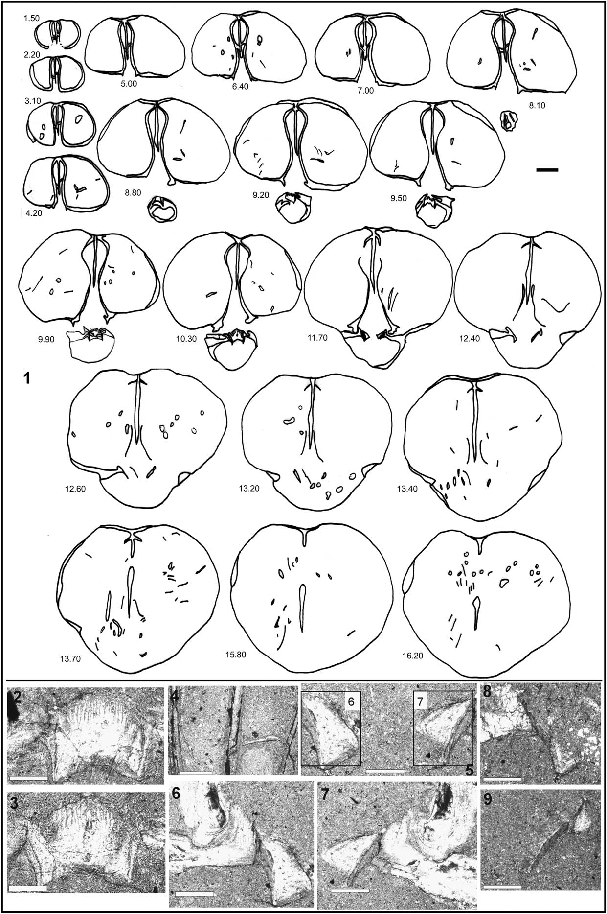
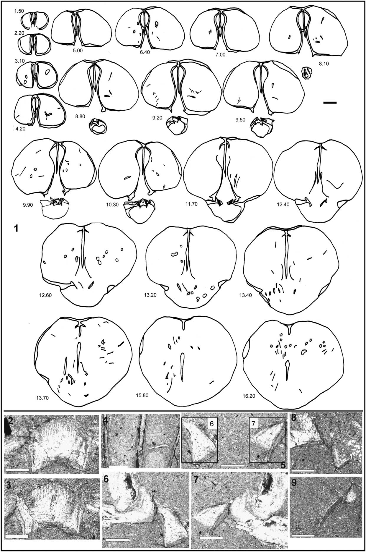
Internal structure of Cisnerospira adscendens (Deslongchamps). (1) Transverse serial sections through the specimen SHN.ad.2 (distance from the beak in millimeters). (2–9) Microphotographs of acetate peels from the same specimen: (2, 3) sections at 9.90 and 10.30 mm from the apex, respectively, showing details of the cardinal process (ctenophoridium); (4) section at 4.20 mm from the apex showing ventral median septum with thin lamellas supported by the dental plates; (5–7) serial section at 11.70 mm from the apex, showing hinge plates tending toward the dorsal valve and crural bases highly inclined dorsally; (6, 7) detailed views; (8, 9) serial sections at 12.40 mm from the apex, showing dorsal crural development. (1) Scale bar = 3.0 mm; (4) scale bar = 0.5 mm; (2, 3, 5–9) scale bars = 1.0 mm.
1852 Spirifer rostratus [A malformation]; Reference DavidsonDavidson, p. 263, pl. 15, fig. 11–11b.
*1858 Spiriferina adscendens (Nobis); Reference DeslongchampsDeslongchamps, p. 165, pl. 4, figs. 7–9.
1878 Spiriferina adscendens Deslongchamps; Reference DavidsonDavidson, p. 227, pl. 29, fig. 20, 20a.
1921a Spiriferina acuta Stur transient S. gibba?; Reference Jiménez de CisnerosJiménez de Cisneros, p. 7, pl. 32, figs. 3, 4.
pars
1921a Spiriferina gibba Seguenza; Reference Jiménez de CisnerosJiménez de Cisneros, p. 7.
pars
1923 Spiriferina adscendens Deslongchamps; Reference Jiménez de CisnerosJiménez de Cisneros, p. 44.
non
1927 Spiriferina tumida V. Buch var. ascendens Deslongchamps; Reference CorroyCorroy, p. 15, fig. 1, pl. 3, figs. 5–12.
2004 Liospiriferina cf. angulata (Oppel); Reference Baeza-Carratalá, Dulai and VörösBaeza-Carratalá et. al., p. 220.
2008 Cisnerospira aff. angulata; Reference Baeza-CarrataláBaeza-Carratalá, p. 366, pl. 27, fig. 2; pl. 28, fig. 5.
Types and material
Cerro de La Cruz (three specimens), Sierra de Orts (five specimens), Tarabillo section (three specimens), supplemented by 24 specimens from the JdC collection, recorded from Sierra de la Espada, Cerro de La Cruz, Rincón de Egea, and Sierra de Quibas. The syntypes originally studied and illustrated by Deslongchamps seem to be lost, as happened with other specimens stored in the Museum of Caen, which was destroyed in World War II (Alméras et al., Reference Alméras, Cougnon, Guibbert and Fauré2014). Hence, it was very helpful to add observations on a plaster cast of a hypotype of Deslongchamps’s species, collected by Jackson from the ‘Junction Bed’ at Thorncombe Beacon, Dorset, SW England (Fig. 3), since this comes from beds of similar facies and age as in Normandy; moreover, it closely matches in morphology the specimen figured by Deslongchamps (Reference Deslongchamps1858, pl. 4, fig. 7–7d), which is herein selected as lectotype (complying with ICZN, 1999, Art. 74.4) to stabilize the interpretation of this species.
Description
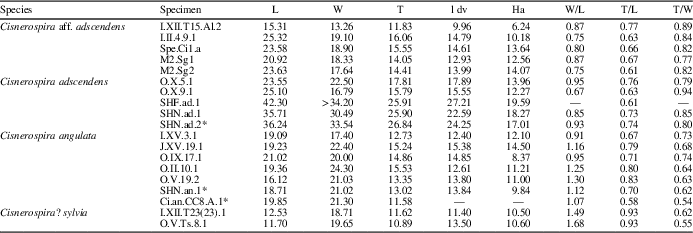
External features: medium-sized ventribiconvex shells (Table 1), with triangular to subpentagonal outline and length greater than width. Lateral commissure is straight, and the anterior one is uniplicate with a low, narrow, parabolic, dorsal median fold. Ventral valve is very large and deeper than the operculiform dorsal one. Dorsal valve shows flat extensions on each flank of the shell near the cardinal margin. Ventral umbo is straight.
Mean biometric measurements in representative specimens of Cisnerospira from the Subbetic area. L = total length; l dv = length of dorsal valve; W = total width; T = total thickness; Ha = height of interarea (in mm). *Specimens sectioned in this work.

The well-developed ventral valve together with the blunt lateral ventral edges give this species a conical shape, instead of pyramidal as is usual in other close species such as C. angulata. Long, triangular interarea is almost catacline (or incipiently apsacline) and shows rounded beak ridges. Delthyrium is narrow and triangular in outline. Smooth shell except for the dense growth lines and microornamentation consisting of very fine and dense spinules with quincuncial arrangement (Fig. 3).
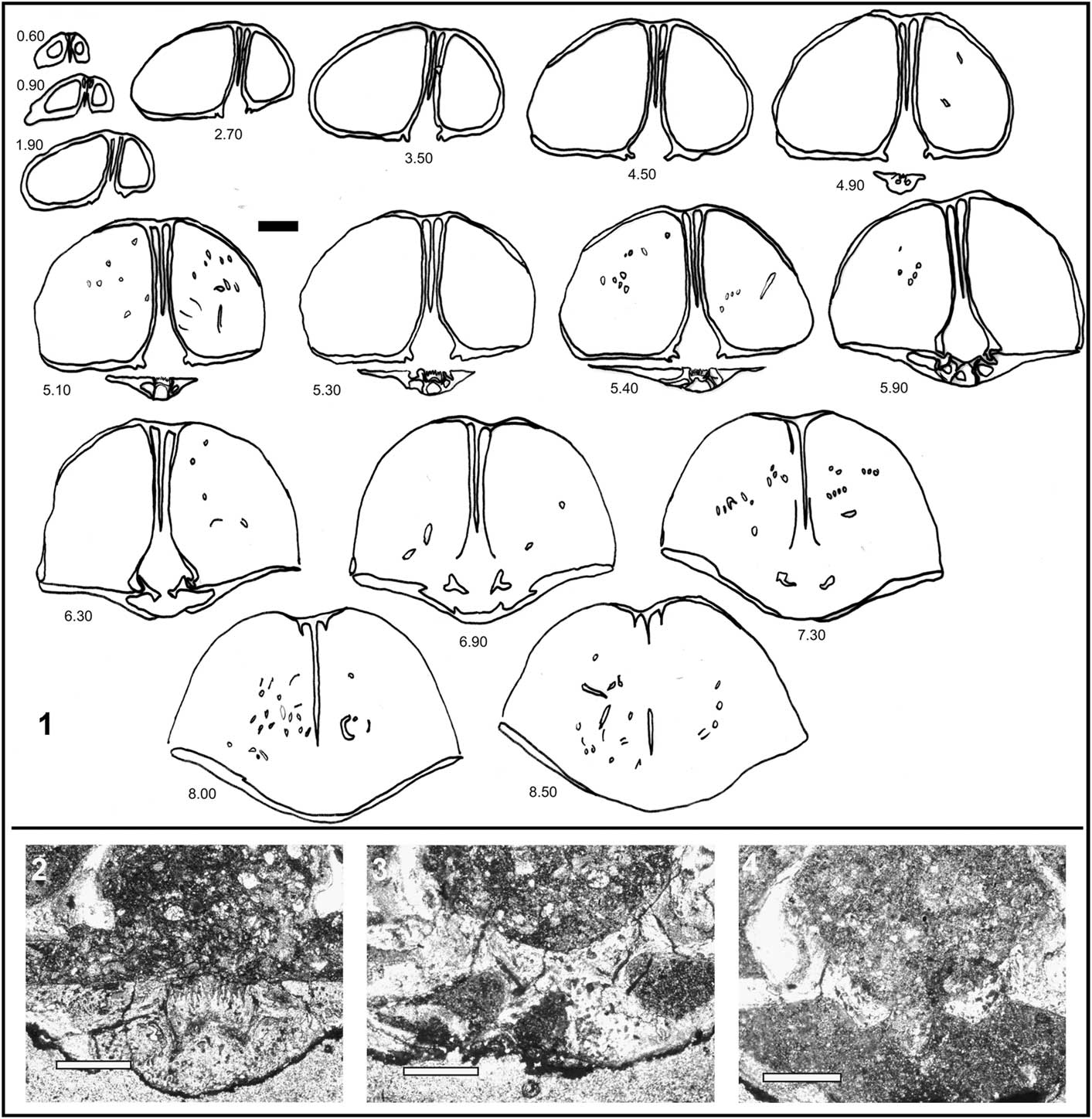
Internal structure (Fig. 5): wide umbonal lateral cavities. A high ventral median septum persists up to half of shell length, without tichorhinum but with thin lamellae supported to the dental plates in the posterior third of the shell (Fig. 5; cf. Ager, Reference Ager1987, text-fig. 2). The central cavity is rather narrow due the close proximity of the dental plates, which are dorsally divergent and rather arcuate. Hinge teeth are massive and inserted in broad and shallow sockets. Slight denticula are present. Outer and inner socket ridges are well developed. Cardinal process is a convex, highly striated ctenophoridium with a wide cardinal platform and a slender myophragm. Dorsal median septum and septalium absent.
Hinge plates are short and clearly tilted toward the dorsal valve, showing crural bases dorsally inclined. Jugum is not developed and, in the Subbetic material, spiralium is not preserved. Manceñido (Reference Manceñido2004), on the basis of the illustrations of Thomas (Reference Thomas1978, also in Ager Reference Ager1987), remarked that Cisnerospira has elongate-conical spiralia featuring both subparallel axes and their apices pointing into the internal cavity of the ventral beak.
Remarks
Spiriferina adscendens Deslongchamps, Reference Deslongchamps1858 was designated by Manceñido (Reference Manceñido2004) as the type species of the genus Cisnerospira. This genus includes smooth shells with very large and subpyramidal ventral valves and operculiform dorsal ones, with the apices of the cone-shaped spiralia pointing into the internal cavity of the ventral beak. This distinctive feature was early predicted by Deslongchamps (Reference Deslongchamps1858, p. 166) and later verified by Thomas (Reference Thomas1978, unpublished text-fig. 14; also in Ager Reference Ager1987, text-fig. 2). This combination of external and internal features agrees with the morpho-grouping scheme of Jiménez de Cisneros (Reference Jiménez de Cisneros1921a), who considered Spiriferina adscendens, S. angulata, and, to a lesser extent, S. acuta as very closely related forms or, at least, a group of morphologically similar species worthy of being split from the remaining spiriferinids.
C. adscendens is similar to C. angulata, this latter species showing a less-developed ventral valve with an acute and straight outline and sharper beak ridges. The similarity with Spiriferina? gibba is also apparent, but the presence of faint costation in S.? gibba led this species to be precautionarily separated from the Cisnerospira stock, awaiting new data on the internal structure of Spiriferina? gibba.
In the Subbetic material, the only variability observed is the presence of oversized specimens and asymmetric shapes in dorsal view.
Occurrence in the Subbetic basin
It was reported from the ‘middle Liassic’ by Jiménez de Cisneros (Reference Jiménez de Cisneros1935). Baeza-Carratalá (Reference Baeza-Carratalá2008, Reference Baeza-Carratalá2013) recorded C. adscendens in latest Pliensbachian–earliest Toarcian deposits.
Cisnerospira aff. adscendens (Deslongchamps, 1921)
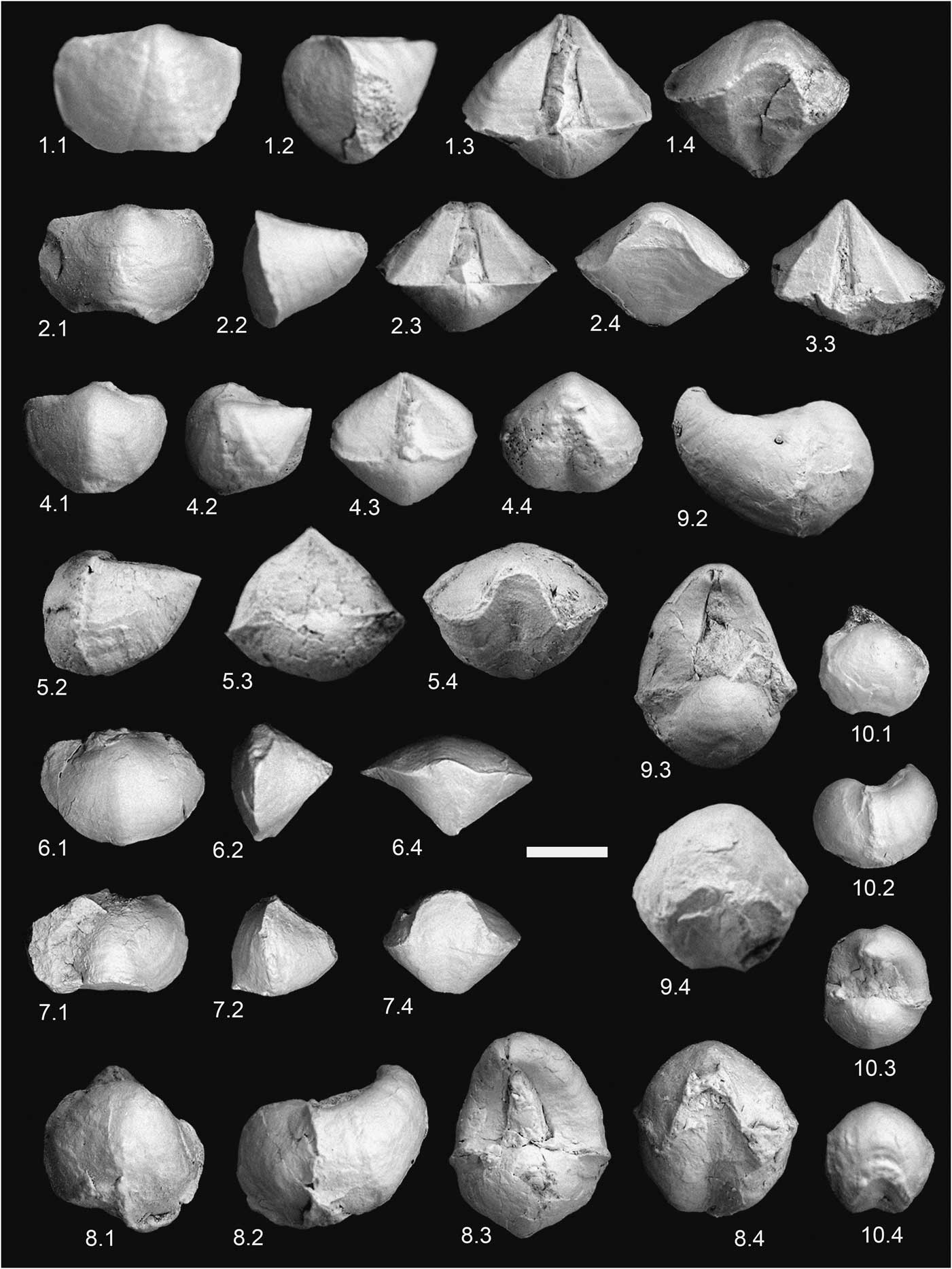
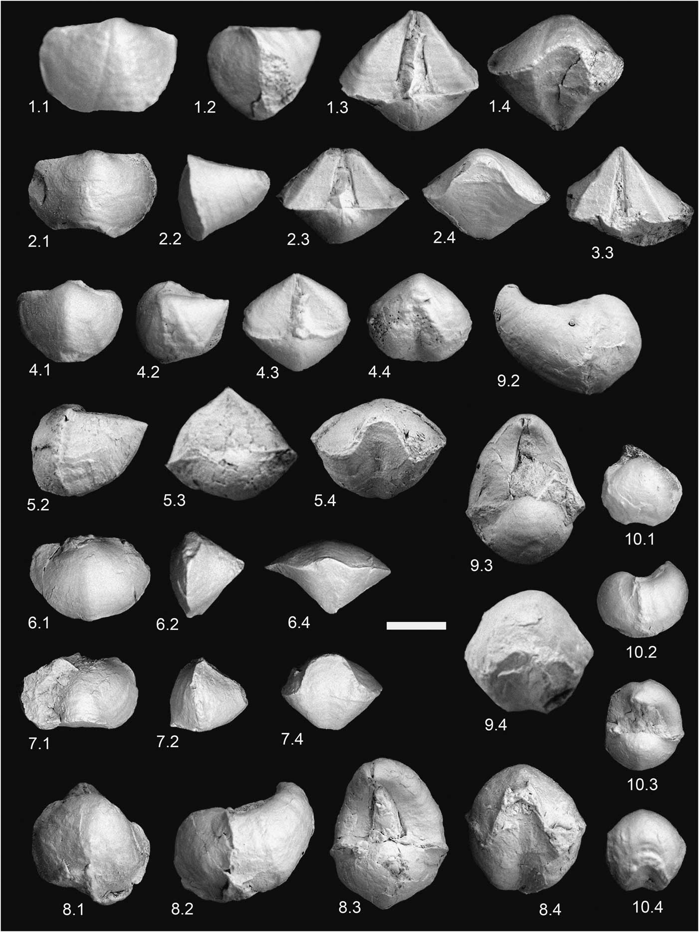
Some representative species of Cisnerospira from the easternmost Subbetic area. Views of each specimen are ordered consecutively in (1) dorsal, (2) lateral, (3) posterior, and (4) anterior views. All specimens were coated with magnesium oxide. (1–5) Cisnerospira angulata (Oppel): (1) specimen J.XV.19.1 from Sierra de Orts; (2) specimen O.V.19.2 from Moleta de Togores; (3) fragmented specimen (CCA.9.Can.1) from Cerro de la Cruz; (4) specimen CCA.9.Can.2 from Cerro de la Cruz; (5) specimen SHN.an.1 from Sierra de Orts, sectioned in Figure 7.1. (6–7) Cisnerospira? sylvia (Gemmellaro): (6) specimen I.XII.t23.23.1 from Sierra de Algayat; (7) Specimen O.V.TS8.1 from Moleta de Togores. (8–10) Cisnerospira aff. adscendens: (8) Specimen I.II.4.9.1 from Cruz de La Algueña; (9) specimen Spe.Ci1.a from Sierra Pelada; (10) specimen I.XII.T15(Al).2 from Sierra de Algayat. Scale bar = 1.0 cm.
1921b Spiriferina adscendens Deslongchamps; Reference Jiménez de CisnerosJiménez de Cisneros, p. 393, pl. 1, fig. 2.
pars
1923 Spiriferina adscendens Deslongchamps; Reference Jiménez de CisnerosJiménez de Cisneros , p. 44.
1935 Spiriferina adscendens Deslongchamps; Reference Jiménez de CisnerosJiménez de Cisneros, p. 24, pl. 1, fig. 4, 4’.
2008 Cisnerospira adscendens (Deslongchamps); Reference Baeza-CarrataláBaeza-Carratalá, p. 371, pl. 27, figs. 3, 4.
Description
Medium- to large-sized Cisnerospira (Table 1), with ventribiconvex shell and rounded subpentagonal dorsal outline. Length greater than width. Ventral valve is elongated and much deeper than the dorsal one. The extremely developed ventral valve and the curvature of the shell, ranging from the posterior umbo to the hinge line, confer to the shell a distinctive incurved subpyramidal shape. Dorsal valve nearly operculiform with an incurved umbo, at either side of which a couple of flat, wing-shaped, lateral expansions are symmetrically arranged adjacent to the posterior edge of each flank. Ventral beak rather incurved and with highly developed, apsacline, triangular interarea, which is bounded by curved, blunt, beak ridges and bisected by a narrow, triangular delthyrium. Lateral commissure is straight and the anterior one uniplicate, with a slight and narrow median fold, which has a rounded to elliptical outline, corresponding with a narrow, well-marked ventral sulcus. Smooth shells, except for a few specimens showing weak striae close to the anterior margin and very fine spinules with quincuncial arrangement. The internal features have not been observed because of the scanty inadequate material; only the large dental plates and ventral median septum are visible on the external surface of specimens.
Materials
Sierra Pelada (three specimens) and Sierra de Los Frailes (two specimens), supplemented by six specimens from JdC collection derived from Sierra de Algayat, Cruz de La Algueña, and Rabillo de Quibas.
Remarks
Intraspecific variability of C. aff. adscendens in the Subbetic area mainly lies in the occasional presence of very faint striae in three specimens, only noticed near the anterior margin. These striae are understood as results of a mature ontogenetic stage, and it is observed only in those individuals with a noticeable folding pattern; the striation therefore is not a costation of ‘Callospiriferina-type’ or related genera. In these latter, the radial ribs, although also slight, are more discernible and more developed in a radial anteroposterior orientation. Variability in the remaining features is rather small, only limited to the dorsal median fold (rounded to more acute in outline) and the incurving of the ventral valve, sometimes asymmetric.
Cisnerospira aff. adscendens shows a high resemblance to C. adscendens except for the greater curvature of its ventral valve and cardinal area, early noticed from the beginning of the ventral beak in C. aff. adscendens, instead of the distinctive straight outline visible in Deslongchamps’ (Reference Deslongchamps1858) species. C. aff. adscendens is also discernible from C. angulata, due to the extreme developement of its rather conical ventral valve and its curving profile. Referring to the ribbed forms, similar conical shapes can be found in Spiriferina gibba Seguenza, Reference Seguenza1885, but this latter taxon shows radial costation on the flanks. Finally, Spiriferina slovenica Siblík, Reference Siblík1965 shows less-developed ventral valve and a ribbing pattern consisting of few, but robust, coarse ribs.
Inasmuch as the analyses of the variability and the resemblance of these forms have been based upon few specimens, which are a unique record in the western Tethys until now, we cautiously consider them very related to C. adscendens under open nomenclature. This could be improved if C. aff. adscendens were subsequently found in further brachiopod faunas, which might justify either erection of a separate species or, interpretation as merely intraspecific variability of a known species.
Occurrence in the Subbetic basin
These endemic Subbetic forms were recorded by Baeza-Carratalá (Reference Baeza-Carratalá2008, Reference Baeza-Carratalá2013) from the uppermost Sinemurian–lowermost Pliensbachian (E. raricostatum-G. aenigmaticum zones).
Cisnerospira angulata (Oppel, 1861)
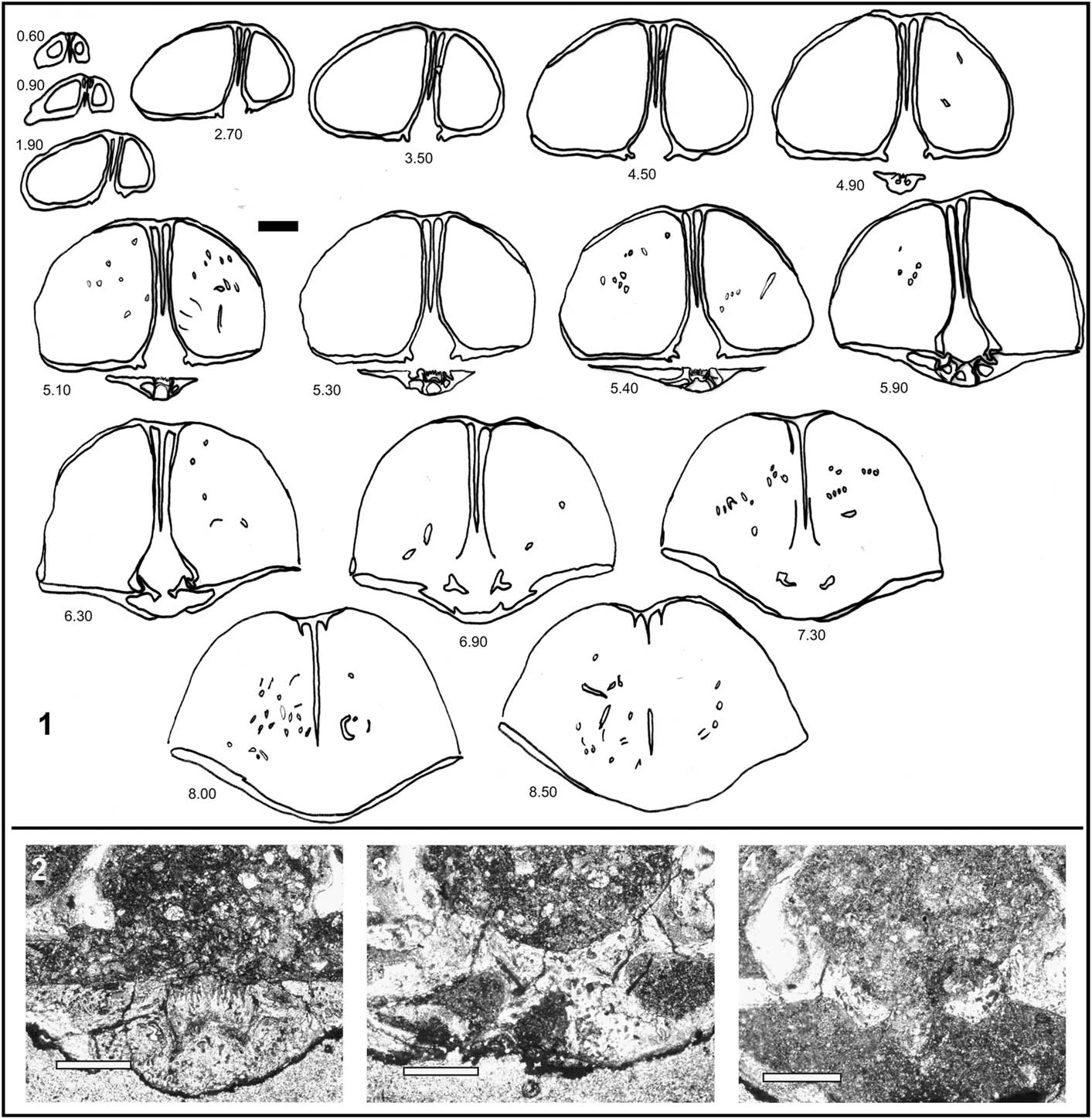
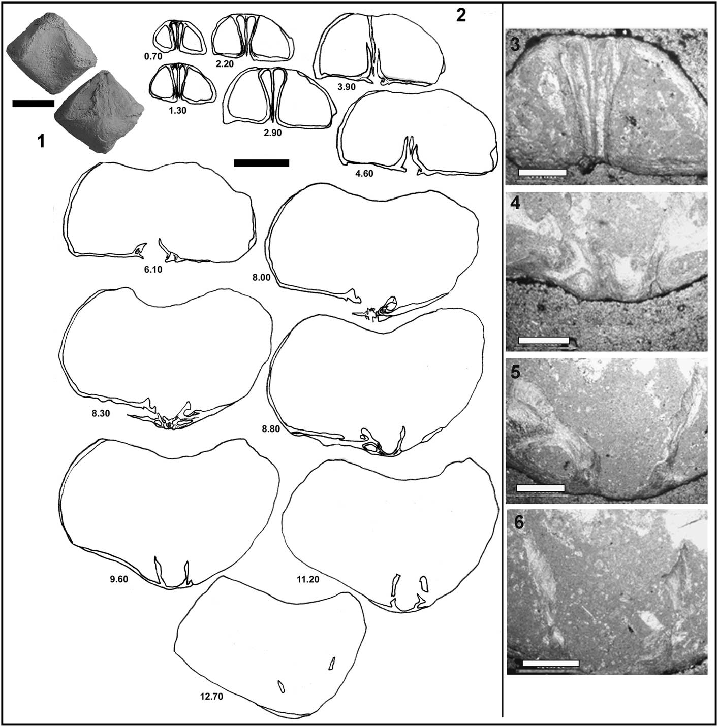
Internal structure of Cisnerospira angulata (Oppel). (1) Transverse serial sections through the specimen SHN.an.1 (distance from the beak in mm). (2–4) Microphotographs of acetate peels from the same specimen: (2, 3) sections at 5.30 and 5.90 mm from the apex, respectively, showing details of the cardinal process (ctenophoridium), hinge plates, and incipient crural bases highly inclined dorsally, nearly fused to the dorsal valve; (4) section at 6.30 mm from the apex, showing the anterior development of the hinge teeth, sockets, and hinge plates. (1) Scale bar = 2.0 mm; (2–4) scale bars = 1 mm.
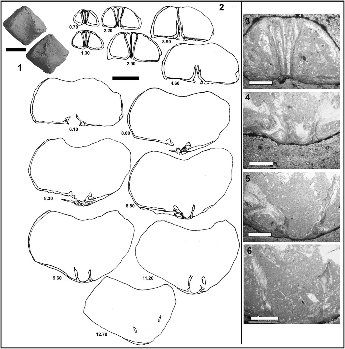
Internal structure of Cisnerospira angulata (Oppel). (1) Specimen Ci.An.CC8.A.1 in which serial sections were performed. (2) Transverse serial sections through the same specimen (distance from the beak in millimeters). (3–6) Microphotographs of acetate peels from the same specimen: (3) section at 0.70 mm from the apex showing dental plates located very close to the ventral median septum; (4) partial section at 8.30 mm from the apex, showing hinge teeth, sockets, and hinge plates; (5, 6) serial sections at 8.80 and 9.60 mm from the apex, showing hinge plates fused to the socket ridges and then developing crural bases attached to the dorsal valve by dorsal adminicula. (1) Scale bar = 1.0 cm; (2) scale bar = 4.0 mm; (3–6) scale bars = 1.0 mm.
*1861 Spiriferina angulata n. sp. Oppel, p. 541, pl. 11, fig. 7a, b.
non
1861 Spiriferina (cf. angulata) obtusa n. sp. Oppel, p. 542, pl. 11, fig. 8.
non
1874 Spiriferina cfr. angulata Oppel; Reference GemmellaroGemmellaro, p. 56, pl. 10, figs. 6, 7a, b.
1879 Spiriferina cf. angualta Oppel [sic]; Reference UhligUhlig, p. 272, pl. 1, fig. 4a–c.
1882 Spiriferina angulata Oppel; Reference GemmellaroGemmellaro, p. 125, pl. 3, figs. 41–46 = p. 412, pl. 31, figs. 41–46.
1885 Spiriferina angulata Oppel; Reference SeguenzaSeguenza, p. 472, 474.
1886 Sp. angulata Oppel; Reference RothpletzRothpletz, p. 160.
1889 Spiriferina angulata Oppel; Reference GeyerGeyer, p. 74, pl. 9, figs. 7–9, 11,12.
non
1889 Spiriferina angulata Oppel; Reference GeyerGeyer, p. 74, pl. 9, fig. 10.
1891 Spiriferina angulata Opp.; Reference Di StefanoDi Stefano, p. 168.
?1893 Spiriferina angulata Oppel; Reference ParonaParona, p. 25, pl. 1, fig. 13.
non
1895 Spiriferina angulata Oppel var. carenata; Reference FuciniFucini, p. 148, pl. 6, fig. 7.
?1900 Spiriferina angulata Oppel; Reference Böse and SchlosserBöse and Schlosser, p. 199, pl. 18, figs. 20, 23, 25.
?1907 Spiriferina gibba Seguenza; Reference Dal PiazDal Piaz, p. 18, pl. 1, fig. 5a–c.
1909 Spiriferina angulata Oppel; Reference Dal PiazDal Piaz, p. 5, pl. 35, fig. 1a–c.
?1910 Spiriferina angulata Oppel; Reference Vinassa De RegnyVinassa de Regny, p. 187, pl. 7, figs. 15, 16.
?1920 Spiriferina angulata Oppel; Reference Dareste de la ChavanneDareste de la Chavanne, p. 46, pl. 3, fig. 13.
?1920 Spiriferina angulata var. obtusa Oppel; Reference Dareste de la ChavanneDareste de la Chavanne, p. 47, pl. 3, fig. 14.
1921a Spiriferina angulata Oppel; Reference Jiménez de CisnerosJiménez de Cisneros, p. 4, text-figs. A, 8, 9; pl. 32, figs. 20, 21.
pars
1921a Spiriferina gibba Seguenza; Reference Jiménez de CisnerosJiménez de Cisneros, p. 7.
1923 Spiriferina angulata Oppel; Reference Jiménez de CisnerosJiménez de Cisneros, p. 43, pl. 6, fig. 21.
1930 Spiriferina angulata Oppel; Reference De GregorioDe Gregorio, p. 43, pl. 12, fig. 6.
1935 Spiriferina angulata Oppel; Reference Jiménez de CisnerosJiménez de Cisneros, p. 24.
?1937 Spiriferina angulata Oppel; Reference OrmósOrmós, p. 18, pl. 1, fig. 3.
?1937 Spiriferina angulata Oppel var. baconica; Reference OrmósOrmós, p. 18, pl. 1, fig. 4.
?1966 Spiriferina angulata Oppel; Reference SiblíkSiblík, p. 144.
1967 Spiriferina angulata Oppel; Reference Sacchi Vialli and CantaluppiSacchi Vialli and Cantaluppi, p. 88, pl. 13, fig. 7.
1992 Liospiriferina angulata (Oppel); Reference DulaiDulai, p. 58, pl. 3, fig. 1.
1993 Liospiriferina angulata (Oppel); Reference IñestaIñesta, p. 11, pl. 1, fig. 3.
1999 Liospiriferina angulata (Oppel); Reference IñestaIñesta, p. 21, pl. 4, fig. 5.
?2000 Liospiriferina angulata (Oppel); Reference Alméras and FauréAlméras and Fauré, p. 214, pl. 23, figs. 2, 3.
2003 Liospiriferina angulata (Oppel); Elmi Reference Elmi, Alméras, Benhamou, Mekahli and Maroket al., p. 698, pl. 4, fig. 2.
?2003 Liospiriferina angulata (Oppel); Vörös Reference Vörös, Szabó, Dulai, Szente, Ebli and Lobitzeret al., p. 74.
2003 Liospiriferina angulata (Oppel); Reference DulaiDulai, p. 53, pl. 8, figs. 7–10; back cover, figs. 8, 9.
?2006 Cisnerospira angulata (Oppel); Comas-Rengifo Reference Comas-Rengifo, García Joral and Goyet al., p. 151, fig. 4.4a–d.
2007 Cisnerospira angulata (Oppel). Reference Vörös and DulaiVörös and Dulai; p. 52, pl. 1, fig. 27.
?2007 Liospiriferina angulata (Oppel); Reference Alméras, Elmi and FauréAlméras et al., p. 37, pl. 1, fig. 4.
2008 Cisnerospira angulata (Oppel); Reference Baeza-CarrataláBaeza-Carratalá, p. 371, pl. 28, figs. 1–5.
2010 Cisnerospira angulata (Oppel); Reference Mandl, Dulai, Schlögl, Siblík, Szabó, Szente and VörösMandl et al., p. 89, pl. 4, fig. 2.
Description
External features: Small- to medium-sized ventribiconvex shells (Table 1), with a triangular to subpentagonal outline. The relationship between the main biometric indices is rather variable; there are shells longer than wide and vice versa. The lateral commissure is straight, and the anterior one is uniplicate with a high dorsal median fold, which is narrow and rounded in outline corresponding with a well-defined ventral sulcus. Ventral valve is deeper than the operculiform dorsal one. Dorsal valve commonly shows flat wing-shaped extensions near the cardinal margin. Ventral beak is straight, strong, and prominent. The ventral cardinal area is catacline and forms a very wide angle with the lateral commissure, thus rendering a typical subpyramidal appearance to the shell. Very wide, flat interareas split by narrow, triangular delthyrium and with conspicuous beak ridges, fairly sharp and straight. Smooth shells except for the dense growth lines.
Internal structure (Figs. 7, 8): wide umbonal lateral cavities. Highly developed ventral median septum without tichorhinum, extending forward almost to midlength. The central cavity is rather narrow due to the position of the dental plates, placed close to each other. These dental plates are subparallel, becoming slightly arcuate toward the anterior margin. Hinge teeth are inserted in broad and shallow sockets and slight denticula present. Outer and inner socket ridges are well developed. It shows a wide cardinal platform, a cardinal process (striated ctenophoridium), and a slender myophragm.
Dorsal median septum and septalium are absent. Hinge plates are short, nearly fused to the socket ridges, and crural bases are developed close (even attached) to the dorsal valve. This is evident since discrete dorsal adminicula extend from the crural bases as supporting crural plates fused to the wall of the dorsal valve (Fig. 8). Jugum is not developed and spiralium is not well preserved. Hence, vestiges of brachidium shown in serial sections performed in the Subbetic specimens of C. angulata (Fig. 7) may perhaps be interpreted as hinting to some ventral development of the spiralia, albeit not conclusively.
Materials
Cerro de la Cruz (42 specimens), complemented with 75 specimens housed in the JdC Collection from Sierra de Algayat, Cruz de La Algueña, Moleta de Togores, Sierra de la Espada, Rincón de Egea, and Collado de La Campana.
Remarks
Spiriferina angulata Oppel, 1861 may be the most widely reported species belonging to Cisnerospira (Manceñido, Reference Manceñido2004; Comas-Rengifo et al., Reference Comas-Rengifo, García Joral and Goy2006, Vörös and Dulai, Reference Vörös and Dulai2007; Baeza-Carratalá, Reference Baeza-Carratalá2008, Reference Baeza-Carratalá2013; Mandl et al., Reference Mandl, Dulai, Schlögl, Siblík, Szabó, Szente and Vörös2010; Baeza-Carratalá et al., Reference Baeza-Carratalá, Giannetti, Tent-Manclús and García Joral2014) although some of the numerous assignments to this species deserve to be discussed. Gemmellaro (Reference Gemmellaro1882, pl. 31, 41–46) clearly showed the extent of the intraspecific variability of this taxon, describing both wider and narrower morphotypes, whereas some specimens without a clear ventral sulcus (Gemmellaro, Reference Gemmellaro1874, pl. 10, figs. 6, 7) appear referable to another species, i.e., C.? darwini (Gemmellaro, Reference Gemmellaro1882, p. 122 = 409), according to Di Stefano (Reference Di Stefano1891, p. 163) and Vörös (Reference Vörös2009, p. 125–126).
On the other hand, Geyer (Reference Geyer1889, pl. 9, fig. 10) figured a ribbed specimen, probably not conspecific with C. angulata, as was noticed by Siblík and Lobitzer (Reference Siblík and Lobitzer2008, p. 71). This weak costation is also visible in the individual depicted by Alméras et al. (Reference Alméras, Elmi and Fauré2007), but the fragmentary specimen does not allow an accurate identification. Likewise, as discussed by Dulai (Reference Dulai2003), S. angulata described by Ormós (Reference Ormós1937) shows weak ribs. In this sense, the variety baconica of this latter author may be assigned to C. aff. adscendens or related forms, due to the curvature of the beak and cardinal area.
On the contrary, the specimen assigned to S. gibba by Dal Piaz (Reference Dal Piaz1907) does not show apparent costation and may have a better matching with C. angulata. In the specimens depicted by Fucini (Reference Fucini1895), the cardinal area is not clearly observed. This author described a ridge in the central area of the dorsal valve, the reason it is excluded from the C. angulata stock herein. This feature was early noticed by Vinassa de Regny (Reference Vinassa De Regny1910). Finally, De Gregorio (Reference De Gregorio1930) illustrated specimens with asymmetric shapes, just as it happens in several individuals of the Subbetic material.
In this sense, in the Subbetic material, the most variable features (in addition to the main biometric ratios) are the depth of the ventral sulcus and, consequently, the height of the dorsal fold. The curvature of the ventral beak also shows slight variations, but always showing a straight ventral interarea (in contrast to other species such as Cisnerospira aff. adscendens with clearly incurved ventral interareas). A stock with wider ventral valve and posterior asymmetry was denominated by Jiménez de Cisneros (Reference Jiménez de Cisneros1921a, pl. 32, figs. 20, 21) as Spiriferina angulata ‘asymmetric form.’ Finally, two specimens show a very slight reticulation due to a gentle striation, far from the costation observed in some taxa ornamented with fine weak ribs (e.g., representatives of the genus Callospiriferina).
Undoubtedly, one of the species most related to Cisnerospira angulata is C. adscendens, but this latter has a more highly developed ventral valve with blunt, effaced beak ridges. On the other hand, Cisnerospira aff. adscendens has a more conical aspect (instead of pyramidal), with both ventral beak and interarea being clearly arched. The smaller species sylvia shows a procline ventral interarea, instead of the catacline orientation observed in C. angulata.
As Manceñido (Reference Manceñido2004) foresaw, material figured as Spiriferina gibba by Jiménez de Cisneros (Reference Jiménez de Cisneros1921a) as well as some other individuals held in the JdC collection formerly attributed to S. gibba actually belong to Cisnerospira, as these specimens have smooth shells; they have been identified herein as C. angulata and C. adscendens.
Occurrence in the Subbetic basin
Jiménez de Cisneros (Reference Jiménez de Cisneros1921a, Reference Jiménez de Cisneros1923, Reference Jiménez de Cisneros1935) reported this species from the ‘middle Lias.’ Iñesta (Reference Iñesta1988, Reference Iñesta1993) and Baeza-Carratalá (Reference Baeza-Carratalá2008, Reference Baeza-Carratalá2013) found it in Pliensbachian sediments, and finally in this work, C. angulata is restricted in the Subbetic basin to the late Sinemurian–late Pliensbachian.
Cisnerospira? sylvia (Gemmellaro, Reference Gemmellaro1882)
*1882 Spiriferina sylvia, Gemmellaro; Reference GemmellaroGemmellaro, p. 123, pl. 3, figs. 27–33 = p. 410, pl. 31, figs. 27–33.
1886 Sp. sylvia Gemmellaro; Reference RothpletzRothpletz, p. 159.
pars
1895 Spiriferina sylvia Gemmellaro; Reference FuciniFucini, p. 158, pl. 6, figs. 11, 14–17.
non
1895 Spiriferina sylvia Gemmellaro; Reference FuciniFucini , p. 158, pl. 6, figs. 12, 13.
1926 Spiriferina sylvia Gemmellaro; Reference Jiménez de CisnerosJiménez de Cisneros, p. 174.
1937 Spiriferina sylvia Gemmellaro; Reference OrmósOrmós, p. 18, pl. 1, fig. 2.
1992 Liospiriferina sylvia (Gemmellaro); Reference DulaiDulai, p. 61, pl. 3, fig. 4.
2003 Liospiriferina sylvia (Gemmellaro); Reference DulaiDulai, p. 65, pl. 11, figs. 1–3.
2008 Cisnerospira sylvia (Gemmellaro); Reference Baeza-CarrataláBaeza-Carratalá, p. 369, pl. 26, figs. 3, 4.
2010 Cisnerospira sylvia (Gemmellaro); Reference Mandl, Dulai, Schlögl, Siblík, Szabó, Szente and VörösMandl et al., p. 91, 94, 97, pl. 4, fig. 6.
Description
Small- to medium-sized ventribiconvex shells (Table 1), with a rounded-subpentagonal dorsal outline. Width greater than length. Lateral commissure straight and frontal one uniplicate, with a semicircular dorsal median fold. Ventral valve deeper than the operculiform dorsal one. The beak is straight and robust with a subpyramidal profile. Very wide, triangular ventral interareas, distinctively with a procline orientation and showing very sharp beak ridges. Delthyrium is triangular and narrow. Smooth shells, except for thin growth lines. The internal structure was not studied by serial sectioning due to the paucity of material.
Material
A single specimen from Cerro de La Cruz and two specimens (JdC collection) from Sierra de Algayat and Moleta de Togores.
Remarks
Cisnerospira? sylvia includes those shells with subpyramidal outline with procline orientation of the ventral interareas and more box-like shape than C. angulata, this latter form typified by catacline interareas. This distinctive feature has been accurately noticed by several previous authors (Fucini, Reference Fucini1895 pro parte: pl. 6, figs. 11, 14–17; Ormós, Reference Ormós1937; Dulai, Reference Dulai1992, Reference Dulai2003; Mandl et al., Reference Mandl, Dulai, Schlögl, Siblík, Szabó, Szente and Vörös2010). Fucini (Reference Fucini1895, pl. 6, figs. 12, 13) considered as belonging to C.? sylvia; also some specimens provided with clear catacline interareas ‘angulata/acuta-type,’ and consequently they have been excluded from the synonymy herein. Some other nominal species, such as Spiriferina depressa Seguenza, Reference Seguenza1885 (p. 468, pl. 21, fig. 3), S. statira Gemmellaro, Reference Gemmellaro1874 (p. 54, pl. 10, fig. 3), or S. zignoi Di Stefano, Reference Di Stefano1891 (p. 171, pl. 1, fig. 7), show procline interareas, but their precise taxonomic status has not been confirmed by means of additional detailed analyses in their regions of origin.
It could be argued that the peculiar procline nature of the interarea shared by these closely similar nominal species might justify erection of a separate subgenus (or genus?) for them. However, since that particular character state is so often correlated with small size of the bearer, it does not yet allow to completely rule out the possibility that they may well represent, at least, a paedomorphic derivative from the main stock, or perhaps, even a young ontogenetic stage of known (larger) species. Under such circumstances, and without excluding the chances of some kind of environmental influence, deliberate adoption of a query serves to adequately reflect the present state of knowledge at generic level.
Occurrence in the Subbetic basin
Baeza-Carratalá (Reference Baeza-Carratalá2008, Reference Baeza-Carratalá2013) assigned the record from the Subbetic area to the Pliensbachian.
Interpretations on the habitat and adaptive strategies of Cisnerospira
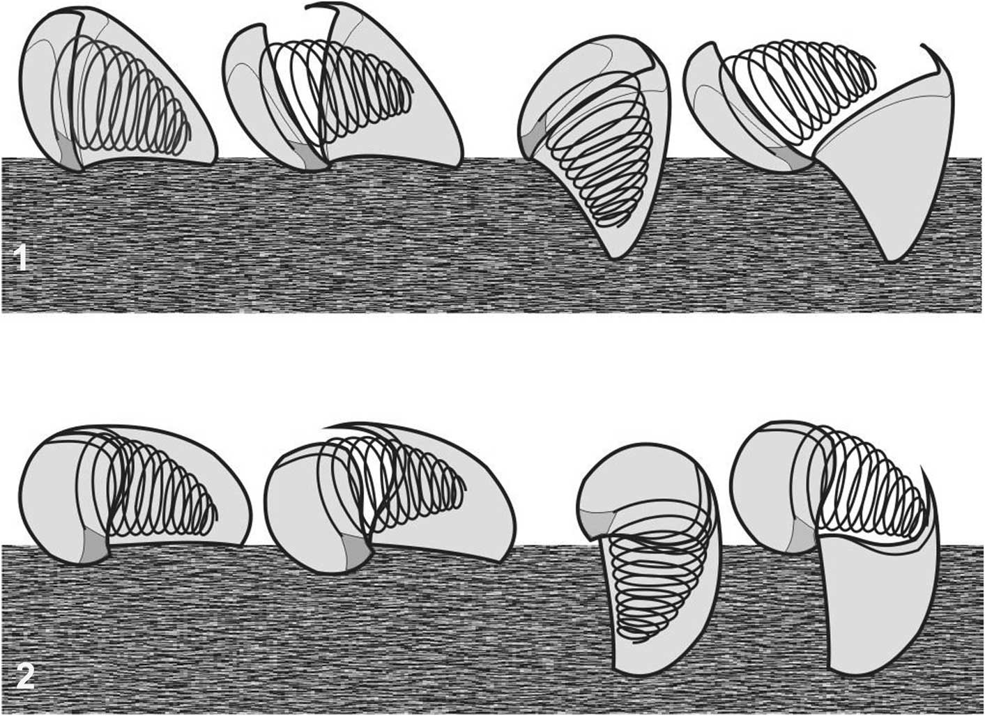
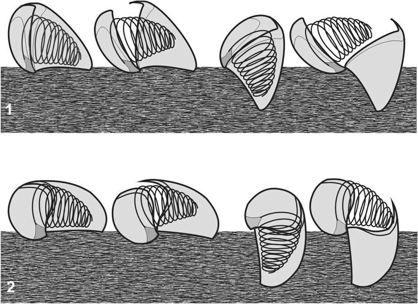
From the perspective of a morphofunctional analysis, the peculiar morphology of Cisnerospira suggests a specialized way of life. Cisnerospira representatives have a subpyramidal form, with very narrow, long, and triangular delthyrium, highly developed, smooth cardinal areas, and nearly operculiform dorsal valve. As envisaged by Manceñido (Reference Manceñido2004, p. 272), these features probably allowed them a mainly benthonic liberosessile adaptive strategy to live resting or at least weakly attached on soft bottoms (Fig. 9). In this context, wide, flat cardinal areas may be highly functional, conferring hydrodynamic stability to the shell according to an epifaunal recliner strategy, resting on a large interarea closely adpressed to the substrate (cf. Ager, Reference Ager1968, fig. 1d; Rudwick, Reference Rudwick1970, fig. 43; Fürsich and Hurst, Reference Fürsich and Hurst1974, p. 893–894, fig. 6; Harper and Moran, Reference Harper and Moran1997, p. 236, fig. 3.9), or as semi-infaunal stickers allowing their heavy umbones and subpyramidal ventral valves to be partially sunk subperpendicular into the sediment (cf. Ager, Reference Ager1965, p. 156, 1968, fig. 1e; Harper and Moran, Reference Harper and Moran1997, p. 237; Seilacher, Reference Seilacher1999, fig. 8; Baeza-Carratalá et al., Reference Baeza-Carratalá, Giannetti, Tent-Manclús and García Joral2014, fig. 11).
Benthonic liberosessile adaptive strategies inferred for Cisnerospira to live resting on or sticking into unconsolidated substrata. (1) Cisnerospira angulata. (2) Cisnerospira adscendens. On the left, a reclining position showing the ventral interarea closely adpressed to substrate and permissible gaping angle between both valves. On the right, a half-sunk, sediment-sticker lifestyle; note the maximum degree of valve gape inferred, thus allowing the spiralia to be entirely exposed.
In the Subbetic material, an apparent relationship linking these subpyramidal shapes with the particular nature of substrate has been revealed. Thus, the onset of the Cisnerospira record is detected in micritic and peloidal wackestone with lateral and upward transitions to oolitic grainstone/packstone beds that contain intraclast- and peloid-rich levels. Baeza-Carratalá et al. (Reference Baeza-Carratalá, Giannetti, Tent-Manclús and García Joral2014) confirmed that this lithological unit was deposited in a soft-ground environment, with the presence of rounded rhynchonellids and terebratulids thickened in the posterior third of the shell, with very incurved beaks and obliterated or minute pedicle foramina (i.e., absence of a functional pedicle capable of resisting currents; sensu Copper, Reference Copper1997), which suggested a primarily free-lying life mode for the brachiopod assemblage. The morphological features characterizing these forms were interpreted to provide improved stability in unconsolidated sediments since they allow the shell to turn with prevailing currents (Surlyk, Reference Surlyk1972; Álvarez and Emig, Reference Álvarez and Emig2005; Richardson et al., Reference Richardson, Aldridge and Endersby2007; Sulser et al., Reference Sulser, García-Ramos, Kürsteiner and Menkveld-Gfeller2010; Baeza-Carratalá et al., Reference Baeza-Carratalá, Giannetti, Tent-Manclús and García Joral2014).
The overlying lithostratigraphical unit where Cisnerospira is recorded consists of red crinoidal grainstone beds with abundant glauconite and occasionally more calcarenite levels interspersed. They can be also considered as sand-grained, unconsolidated, and mobile substrates. Finally, the last occurrences of Cisnerospira in the Subbetic area are established in the marly soft bottoms consisting of marls and sandy marlstone beds of the Zegrí Formation (Fig. 1.3).
Consequently, a feasible relationship between the morphological functionality of Cisnerospira and the presence of generally unconsolidated sediments is rendered evident. Both epibenthonic adaptive strategies inferred in this work (Fig. 9) enabled the adaptation to substrates with different degree of consolidation and firmness, yet provide a notable hydrodynamic stability to the shell while keeping the frontal margin away from the botttom. This strategy allowed a feeding pattern whereby the inhalant current was located in a broad inhalant area coincident with the median deflection of the anterior commissure, through which the water flow entered medially, passed through the spirolophe, and exited posterolaterally (Manceñido and Gourvennec, Reference Manceñido and Gouvernec2008; Shiino et al., Reference Shiino, Kuwazuru and Yoshikawa2009; Shiino and Kuwazuru, Reference Shiino and Kuwazuru2010).
The adaptive strategy of the inferred sediment-sticker lifestyle (Fig. 9) involves the feasibility of a wider range of valve gaping. Such a widely open gape (as depicted) makes it possible that the spiralium may emerge even entirely, thus presumably facilitating more effective suspension feeding and breathing in a low-energy environment. The wide opening of the valves is also congruent with the pseudodeltidiodont, noninterlocked articulation of the spiriferinides, weaker than the cyrtomatodont articulation of rhynchonellides and terebratulides (Sheehan, Reference Sheehan1978; Williams and Carlson, Reference Williams and Carlson2007; Baeza-Carratalá et al., Reference Baeza-Carratalá, Giannetti, Tent-Manclús and García Joral2014). This fact is supported by the recurrent record of isolated disarticulated valves in Cisnerospira (Gemmellaro, Reference Gemmellaro1874, pl. 10, figs. 6, 7, 1882, pl. 31, figs. 41–46; Uhlig, Reference Uhlig1879, pl. 1, fig. 4; Dal Piaz, Reference Dal Piaz1907, pl. 1, fig. 5; Reference Dal Piaz1909, pl. 35, fig. 1; Dareste de la Chavanne, Reference Dareste de la Chavanne1920, pl. 3, figs. 13–14; Dulai, Reference Dulai1992, pl. 3, fig. 1; Vörös, Reference Vörös2009, pl. 14, figs. 2, 3; Mandl et al., Reference Mandl, Dulai, Schlögl, Siblík, Szabó, Szente and Vörös2010, pl. 4, fig. 2; Fig. 6.3 herein, among others), being that this genus more prone to disarticulation than several other brachiopod taxa recorded together in the same assemblage. The exposure of the spiralia may also help explain why among the Cisnerospira individuals it is difficult to find this delicate internal structure in its original arrangement, being often subjected to the biostratinomic factors that tend to break down or disjoint it.
Several morphological features displayed by Cisnerospira fit the paradigm for subconical ‘sediment stickers,’ a soft-bottom guild convergently evolved by representatives of different animal phyla along Phanerozoic times (Ager, Reference Ager1968; cf. Seilacher, Reference Seilacher1999). From this viewpoint, the existence of some specimens with their ventral umbos bent sideways to various degrees (but otherwise displaying normal bilateral symmetry) could be explained as the result of accidental tilting or toppling on a muddy bottom, followed by upward growth to restore vertical attitude.
Conclusions
On the basis of a detailed revision of the species recorded in the Subbetic area (SE Spain) and their comparison with those assigned by previous authors to the genus Cisnerospira in several western Tethyan basins, the diagnosis for the genus Cisnerospira Manceñido, Reference Manceñido2004 is broadened and emended to include taxa that, besides having their very large diagnostic subpyramidal ventral valve, reveal an internal structure consisting of dental plates placed close to each other, high ventral median septum, hinge plates nearly fused to the socket ridges, and crural bases developed very close (even attached) to the dorsal valve. The absence of the dorsal median septum and septalium and the presence of a ventrally oriented spiralium also discriminate Cisneropira from some comparable spiriferinids.
On the basis of the cyrtiniform shape and the internal structure mainly concerning the hinge area and cardinalia, absence of dorsal median septum and septalium, and crural bases developed very close to the dorsal valve, as most outstanding features, this genus is allocated into the subfamily Paralaballinae.
The stratigraphical distribution of the Subbetic representatives (Cisnerospira adscendens, C. aff. adscendens, C. angulata, and C.? sylvia) ranges from the late Sinemurian to the earliest Toarcian, thus comprising the most usual timespan in which Cisnerospira is recorded in the remaining western Tethyan basins. The Subbetic occurrences together with the Sinemurian (perhaps even Hettangian) records from the most intra-Tethyan areas and the early Toarcian cases from the NW European bioprovince support that Cisnerospira had a distribution restricted to the Early Jurassic, becoming extinct during the global crisis of the ETOAE.
The analysis of its geographical distribution corroborates that the genus exhibits relatively widespread occurrences across southern Europe and northern Africa, with a high fidelity for western Tethys (though occasionally reaching northwestward to SW England and eastward to Indonesia).
From a morphofunctional standpoint, the unusual shape, with subpyramidal ventral valve and lid-like dorsal valve, suggests a liberosessile adaptive strategy for living on unconsolidated sediment. This could be attained as recliners resting on their large, flat interareas closely adpressed onto the substrate, or even as sediment stickers with their ventral umbones sunk subperpendicular into the soft bottom, perhaps allowing their conical spiralia to be entirely exposed to surrounding seawater at maximum degree of valve gaping.
Acknowledgments
This study is partially supported by the Research Group VIGROB-167 (University of Alicante) and is a contribution to the project CGL2015-66604-R (Ministry of Science and Innovation, Government of Spain). MOM’s activities were financed by several grants from CONICET (Argentina), and his visit to examine the JdC collection was funded by the University of Alicante (Spain). All these institutions are gratefully acknowledged for their support. JFBC thanks the late D. Simeón Peiró, who provided him some specimens from his collection. Useful comments from two reviewers (A. Vörös and A.T. Halamski) designated by the journal editors are also deservedly acknowledged.