Background
Hypereosinophilic syndrome is characterised by sustained peripheral blood eosinophilia more than 1500 Eos/mm3 persisted for more than 6months associated with end-organ involvement. Hypereosinophilic syndrome is idiopathic in most cases; although it may be familial. Reference Ogbogu, Rosing and Horne1
Cardiovascular complications of HES, with the approximate prevalence of 40–50%, are a substantial source of disease-related morbidity and mortality. Reference Ogbogu, Rosing and Horne1
The spectrum of coronary artery involvement includes ectasia and aneurysm formation, spontaneous coronary artery dissections, and perivascular eosinophilic infiltration, which may sometimes lead to sudden cardiac death in otherwise healthy individuals. Reference Robinowitz, Virmani and McAllister2 Coronary ectasia in HES is related to procoagulant, vasoactive, and inflammatory properties of eosinophils. Reference Demir, Keceoglu and Melek3
Although without endomyocardial biopsy, it is not easy to recognise HES cardiac involvement, modalities such as cardiac MRI and CT imaging could identify left ventricular thrombus and concurrently determine the coronary artery involvement. Reference Mankad, Bonnichsen and Mankad4
Herein, we describe a previously known case of HES who presented with acute coronary syndrome. To our knowledge, this association which occurs rarely in adults is reported for the first time in a child.
Case presentation
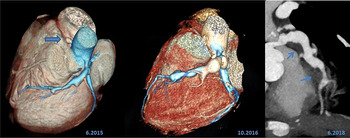
A 14-year-old boy was presented to the emergency department with the complaint of typical retrosternal chest pain radiated to the left arm and was associated with nausea and vomiting. He was a known case of HES from 4 years ago. Since his diagnosis, he has received 12 courses of chemotherapy. Also, he underwent bone marrow transplantation during the last year. One year after disease diagnosis, coronary CT angiography had shown progressive coronary dilatation and right coronary artery occlusion, but he had never experienced a similar episode of chest pain before his presentation to us (Fig 1).
CT angiograms show the progressive trend of coronary ectasia and aneurysm formation during 3 years. Complete thrombotic occlusion of the proximal right coronary artery (left image, blue arrow in the left image) and mural thrombosis development along with the left anterior descending (blue arrows on the right image).

At the time of admission, the patient was haemodynamically stable with a blood pressure of 100/70 mmHg, a regular heart rate of 88bpm, and a normal heart and lung auscultation.
His physical examination revealed significant periocular hyperpigmentation and multiple small vascular aneurysms in his scalp, left arm, and right forearm.
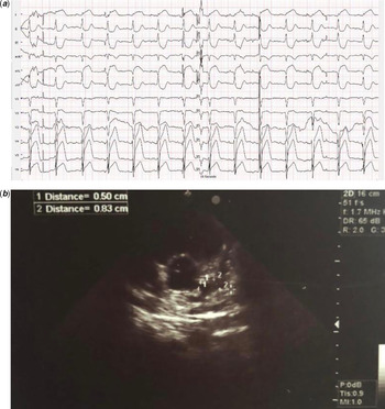
Electrocardiogram had normal sinus rhythm and axis with ST-segment elevation I, AVL, V4, V5, and V6 leads (Fig 2a).
Laboratory data indicated elevated creatinine phosphokinase-myocardial band, troponin I, and pro-brain natriuretic peptide with values of 2294 IU/L (with a normal range between 25 and 225 IU/L), 20,279 ng/ml (with a normal range less than 19 ng/ml), and 809 pg/ml (with an NL range less than 125 pg/ml), respectively.
Lab tests showed normal coagulation tests, lactate, blood sugar, electrolytes, blood gases and erythrocyte sedimentation rate, haemoglobin level (15.1 g/dl), and platelet count (208,000/mm3) with mild leukocytosis of 11,500 cells/mm3 consisting of 70.7% polymorphonuclear cells, 27.2% lymphocytes, 1.2% monocytes, 0.4% basophils, 0.5% eosinophils (absolute eosinophil counts of 57) as well as mild increase in serum glutamic-oxaloacetic transaminase (183 IU/ml) (with a normal range between 5 and 40 IU/ml), serum glutamate-pyruvate transaminase (45 IU/ml) (with a normal range between 5 and 40 IU/ml), and C-reactive protein (10.1 mg/L) (normal range <6 mg/L).
Chest X-ray was normal and transthoracic echocardiography showed notably reduced left ventricular ejection fraction 30%–35%, septal dyskinesia, and posterior wall hypokinesia, aortic velocity time integral of 14 cm, moderate mitral regurgitation, and dp/dt of 700. Dilatation of proximal RCA (size: 0.5 cm) with aneurysmal left main coronary artery, left anterior descending, and left circumflex artery (size: 0.8 cm) were also noted (Fig 2b).
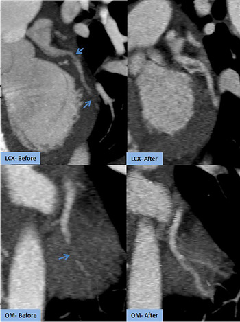
CT angiography (Fig 3 left column) revealed thrombotic stenosis and occlusion of the LCX and first obtuse marginal branch, which prompted starting intravenous thrombolytic therapy with Alteplase 0.5 mg/kg. Subsequently, his symptoms were resolved. The troponin I and pro-BNP levels dropped to 1125 IU/L (with a normal range of 25–225 IU/L) and 345 pg/ml (with a normal range less than 125 pg/ml), respectively.
(a) ECG showing ST elevation in anterolateral leads with reciprocal changes. (b) Echocardiogram image of aneurysmal left circumflex artery and dilated left main coronary artery and left anterior descending artery.

Acute thrombosis of the proximal left circumflex and obtuse marginal branch before and after thrombolytic therapy.

Coronary CT angiography was taken as control 2 days after admission (Fig 3 right column) and demonstrated a noticeable resolution of the thrombosis.
The patient was discharged 7 days later with a stable condition while his last echocardiography displayed improved ejection fraction (45–50%), mitral regurgitation, aortic velocity time integral (17 cm) with mild residual septal hypokinesia. Warfarin (labelled 5 mg of warfarin per tablet, Orion Co., Espoo, Finland) and Aspirin (A.S.A, Jalinus Co., Tehran, Iran) were started after Alteplase as maintenance therapy and the INR was preserved between 2.5 and 3.5. He is doing well during a 1-year follow-up.
Discussion
Cardiovascular complications occur frequently in HES. Various cardiac presentations of HES range from an asymptomatic myocardial involvement to ischaemic events, stroke, heart failure, fatal arrhythmia, and sudden cardiac death. Coronary aneurysm presenting as acute coronary syndrome may also occur; however it is rare. Reference Mankad, Bonnichsen and Mankad4,Reference Al Ali, Straatman, Allard and Ignaszewski5
Eosinophilic vasculitis has been identified at autopsy in otherwise healthy individuals with spontaneous coronary dissection or rupture. It has been proposed that perivascular eosinophils and their cytotoxic substances might be the mechanism that predisposes these patients to aneurysm formation or spontaneous dissection. Reference Carreon and Esposito6
ECG and echocardiography are simple modalities for the evaluation of cardiac involvement in HES. There are non-specific ECG changes such as ST-segment and T-wave changes or left ventricular hypertrophy. Although these signs are not specific ECG abnormalities for HES. The most important echocardiographic findings in HES are thrombosis in the left or right ventricle and endomyocardial thickening. Reference Ogbogu, Rosing and Horne1
Coronary aneurysms and their increase in diameter are possible in HES patients, Reference Patanè, Marte, Sturiale, Grassi and Patanè7 furthermore, there are some reports of peripheral aneurysms such as radial and ulnar arteries and hepatic arteries in these patients that rationalised evaluation of peripheral arteries in HES. Reference Demir, Keceoglu and Melek3
Due to these reports, serial follow-up imaging seems to be reasonable for prevention and prophylactic therapy of the probable subsequent adverse outcomes in HES patients. The differential diagnoses of our patient include Kawasaki disease, systemic lupus erythematous, polyarteritis nodosa, Takayasu disease, and vasculitis involving coronary artery disease. Our patient did not have a history of fever or any other symptom suggestive of Kawasaki disease. The complement and inflammatory markers were all normal that could rule out vasculitis. A thorough rheumatological consult was performed and no sign of the rheumatological disease was observed.
There are limited reports of adult HES patients with coronary aneurysm thrombosis. To our knowledge, this is the first report of thrombotic coronary occlusion in a child.
Acknowledgements
None.
Financial support
This research received no specific grant from any funding agency, commercial, or not-for-profit sectors.
Conflict of interest
None.
Ethical standards
All procedures performed in this study were per the ethical standards of the institutional review board of Rajaie Cardiovascular Center and with the 1964 Helsinki declaration and its later amendments.