INTRODUCTION
Lung cancer has the highest mortality rate among all malignant cancers throughout the world. Over the past decade, its mortality rate has increased due to several factors, such as population ageing, smoking and environmental pollution (Siegel et al. Reference Siegel, Ward, Brawley and Jemal2011). However, the majority of lung cancers can be diagnosed definitively only at the terminal stage, with the 5-year survival rate lower than 15% (Govindan et al. Reference Govindan, Page, Morgensztern, Read, Tierney, Vlahiotis, Spitznagel and Piccirillo2006; Cazzoli et al. Reference Cazzoli, Buttitta, Di Nicola, Malatesta, Marchetti, Rom and Pass2013). Small cell lung cancer (SCLC) accounts for approximately 15% of all lung cancers and, due to its short multiplication time, has a high degree of malignancy, early metastasis and a generally unfavourable prognosis (Byers and Rudin, Reference Byers and Rudin2015; Sharp et al. Reference Sharp, Bhosle, Abdelraouf, Popat, O'Brien and Yap2016). Platinum-based chemotherapy remains the main therapeutic method for advanced SCLCs, but has significant adverse effects, and the cancer may easily reoccur (Hann and Rudin, Reference Hann and Rudin2008; Sharp et al. Reference Sharp, Bhosle, Abdelraouf, Popat, O'Brien and Yap2016). Biological therapy, in contrast, is regarded as a safe and efficient therapeutic method. Consequently, many experimental studies have been conducted to explore the inhibition of cancers through biological therapy (Altundag et al. Reference Altundag, Esteva and Arun2005; Eng and Shalan, Reference Eng and Shalan2006; Di Marco et al. Reference Di Marco, Grassi, Durante, Vecchiarelli, Palloni, Macchini, Casadei, Ricci, Panzacchi, Santini and Biasco2016), including the antineoplastic effects of parasites, another research hotspot (Seyedeh et al. Reference Seyedeh, Nahid, Nahid, Shima, Morteza and Hossein2015; Ubillos et al. Reference Ubillos, Freire, Berriel, Chiribao, Chiale, Festari, Medeiros, Mazal, Rondán, Bollati-Fogolín, Rabinovich, Robello and Osinaga2016).
Studies show that parasites may inhibit tumour cell growth or shrink tumour cells in animals. Trichinella spiralis is an obligate intracellular parasite, which can infect people and many kinds of animals through the digestive tract, causing trichinosis (Lee et al. Reference Lee, Yoo, Kim, Lee and Seo2013). Trichinella spiralis excretory–secretory proteins (ESPs) are complex proteins that are excreted and secreted by T. spiralis during host infection. Since 1975, when it was reported that infection by T. spiralis could inhibit sarcoma growth (Lubiniecki and Cypess, Reference Lubiniecki and Cypess1975), researchers have been studying its antineoplastic effects. Parasite crude proteins and their ESPs inhibit the proliferation of tumour cells in vitro and in vivo and may induce tumour cell apoptosis (Kallinikova et al. Reference Kallinikova, Matekin, Ogloblina, Leĭkina, Kononenko, Sokolova and Pogodina2001; Kim et al. Reference Kim, Jung, Kim, Kim, Shin, Lee and Lee2007; Darani et al. Reference Darani, Shirzad, Mansoori, Zabardast and Mahmoodzadeh2009). Prior research has shown that T. spiralis or its active proteins may inhibit sarcoma, melanoma, colon cancer, lung cancer, stomach cancer, leukaemia, myeloma, breast cancer and other cancers (Deng et al. Reference Deng, Gong, Li, Cheng, Ren, Yang, Li, Zhang and Zhang2013; Kang et al. Reference Kang, Jo, Cho, Yu, Leem, Song, Ock and Cha2013; Vasilev et al. Reference Vasilev, Ilic, Gruden-Movsesijan, Vasilijic, Bosic and Sofronic-Milosavljevic2015), and induce apoptosis in certain cancer cells (Wang et al. Reference Wang, Fu, Yang, Wu, Cui, Liu, Zhao, Yu, Liu, Deng, Chen and Liu2009, Reference Wang, Liu, Sun, Liu, Yu, Wang, Chu, Rosenthal, Shi, Boireau, Wang, Zhao and Wu2013; Vasilev et al. Reference Vasilev, Ilic, Gruden-Movsesijan, Vasilijic, Bosic and Sofronic-Milosavljevic2015). However, no specific mechanism of action has been found, nor have there been reports of the effect of T. spiralis on SCLC. To study the action of T. spiralis ML ESPs on inhibiting the growth of H446 SCLC cells and the ESP mechanism of action, we tested inhibition and apoptosis of H446 cells by ESPs using the [3-(4,5-dimethylthiazol-2-yl)-2,5-diphenyltetrazolium bromide] MTT colorimetric method and flow cytometry (FCM). We conducted reverse transcription polymerase chain reaction (RT–PCR) and Western blotting to detect expression of the relevant genes to determine whether apoptosis occurred via a mitochondrial pathway and to interpret a possible antineoplastic mechanism on the basis that ESPs may inhibit human H446 SCLC cells and promote apoptosis.
The life history of T. spiralis includes three stages, including the adult worm (AD), newborn larva (NBL) and muscle larva (ML), in which polypeptide proteins and ESPs may be effective in inhibiting tumour growth. Because the ML lives longer than AD and NBL, it has a greater effect on host immunization (Vasilev et al. Reference Vasilev, Ilic, Gruden-Movsesijan, Vasilijic, Bosic and Sofronic-Milosavljevic2015); it is also easier to obtain ML than AD or NBL. We therefore selected ML ESPs to explore their effects on H446 SCLC cells.
MATERIALS AND METHODS
Preparation of T. spiralis ML ESPs
Mice infected with T. spiralis for 40 days were terminated by the cervical dislocation method. Pure T. spiralis excystation MLs were collected as per the methods specified by Beiting et al. (Reference Beiting, Bliss, Schlafer, Roberts and Appleton2004). ESPs were prepared according to the methods of Appleton et al. (Reference Appleton, Schain and McGregor1988). The MLs were cleaned repeatedly by rinsing three times with stroke-physiological saline solution containing 500 U mL−1 mycillin and then adding them at 6000 larvae mL−1 density to Roswell Park Memorial Institute (RPMI) 1640 medium containing 500 U mL−1 mycillin. The medium was then placed in a 5% CO2 incubator at 37 °C constant temperature for 24 h. The culture solution was collected after centrifuging at 12 000 g at 4 °C for 30 min, and the supernatant (ESPs) was gathered. The ESPs were filtered using 0·22-μm filter paper, packaged separately, and stored at −80 °C.
Recovery and culture of H446 cells
Cryogenic vials containing H446 cells were removed from the −80 °C refrigerator and placed into warm (37 °C) water; rapid dissolution was promoted by shaking to avoid cell damage due to crystallization. Upon dissolution, the cryogenic vials were centrifuged at 120 g for 2 min. The cells were then moved into the complete RPMI 1640 medium (10% fetal calf serum, 1% penicillin–streptomycin solution) for culturing. The culture media was replaced daily. Once the cells had grown to 80% of the culture dish, the cells were removed using 0·25% trypsin. After three generations, cells in the logarithmic phase were divided into three groups: experimental (adding ESPs of different doses), positive control [adding different concentrations of cis-platinum (DDP) cells] and negative control (adding serum-free RPMI 1640 medium).
Determination of proliferation inhibition rate
H446 cells were collected in the logarithmic phase, counted using an optical microscope and inoculated into a 96-pore culture plate at 1 × 10⁴ cells per pore. We set three concentrations for the experimental group: 0·3, 0·6 and 1·2 mg mL−1 and five concentrations for the positive control group: 0·8, 1·6, 3·2, 6·4 and 12·8 µg mL−1, with each concentration having five double-pores. Five double-pores were used for the negative control group (adding serum-free RPMI 1640 medium). Used culture media was removed once cells were adherent. We added 200 µL of ESPs of corresponding concentration into each pore of the experimental group, 200 µL of DDP of corresponding concentration into each pore of the positive control group, and 200 µL of serum-free medium into the negative control group. The three groups were cultured for 12 h, 24 h and 48 h, and then 10 µL of MTT (5 mg mL−1) was added. Used culture media was removed after 4 h, and then 150 µL of dimethyl sulfoxide was added. The optical density (OD) of each pore was measured with a microplate reader (BioTek, USA). The proliferation inhibition rate was calculated using the following formula: Proliferation inhibition rate = (1 − ODexperimental group or ODpositive control group/ODnegative control group) × 100% IC50, where IC50 is the half-inhibition rate. This value was then used to determine the optimal drug concentration.
Detection of apoptosis
H446 cells were collected in the logarithmic phase and digested using trypsin. The cell density was adjusted to 5 × 10⁴ mL−1. The cells were inoculated in a 60-mm culture dish at 3 mL per dish. There were three experimental concentrations, three positive control concentrations and one negative control concentration, with two dishes per treatment. Cells were blended and incubated for 12 h until they were adherent and had favourable growth states. ESPs were prepared in the above mentioned and the DDP was used at final concentration of 1·6, 3·2 and 6·4 µg mL−1, respectively. ESPs and DDP were then added in corresponding concentrations. Fetal bovine serum-free medium was added to the negative control group. Cells were cultured for each group for 24 h, flushing twice softly using phosphate-buffered saline (PBS) solution. Cells were collected by trypsinization without EDTA and transferred into a 5-mL flow tube for centrifuge processing (440 g ; 4 °C) for 5 min to remove the supernatant. The cells were washed twice with pre-cooling PBS and then counted. Each group was adjusted so that it contained 5 × 10⁴ cells. The cells were re-suspended using 100 µL of 1 × binding buffer, to which 5 µL of fluorescein isothiocyanate Annexin V and propidium iodide were added; the cells were dyed for 30 min. To this we added 400 µL of 1 × binding buffer. The samples were then filtered using a 300-mesh nylon net for apoptosis rate testing with FCM (BD, USA).
RT–PCR
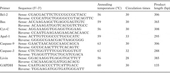
Cells were collected in the logarithmic phase and cultured in 60-mm culture dishes at 5 × 10⁴ cells per dish for 24 h; 3 mL of 1·2 mg mL−1 ESPs was added to the experimental group, 3 mL of 3·2 µg mL−1 DDP was added to the positive control group, and a serum-free medium was added to the negative control group. After 24 h, all RNA was extracted from the cells as per the instructions for the TRIzol reagent (Invitrogen, USA). The RNA concentration was tested using ultraviolet spectrometry photometers (Beckman Coulter, USA). The OD value below 260/280 nm was 1·85–2·0, indicating that the RNA purity of each group was relatively high. cDNA was synthesized from RNA via reverse transcription as per the RT–PCR kit instructions (Takara, Japan). The reverse transcription preparation was as follows: 3·75 µL of RNase-free dH2O, 2 µL of MgCl2, 1 µL of 10 × RT buffer, 1 µL of dNTP Mixture, 0·5 µL of AMV Reverse Transcriptase, 0·25 µL of RNase inhibitor and 1 µL of experimental sample RNA. The primer design is given in Table 1. The PCR total reaction system was 20 µL, including 4 µL of cDNA, 4 µL of 5 × PCR buffer, 11·5 µL of sterile double-distilled water, 0·1 µL of Taq polymerase, 0·4 µL of sense primer and 0·4 µL of reverse primer. Reaction conditions were: initial denaturation at 94 °C for 2 min, denaturation at 94 °C for 30 s, annealing for 30 s, extension at 72 °C for 45 s and re-extension at 72 °C for 5 min; 10 µL of amplification products were used in electrophoresis measurements in 1·5% sepharose gel. To obtain the relative expression quantity of mRNA, products were placed into the gel imaging system (Bio-Rad, USA) to analyse the grey value and level of each group.
Primer description

Western blot analysis
We collected cells in the logarithmic phase, cultured them in 100-mm culture dishes at 1·5 × 106 cells per dish for 24 h, and added 1·2 mg mL−1 ESPs to the experimental group, 3·2 µg mL−1 cis-platinum to the positive control group and serum-free RPMI 1640 medium to the negative control group. After 24 h, 300 µL of cell lysis buffer was added to each culture dish. Cells were split and centrifuged at 12 000 g and 4 °C for 5 min; the supernatant was collected and the protein concentrations were tested using a BCA protein assay kit (Solarbio, China). The proteins were blended with 6 × sodium dodecyl sulphate polyacrylamide gel electrophoresis (6 × SDS–PAGE) loading buffer solution at a 5:1 ratio and boiled in water for 5 min until denaturation. A mixture of 5% polyacrylamide spacer gel and 12% separated polyacrylamide was prepared, in which the loading quantity of proteins in each group was 90 µg. The voltage was varied from 80 to 120 V during SDS–PAGE electrophoresis. A polyvinylidene difluoride (PVDF) membrane (0·45 µm, Millipore, USA) was transferred with a membrane transfer buffer containing 2·05% glycine, 0·34% tris and 20% methanol for 1·5–2 h. The membrane was sealed in block buffers containing 0·88% NaCl, 0·24% tris, 0·05% Tween-20 and 5% skim milk powder at room temperature for 3 h; incubated at room temperature with rabbit-anti-human Cyt-C, Apaf-1, caspase-9, Bax, Bcl-2, caspase-3 and Livin monoclonal antibodies (the antibodies were both purchased from Bioworld, USA and both diluted 1:500) or mouse-anti-human β-actin monoclonal antibodies (Santa Cruz Biotechnology, USA, diluted 1:200) for 2 h at room temperature; and left overnight at 4 °C. We washed the membrane with tris-buffered saline (TBST) containing 0·88% NaCl, 0·24% tris and 0·05% Tween-20 three times and incubated it with horseradish peroxidase-conjugated goat anti-rabbit IgG or goat anti-mouse IgG (Bioworld, USA, both diluted to 1:5000) at room temperature for 1·5 h. The membrane was washed with TBST three times and then visualized with an enhanced luminol-based chemiluminescent substrate (ECL Kit, Thermo Scientific, USA); the membrane was photographed using a Gel Imaging System (Bio-Rad, USA).
Statistical analysis
The results shown in the figures are expressed as means ± s.d. All data were analysed using SPSS 19·0 statistical software. The normality assumption was satisfied; we used one-way analysis of variance (ANOVA) to compare means, followed by the least-significant difference (LSD) test for multiple comparisons. Differences were defined as significant at P < 0·05.
RESULTS
Trichinella spiralis ML ESPs inhibited H446 cell proliferation
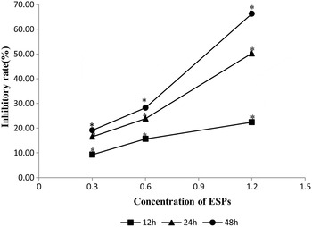
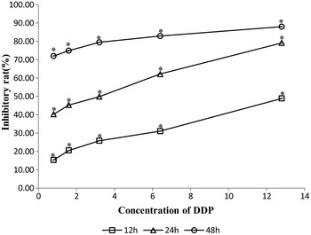
Trichinella spiralis ML ESPs of different concentrations were cultured together with H446 cells for 12, 24 and 48 h in the experimental group; the proliferation activity of H446 cells was clearly inhibited in the experimental group, compared with the negative control group (P < 0·01), indicating dose- and time-dependence (Fig. 1). DDP of different concentrations were cultured together with H446 cells for 12, 24 and 48 h; compared with the negative control group, the growth of H446 cells in the positive control group was likewise inhibited and was positively correlated with action time; a significant dose–effect relationship was shown in the DDP 12-h group and the DDP 24-h group (Fig. 2).
Proliferation inhibition rate of ESPs on H446 Cells. Different concentration of ESPs co-cultured with H446 cells for 12, 24 and 48 h in vitro, and the proliferation inhibition rate were detected by MTT assay. The values shown are the means ± s.d. *P < 0·01 relative to negative controls (n = 6).

Proliferation inhibition rate of DDP on H446 Cells. Different concentration of DDP co-cultured with H446 cells for 12, 24 and 48 h in vitro, and the proliferation inhibition rate were detected by MTT assay. The values shown are the means ± s.d. *P < 0·01 relative to negative controls (n = 6).

Trichinella spiralis ML ESPs induce H446 cell apoptosis in vitro
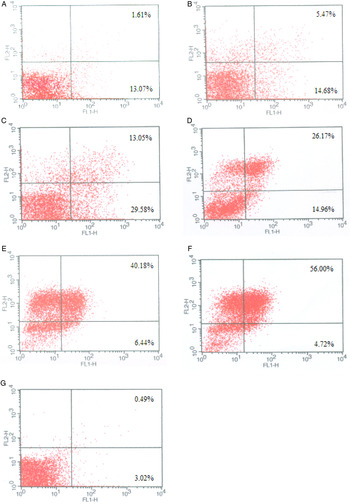
After co-culturing with H446 cells for 24 h, the apoptosis level of each group was tested by FCM (Fig. 3). The apoptosis rate of negative control group cells was 3·51%. In the experimental group, ESPs with 0·3, 0·6 and 1·2 mg mL−1 concentrations had all induced apoptosis in H446 cells, with apoptosis rates of 14·68, 20·15 and 42·63%, respectively, all higher than that of the negative control group (P < 0·05), indicating dose dependence (Fig. 3A–C). In the positive control group, DDP at concentrations of 1·6, 3·2 and 6·4 µg mL−1 all induced apoptosis in H446 cells, with apoptosis rates of 41·63, 46·62 and 60·72%, respectively, higher than the negative control group (P < 0·05), thus indicating dose-dependence (Fig. 3D–F).
Effect of ESPs or DDP on the Apoptosis of H446 cells. Different concentration of ESPs co-cultured with H446 cells for 24 h (A–C), (A) ESPs 0·3 mg mL−1; (B) ESPs 0·6 mg mL−1; (C) ESPs 1·2 mg mL−1. Fig. 3G was the negative control. Different concentration of DDP co-cultured with H446 cells for 24 h (D–F), (D) DDP 1·6 µg mL−1; (E) DDP 3·2 µg mL−1; (F) DDP 6·4 µg mL−1. The cells were collected, stained with Annexin V/PI and then analysed by FCM. *P < 0·01 relative to negative control (n = 3).

Trichinella spiralis ML ESPs induce apoptosis in H446 cells via mitochondrial pathway
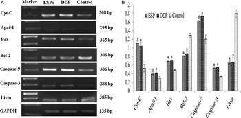
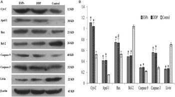
To determine whether ESPs induce apoptosis in H446 cells through a mitochondrial pathway, we tested the change in expression levels of mitochondria-related genes, using a positive control and negative control groups for comparison, with RT–PCR and Western blotting. Compared with the negative control group, the expression levels of pro-apoptosis genes Bax, Cyt-C, Apaf-1, caspase-9 and caspase-3 mRNA and their proteins all showed an increase, while the expression levels of anti-apoptosis genes Bcl-2 and Livin gene mRNA and their proteins had all decreased (P < 0·05) in the experimental and positive control groups (Figs 4 and 5). There was no significant difference between expression in the experimental and positive control groups (P > 0·05). Thus, the mitochondrial pathway may be a means for ESPs to induce apoptosis in H446 cells; this result is basically consistent with the DDP-induced apoptosis mitochondrial pathway.
Expression of seven factors mRNA in H446 cells. H446 cells were collected and treated with ESPs (1·2 mg mL−1) or DDP (3·2 µg mL−1) for 24 h. Expression of each gene were measured by RT–PCR using the GAPDH gene as a reference for normalization of gene expression. Fig. 4B shows means ± s.d. (n = 3). # P < 0·05; *P < 0·01 compared with control.

Expression of seven proteins in H446 cells by Western blot. The ESPs (1·2 mg mL−1) or DDP (3·2 µg mL−1) were co-cultured with H446 cells for 24 h. Then total proteins of H446 cells were obtained and detected by Western blot using β-actin gene as a reference for normalization of protein expression. Fig. 5B represents the means ± s.d. on different assays (n = 3). The H446 cells without co-culture of ESPs and DDP were used as a control. # P < 0·05; *P < 0·01 compared with control.

DISCUSSION
In this study, ML ESPs, DDP and serum-free RPMI 1640 were used as test, positive control and negative control groups, respectively, to act on H446 cells. The results clearly showed that both ESPs and DDP inhibited H446 cell proliferation and induced apoptosis. According to our apoptosis gene expression results, both ESPs and DDP can induce H446 cell apoptosis via a mitochondrial pathway; no significant difference was found between these gene expressions. The results clearly showed that ML ESPs inhibit growth and induce apoptosis in SCLC cells; this is comparable with DDP, a chemotherapeutic drug commonly used in clinics. As DDP has toxic side-effects and is prone to secondary drug resistance, ESPs have the potential to be a targeted therapy in the future. However, although our experiment has preliminarily verified the effects of ESPs on the inhibition of H446 cells, a toxicity test has not yet been completed; also the effects of ESPs should be tested on animals.
According to several studies, the antineoplastic mechanism of T. spiralis is divided into two types. First, T. spiralis may motivate an immunized response in the host through its tumour antigens upon parasitism, thus killing cancer cells. Second, T. spiralis may contain active anti-tumour substances, which directly participate in elimination, inducing cancer cell apoptosis (Molinari and Ebersole, Reference Molinari and Ebersole1977; Molinari et al. Reference Molinari, Carrick and Lubiniecki1979; Duan et al. Reference Duan, Li, Cheng, Lv, Gong, Su, Cai and Zhang2013). A proteome analysis on the composition of ML ESPs revealed that 162 protein ingredients in ML ESP, of which six are clearly relevant to antitumour action, and some unknown proteins, may be effective in tumour inhibition (Luo et al. Reference Luo, Cheng, Guan, Li, Yu and Du2016). We selected T. spiralis ML ESPs to act on human SCLC H446 cells and showed that T. spiralis ML ESPs inhibit SCLC proliferation with time- and dose-dependence and possibly induce apoptosis. ESP is a mixed protein; further studies are required to explore the specific protein ingredient that plays a role in its antitumour action. Our in vitro experiment lays a preliminary basis for in vivo experiments.
The mitochondrial apoptotic pathway, also known as the endogenous apoptotic pathway, is one of two classical cell apoptosis pathways. Various apoptosis stimulating signals motivate Bcl-2-asslciated protein X (Bax) proteins to invert from the cytoplasm into the outer mitochondrial membrane with multimerization, under the action of BH3 (Bcl-2 homology domain 3)-only proteins. These changes could be blocked by Bcl-2 overexpression, thus inhibiting the apoptosis of Bax. Once Bax is activated, it would result in the breakage of the outer mitochondrial membrane and formation of membrane channels, which may motivate mitochondria to release cytochrome C (Cyt-C) to the cytoplasm. Cyt C and apoptosis protease-activating factors-1 (Apaf-1) in the cytoplasm could combine and form oligomers and procaspase-9, thus forming the apoptosome (Zou et al. Reference Zou, Li, Liu and Wang1999). Procaspase-9 in the apoptosome would activate into caspase-9, which could effectively cut and activate reverse effector caspase, such as caspase-3, resulting in a reverse cascade reaction (Rodriguez and Lazebnik, Reference Rodriguez and Lazebnik1999). Finally, the substrate protease would hydrolyse, leading to the apoptosis of cells. Meanwhile, IAPs (inhibitors of apoptosis protein) could control and adjust the apoptosis of cells through cascade reaction. Currently, the apoptosis pathways of cancer cells induced by T. spiralis have yet to be clarified. Some researchers have found that phase 1 T. spiralis muscle excretory–secretory (ES) antigens could induce and motivate the caspase-dependent external apoptotic pathways (Vasilev et al. Reference Vasilev, Ilic, Gruden-Movsesijan, Vasilijic, Bosic and Sofronic-Milosavljevic2015). Other researchers have suggested that T. spiralis promotes apoptosis in human liver HepG2 cancer cells, the mechanism of which might be as follows. By activating caspase-9, T. spiralis could further activate caspase-3, thus resulting in cell apoptosis through mitochondria apoptosis pathways (Liu et al. Reference Liu, Xu, Huang and Xu2015). By testing the change of expression levels of mitochondrial apoptosis-related genes, this study discovered that ESPs could upregulate the expression of such pro-apoptosis genes as Bax, Cyt-C, Apaf-1, caspase-9 and caspase-3, while downregulating the expression of anti-apoptosis genes Bcl-2 and Livin. Hence, it could be inferred that ESPs weaken the inhibition and activation of Bcl-2 to Bax by inhibiting the expression of Bcl-2, so as to thoroughly activate Bax; this would, in turn, promote mitochondria to release a large quantity of Cyt-C into the cytoplasm. The combination of Cyt-C with Apaf-1 and procaspase-9, i.e. polymer formation, could further motivate the promoter caspase-9 and caspase-3, cutting substrate proteins in the cell. Meanwhile, ESPs may inhibit the expression of the anti-apoptosis protein Livin, hinder its apoptosis regulation functions on the cascade reaction, and finally promote apoptosis in H446 cells. Thus, T. spiralis ML ESPs activate mitochondrial intrinsic pathways in the process of inducing apoptosis in H446 SCLC cells. In summary, we discovered that T. spiralis ML ESPs may inhibit the proliferation of human H446 SCLC cells and induce their apoptosis, by activating mitochondria apoptosis pathways.
FINANCIAL SUPPORT
This study was supported by a grant from the important project of Education fund from Hebei Province of China No. ZD2014049 (to Du Luanying).