INTRODUCTION
Frontotemporal dementia (FTD) is a clinical syndrome characterized by progressive decline in behavior, language, and cognitive function with underlying frontotemporal lobar degeneration (FTLD) (Neary et al., Reference Neary, Snowden, Gustafson, Passant, Stuss, Black and Benson1998; Rascovsky et al., Reference Rascovsky, Hodges, Knopman, Mendez, Kramer, Neuhaus and Miller2011). Three clinical syndromes occur in FTD: behavioral variant FTD (bvFTD), non-fluent/agrammatic variant of primary progressive aphasia (nfvPPA), and semantic variant of primary progressive aphasia (svPPA) (Gorno-Tempini et al., Reference Gorno-Tempini, Hillis, Weintraub, Kertesz, Mendez, Cappa and Grossman2011; Neary et al., Reference Neary, Snowden, Gustafson, Passant, Stuss, Black and Benson1998; Rascovsky et al., Reference Rascovsky, Hodges, Knopman, Mendez, Kramer, Neuhaus and Miller2011). Approximately 30–50% of FTD cases have a positive family history, often with an autosomal dominant pattern of inheritance (Chow, Miller, Hayashi, & Geschwind, Reference Chow, Miller, Hayashi and Geschwind1999; Mackenzie et al., Reference Mackenzie, Neumann, Bigio, Cairns, Alafuzoff, Kril and Mann2009; McKhann et al., Reference McKhann, Albert, Grossman, Miller, Dickson and Trojanowski2001; Rabinovici & Miller, Reference Rabinovici and Miller2010; Stevens et al., Reference Stevens, van Duijn, Kamphorst, de Knijff, Heutink, van Gool and van Swieten1998). One of the most common causes of familial FTD is mutations of the progranulin gene (GRN) that result in haploinsufficiency, associated with TDP-43 pathology (FTLD-TDP) (Arai et al., Reference Arai, Hasegawa, Akiyama, Ikeda, Nonaka, Mori and Oda2006; Baker et al., Reference Baker, Mackenzie, Pickering-Brown, Gass, Rademakers, Lindholm and Hutton2006; Benussi et al., Reference Benussi, Ghidoni, Pegoiani, Moretti, Zanetti and Binetti2009; Mackenzie et al., Reference Mackenzie, Baker, Pickering-Brown, Hsiung, Lindholm, Dwosh and Feldman2006; Neumann et al., Reference Neumann, Sampathu, Kwong, Truax, Micsenyi, Chou and Lee2006; Yu et al., Reference Yu, Bird, Bekris, Montine, Leverenz, Steinbart and Van Deerlin2010). Previous studies indicate GRN is responsible for 20% of familial FTD (FTD-GRN) with a clear pattern of autosomal dominant inheritance (Gijselinck, Van Broeckhoven, & Cruts, Reference Gijselinck, Van Broeckhoven and Cruts2008).
Although penetrance is high with 90% of mutation carriers manifesting symptoms by age 75 (Cruts et al., Reference Cruts, Gijselinck, van der Zee, Engelborghs, Wils, Pirici and Van Broeckhoven2006; Gass et al., Reference Gass, Cannon, Mackenzie, Boeve, Baker, Adamson and Rademakers2006), the phenotypes associated with GRN mutations are remarkably variable, both between families with different GRN mutations and among members of individual families (Baker et al., Reference Baker, Mackenzie, Pickering-Brown, Gass, Rademakers, Lindholm and Hutton2006; Behrens et al., Reference Behrens, Mukherjee, Tu, Liscic, Grinberg, Carter and Cairns2007; Bruni et al., Reference Bruni, Momeni, Bernardi, Tomaino, Frangipane, Elder and Rogaeva2007; Gass et al., Reference Gass, Cannon, Mackenzie, Boeve, Baker, Adamson and Rademakers2006; Hsiung & Feldman, Reference Hsiung and Feldman1993; Josephs et al., Reference Josephs, Ahmed, Katsuse, Parisi, Boeve, Knopman and Dickson2007; Lopez de Munain et al., Reference Lopez de Munain, Alzualde, Gorostidi, Otaegui, Ruiz-Martinez, Indakoetxea and Marti Masso2008; Masellis et al., Reference Masellis, Momeni, Meschino, Heffner, Elder, Sato and Rogaeva2006; Mesulam et al., Reference Mesulam, Johnson, Krefft, Gass, Cannon, Adamson and Graff-Radford2007; Moreno et al., Reference Moreno, Indakoetxea, Barandiaran, Alzualde, Gabilondo, Estanga and Lopez de Munain2009; Mukherjee et al., Reference Mukherjee, Pastor, Cairns, Chakraverty, Kauwe, Shears and Goate2006; Rademakers et al., Reference Rademakers, Baker, Gass, Adamson, Huey, Momeni and Hutton2007; Spina et al., Reference Spina, Murrell, Huey, Wassermann, Pietrini, Baraibar and Grafman2007). There is a broad range in age of onset (48–83 years; mean=59±7), as well as mean age at death (53–87 years, mean=65±8) (Gass et al., Reference Gass, Cannon, Mackenzie, Boeve, Baker, Adamson and Rademakers2006). The most common clinical diagnoses of FTD-GRN are bvFTD and nfvPPA (Bruni et al., Reference Bruni, Momeni, Bernardi, Tomaino, Frangipane, Elder and Rogaeva2007; Chen-Plotkin et al., Reference Chen-Plotkin, Martinez-Lage, Sleiman, Hu, Greene, Wood and Van Deerlin2011).
Cross-sectional neuropsychological studies of fully symptomatic FTD-GRN subjects have revealed heterogeneous cognitive profiles. Language dysfunction in the form of word-finding difficulties, paraphasic errors, and impaired comprehension (van Swieten & Heutink, Reference van Swieten and Heutink2008) is prominent early in the disease process and affects 20–25% of FTD-GRN patients. Individuals often have deficits in verbal fluency, working memory, problem solving, response inhibition, and mental flexibility (Le Ber et al., Reference Le Ber, Camuzat, Hannequin, Pasquier, Guedj and Rovelet-Lecrux2008; van Swieten & Heutink, Reference van Swieten and Heutink2008). Prominent parietal dysfunction is common, with evidence of corticobasal syndrome, dyspraxia, alien-hand, and dyscalculia (Kelley et al., Reference Kelley, Haidar, Boeve, Baker, Shiung, Knopman and Petersen2010; van Swieten & Heutink, Reference van Swieten and Heutink2008; Yu et al., Reference Yu, Bird, Bekris, Montine, Leverenz, Steinbart and Van Deerlin2010). A single longitudinal, neuropsychological case study from Rohrer and colleagues (Rohrer, Warren, Barnes, et al., Reference Rohrer, Warren, Barnes, Mead, Beck, Pepple and Fox2008) documented the progression of an FTD-GRN family member from the asymptomatic phase with progressive atrophy in frontal lobe (word retrieval and fluency), parietal lobe (praxis, speech repetition, and calculation), and orbito-frontal and anterior temporal lobes (facial emotion recognition).
Studies of the prodromal neuropsychological changes in asymptomatic GRN mutation carriers are limited. In one cross-sectional study (Borroni et al., Reference Borroni, Alberici, Premi, Archetti, Garibotto, Agosti and Padovani2008), presymptomatic GRN mutation carriers and non-carriers from a single family underwent clinical assessments with neuropsychological measures, which did not reveal neuropsychological dysfunction. However, comparisons between mutation carrier and non-carrier groups were not reported, and the neuropsychological battery included many dementia screening measures, likely insensitive to subtle cognitive changes in early disease expression. In another study, the asymptomatic siblings of FTD-GRN patients underwent neuropsychological assessment. Asymptomatic mutation carriers as compared to non-carriers revealed no group differences on one dementia screening measure and four neuropsychological measures (Borroni et al., Reference Borroni, Alberici, Cercignani, Premi, Serra, Cerini and Bozzali2012). However, the neuropsychological battery was not comprehensive and the neuropsychological assessment of asymptomatic siblings was a secondary analysis, not central to the study’s aims. Accordingly, it was likely insensitive to subtle cognitive changes in early disease expression. In another cross-sectional study (Barandiaran et al., Reference Barandiaran, Estanga, Moreno, Indakoetxea, Alzualde, Balluerka and de Munain2012), subtle cognitive changes were noted in domains of attention, mental flexibility, and language (confrontation naming) in GRN mutation carriers as compared to non-carriers. The authors argued that their study provided evidence of prodromal cognitive dysfunction preceding dementia. As opposed to dementia screening measures used in most of the previous studies, we administered a comprehensive neuropsychological battery likely to be more sensitive to early cognitive changes in mutation carriers.
The present study aims to add to the limited body of literature investigating the earliest cognitive features in a cohort of GRN mutation carriers before the onset of frank dementia. We administered a comprehensive neuropsychological battery to mutation carriers and non-carrier members from the same families. We hypothesized that mutation carriers would have more frequent low scores than non-carriers. We also hypothesized that at least some of the mutation carriers would perform more poorly than non-carriers on domain scores on language, working memory and executive functions. Within the GRN mutation carrier group, we hypothesized that older subjects (closer to the mean age of onset of dementia for their family) would perform more poorly than younger mutation carriers. Given the variable phenotype in FTD-GRN, we also anticipated that neuropsychological symptoms might be heterogeneous among subjects.
METHODS
Study Overview
The University of British Columbia (UBC) FTD research program is a multidisciplinary, prospective study of familial FTD. The broad goals of the study are to characterize the neurological, neuropsychological, neuropsychiatric, pathological, and molecular genetic features in these families. The present study focuses on the preclinical and early stage of disease, before the onset of dementia, by investigating the neuropsychological profile of mutation carriers from families known to have FTD caused by GRN mutations.
Subjects
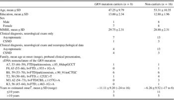
The 24 subjects were recruited from six different families with autosomal dominant FTD in which a deceased proband was clinically diagnosed with FTD at the UBC Clinic for Alzheimer’s Disease and Related Disorders (Table 1). Furthermore, probands were pathologically confirmed to have FTLD-TDP and subsequent genetic testing identified a pathogenic GRN mutation as the cause of disease in the family. For inclusion in this study, subjects had to (i) have no evidence of dementia on neurological examination, (ii) be considered at-risk for developing disease by having a parent or sibling affected with dementia, and (iii) be able to provide consent to participate in the study (age 19 or older). There was no upper age limit for subjects included in the study. Subjects were excluded from the study if they had evidence of an acquired medical condition affecting cognitive function (e.g., acquired brain injury). The mutation status of the subjects (carrier or non-carrier) was not determined until after enrollment and was not made known to the subjects or the investigators, except to the database manager and statistical analyst. DNA was analyzed for GRN mutations as previously described (Baker et al., Reference Baker, Mackenzie, Pickering-Brown, Gass, Rademakers, Lindholm and Hutton2006).
Demographic Information for Carriers and Non-carriers of the GRN mutation

Note. GRN=progranulin; MMSE=Mini Mental State Examination; CSND=clinically symptomatic, no dementia; bvFTD=behavioural variant frontotemporal dementia; CBS=corticobasal syndrome; PSP=progressive supranuclear palsy.
a The t test for continuous variables, χ 2 test for categorical variables
b Years to estimated onset=the difference between the subject’s age and the mean age of onset of frontotemporal dementia in the subject’s family; negative numbers indicate ages younger than mean age at onset, positive numbers ages older than mean age at onset.
Consensus conferences consisted of a neurologist, neuropsychologist, and research assistant, all blind to subject mutation status. Clinical diagnoses of normal and clinically symptomatic no dementia (CSND) were assigned in a hierarchical manner. CSND was assigned if the neurologist or neuropsychologist identified by clinical impression or on rating scales evidence of behavioral or cognitive symptoms or findings suggestive of FTD not yet impairing everyday function or by severity not yet meeting dementia diagnostic criteria. As previously described, clients with dementia were excluded from the study. First, the neurologist provided a clinical diagnosis based on results from the neurologist’s neurological examination, the research assistant’s cognitive screening, and functional rating forms completed by collateral sources. The neurologist remained blind to neuropsychological assessment results. Next, the neuropsychologist provided a clinical diagnosis based solely on cognitive performance on the standardized neuropsychological battery. A cut-point was established at 1 standard deviation below the mean for cognitive domain performance (e.g., Attention), with subjects performing below the cut point on any single domain being classified as CSND and subjects performing above this cut point for all domains being classified as normal. The final consensus clinical diagnosis was the most clinically symptomatic diagnosis from either the neurologist or neuropsychologist.
This study was conducted in accordance with the Helsinki Declaration and was approved by the institutional ethics committee. The subjects provided written consent.
Neuropsychological Assessment
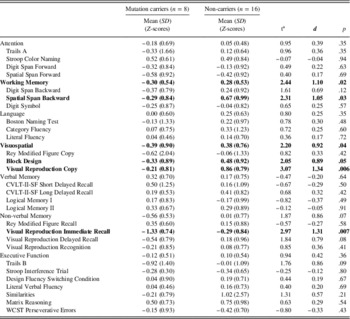
Given that the clinical phenotype of FTD-GRN is still not fully known, the neuropsychological battery was designed to be comprehensive, assessing seven cognitive domains including attention, working memory, language, visuospatial function, verbal memory, non-verbal memory and executive function (Table 2). Whereas many dementia screening batteries use a limited number of tests with selective sampling of cognitive domains known to be affected by a particular disease, the current neuropsychological battery was chosen to sample all domains of cognitive function without a priori assumptions regarding the cognitive profile of early neuropsychological characteristics of GRN mutation carriers. The test battery was comprised of well validated and reliable standardized neuropsychological measures used in clinical settings to better inform the generalizability of findings to clinical practice. To the extent possible, large, demographically-corrected normative samples were chosen to minimize ceiling or floor effects, thus increasing the sensitivity of these measures in detecting subtle cognitive changes. Measures were grouped by domain in accordance with current neuropsychological clinical practices (Lezak, Howieson, & Loring, Reference Lezak, Howieson and Loring2004).
Standardized Neuropsychological Test Scores of GRN Mutation Carriers and Non-carriers

Note. GRN=progranulin gene; CVLT-II-SF=California Verbal Learning Test, Second Edition, Short Form.
a Statistical comparisons were performed between mutation carriers and non-carriers using Univariate Analyses of Variance in a hierarchical manner. First, domain scores were compared for group differences. Subsequently, individual neuropsychological measures within significant domains were compared for group differences, and secondary neuropsychological measures/indices were compared for group differences. For completeness and heuristic value, all individual neuropsychological measure group differences are reported, even within non-significant domains. Results that reach statistical significance at p≤.05 are indicated in bold.
All tests were administered and scored according to standardized procedures. Individual test scores were converted to Z-scores using demographically corrected normative data from test publisher manuals and published studies (Delis, Kramer, Kaplan, & Ober, Reference Delis, Kramer, Kaplan and Ober2000; Heaton, Chelune, Talley, Kay, & Curtiss, Reference Heaton, Chelune, Talley, Kay and Curtiss1993; Kramer et al., Reference Kramer, Jurik, Sha, Rankin, Rosen, Johnson and Miller2003; Mitrushina, Boone, Razani, & D’Elia, Reference Mitrushina, Boone, Razani and D’Elia2005; Ruff, Light, Parker, & Levin, Reference Ruff, Light, Parker and Levin1996; Wechsler, Reference Wechsler1997a, Reference Wechsler1997b). Domain scores (e.g., attention) were calculated by averaging individual test Z-scores within that domain. Because domains scores represent the average of several individual test scores, analyses were included to determine whether low domain scores indicated convergence of several low test scores or marked impairment on a single test. Inter-rater reliability for complex visuospatial measures was established by having testing clinicians independently score a random sample of five subjects (approximately 20% of sample) for the WMS-III Visual Reproduction subtests (Wechsler, 1997b) and Rey Modified Figure (Kramer et al., Reference Kramer, Jurik, Sha, Rankin, Rosen, Johnson and Miller2003). Intra-class correlation coefficients (ICC) for inter-rater reliability were moderate for WMS-III Visual Reproduction Copy (ICC=0.54), and strong for WMS-III Visual Reproduction I (ICC=0.89), WMS-III Visual Reproduction (ICC=0.973), Rey Modified Figure Copy (ICC=0.77), and Rey Modified Figure Recall (ICC=0.88) (Kramer et al., Reference Kramer, Jurik, Sha, Rankin, Rosen, Johnson and Miller2003; Wechsler, 1997b).
Analyses
The Kolmogorov-Smirnov test of normality was conducted for both mutation carrier and non-carrier groups by neuropsychological domain to examine for the presence of outlier scores, defined as scores greater than 1.5 the interquartile range from box plot inspection. Levene’s test was also conducted to assess the equality of variance of neuropsychological data by domain to inform the selection of parametric or non-parametric tests for group comparison.
Due to the large number of neuropsychological measures, data analyses were conducted in a hierarchical manner. First, Student’s t tests (or Mann Whitney test) were conducted to compare the mean scores by neuropsychological domain between mutation carriers and non-carriers. Subsequently, Student’s t tests (or Mann Whitney test) were conducted between mutation carriers and non-carriers for individual neuropsychological measures within significant neuropsychological domains. Significance was set at p<.05 for all group comparisons. Cohen’s d values were computed to measure effect sizes of group comparisons.
Given that we expected only a subset of the mutation carriers to have early clinical symptoms of disease, it was determined that group analyses might not inform the presence of low cognitive scores in individual subjects. Accordingly, qualitative inspection of individual low score counts (1 standard deviation below the mean) was conducted by domain for individual subjects within groups.
The association between age and neuropsychological performance among mutation-carriers was tested using the Spearman product–moment correlation of the ranks. Receiver operating characteristic (ROC) curve was based on the sensitivity and specificity of significant neuropsychological domains and individual neuropsychological measures in identifying cognitive symptoms in the mutation carrier group. All statistical analyses were performed using SPSS package (v 17.0, Chicago, IL).
RESULTS
Genetic analysis revealed that eight subjects were carriers of pathogenic GRN mutations and 16 subjects were non-carriers (Table 1). Subjects came from six different families, each with a different GRN mutation. Fifty percent of total subjects (12/24 GRN mutation carriers and non-carriers) and 75% (6/8) of GRN mutation carriers came from a single family (Family R8), in which the clinical phenotype in the proband was bvFTD with parkinsonism (Table 1). Although not statistically significant, mutation carriers were younger than non-carriers (47±10 vs. 53±10 years) and further below the mean age from disease onset for their respective families (11±9 vs. 6±10 years). The mutation carrier group included only one male subject (13%) while there was equal sex distribution of non-carriers. The groups had similar education and Mini-Mental State Examination scores (Folstein, Folstein, McHugh, & Fanjiang, Reference Folstein, Folstein, McHugh and Fanjiang2001).
Clinical classifications from neurologists blind to both genetic status and neuropsychological test data resulted in similar rates of CSND versus normal in both group. Specifically, of mutation carriers, 87% (7/8) were classified as normal (e.g., clinically asymptomatic) and 13% (1/8) as CSND (e.g., evidence of clinical symptoms not significantly impacting daily function). Of non-carriers, 81% were clinically classified as normal and 19% as CSND. When neuropsychological data was combined with neurological examination, an additional 3 mutation carriers were identified as CSND. Specifically, of mutation carriers, 50% (4/8) were classified as CSND and 50% as normal. There was no change in classification of non-carriers.
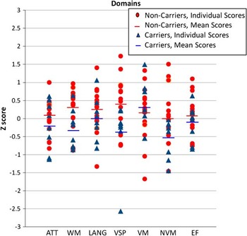
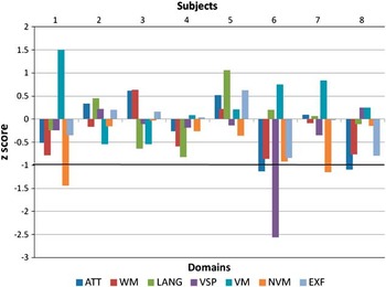
Within the mutation carrier group, four subjects (50%) had low scores (≤1 SD) on at least one neuropsychological domain. Heterogeneous cognitive profiles were noted for individuals within the mutation carrier group (Figures 1 and 2); subjects 1 & 7 both scored low on tests of non-verbal memory; subject 6 scored low on both attention and visuospatial function, and subject 8 scored low on attention. Within the non-carrier group, two subjects (13%) had low scores on at least one domain (Figure 1); subject 11 scored low on language, verbal memory, and nonverbal memory; and subject 24 scored low on verbal memory.
Scatter plots of individual neuropsychological domain standard scores (Z-scores) of GRN mutation carriers and non-carriers. GRN=progranulin gene; ATT=Attention; WM=Working Memory; LANG=Language; VSP=Visuospatial; VN=Verbal Memory; NVM=Non-verbal Memory; EXF=Executive Function.

Neuropsychological performance profiles of individual GRN mutation carriers (domain composite Z-scores). GRN=progranulin gene; ATT=Attention; WM=Working Memory; LANG=Language; VSP=Visuospatial; VN=Verbal Memory; NVM=Non-verbal Memory; EXF=Executive Function.

There were no outliers in the non-carrier group. In the mutation carrier group, there was a single subject (subject 6) with an outlier score in the visuospatial domain but the group difference in the visuospatial domain remained significant even when excluding this subject’s visuospatial performance (p<.04). However, it seemed unreasonable to exclude this subject’s neuropsychological performance from analysis given that the subject did not have outlier scores in any other domain. Furthermore, it also seemed unreasonable to exclude impaired scores when the aim of the study was to investigate the presence of cognitive dysfunction in mutation carriers. Rather, the subject’s scores were retained and nonparametric statistical measures (Mann Whitney) were used to examine group differences in the visuospatial domain since equal variance could not be assumed. Tests of normality confirmed normal distribution of test scores in other cognitive domains, and parametric statistical measures (Student’s t tests) were used to examine all other between group domain scores.
Mutation carrier neuropsychological mean scores by domain were within the normal range (±1 SD) (Table 2) with an overall trend for mutation carriers to perform worse than non-carriers on neuropsychological testing. Compared to non-carriers, mutation carriers demonstrated significantly lower domain scores with large effect sizes (d>.08) for working memory (p<.02; d=1.10) and visuospatial function (p<.04; d=0.92), and approached significance for the domain of non-verbal memory (p<.07; d=0.86). When examining performance on individual neuropsychological measures within domains, mutation carriers demonstrated significantly lower scores (p<.05) with large effect sizes on the following measures: WMS-III Spatial Span Backward in the working memory domain (p<.03; d=1.05); WMS-III Visual Reproduction Copy in the visuospatial domain (p<.006; d=1.34); and WMS-III Visual Reproduction Immediate Recall (p<.007; d=1.31) in the non-verbal memory domain (Table 2; Wechsler, 1997b). The Block Design subtest in the Visuospatial Domain approached significance (p =.05; d=0.89) (Wechsler, 1997a). Mutation carriers did not perform significantly better than non-carriers on any neuropsychological domains or individual measures.
Within the mutation carrier group, there was an overall trend for positive correlations between neuropsychological domain performance and age, with significant correlations in the domains of Attention (r=0.84; p<.01), Working Memory (r=0.83; p<.05), and Executive Function (r=0.78; p<.05). The correlations were similar when comparing neuropsychological domain performance with the mean age of onset of disease for one’s family as a variable, instead of age. Accordingly, worse performance was associated with younger age.
ROC curve analysis suggested that neuropsychological domain performance and individual neuropsychological test performance were both able to accurately separate mutation carriers from non-carriers (area under the curve >.70). However, due to cut-scores falling within the average range (as opposed to impaired performances), sensitivity and specificity was generally poor (<.80) for neuropsychological domain scores (Working Memory, 75% sensitivity, 75% specificity; Visuospatial, 63% sensitivity, 75% specificity; Non-verbal Memory, 63% sensitivity, 88% specificity) and individual neuropsychological measures (WMS-III VR1, 65% sensitivity, 67 specificity; WMS-III VR2, 71% sensitivity, 56 specificity; WMS-III VR Copy, 94% sensitivity, 33 specificity; WAIS-III Blocks, 60% sensitivity, 54% specificity; WMS-III Spatial Span Backward, 53% sensitivity, 63% specificity).
DISCUSSION
This study has revealed measurable group differences between GRN mutation carriers and non-carriers on standardized neuropsychological measures. Furthermore, a larger proportion of GRN mutation carriers (4/8 subjects, 50%) evidenced more frequent low domain scores (1 SD below the mean) as compared to non-carriers (2/15 subjects, 13%). Although a certain percentage of low scores would be expected in a large neuropsychological battery, low scores in more than 45% of healthy subjects with average intelligence would be unusual (Brooks, Iverson, & White, Reference Brooks, Iverson and White2007, Reference Brooks, Iverson and White2009; Brooks, Holdnack, & Iverson, Reference Brooks, Holdnack and Iverson2011). These findings provide evidence for the presence of early cognitive dysfunction in mutation carriers, consistent with present models of neurodegenerative disease in which a prodromal phase of mild cognitive changes precede dementia (Petersen, Reference Petersen2004).
Whereas psychobehavioral symptoms may be the hallmark of early disease expression in FTD-GRN (particularly bvFTD), findings from this study underscore the importance of neuropsychological assessment as part of a comprehensive clinical evaluation for identifying early disease expression in FTD-GRN. For example, in our study, only 1 mutation carrier (see Figure 2, subject 6) was identified as being CSND during diagnostic consensus conferences. When integrating neuropsychological data into consensus conferences, 3 additional mutation carriers were identified as CSND due to measurable cognitive changes (see Figure 2, subjects 1, 7, & 8).
The neuropsychological profile of mutation carriers in our sample suggests early asymmetric, right hemisphere brain dysfunction before the onset of dementia. This is supported by group differences within the visuospatial domain with measures that are highly correlated with right parietal lobe function (Lezak, Howieson, & Loring Reference Lezak, Howieson and Loring2004; Wechsler, 1997a). There was also evidence of selective non-verbal task dysfunction with relative sparing of analogous verbal tasks in other domains. For example, group differences of individual measures comprising the working memory domain revealed significant difference for a non-verbal working memory measure (Spatial Span Backward) but relative preservation of a verbal working memory measure (Digit Span). Similarly, group difference in the domain of non-verbal memory approached significance (p<.07) in contrast to no evidence of group difference in verbal memory function.
Neuropsychological data from the present study align with imaging data we have published previously on a subset of this same cohort (Jacova et al., Reference Jacova, Hsiung, Tawankanjanachot, Dinelle, McCormick, Gonzalez and Mackenzie2013). Imaging data from this study did not reveal clearly marked patterns of lateralized asymmetries in individual subjects but rather demonstrated reduced glucose metabolism in right anterior brain regions of GRN mutation carriers as a group. This would be consistent with current structure-function correlates of non-verbal working memory dysfunction documented in the present study. Given the clinical heterogeneity of fully affected FTD-GRN, right-hemisphere cognitive dysfunction during the prodromal phase may herald the beginning of the disease process but may not necessarily reflect the brain regions likely to be most severely affected during full expression of disease. The present neuropsychological data are also consistent with neuroimaging studies of FTD-GRN outside our research group, that have documented asymmetric atrophy, a finding that distinguishes FTD-GRN from FTD-MAPT in which atrophy tends to be more symmetric (Beck et al., Reference Beck, Rohrer, Campbell, Isaacs, Morrison, Goodall and Mead2008; Rohrer et al., Reference Rohrer, Ridgway, Modat, Ourselin, Mead, Fox and Warren2010; Whitwell et al., Reference Whitwell, Xu, Mandrekar, Boeve, Knopman, Parisi and Josephs2013).
In the absence of visuospatial tasks that parse component skills, it is challenging to determine whether visuospatial performance in GRN mutation carriers is more owing to primary visuospatial dysfunction or to the executive function demands (e.g., planning) inherit to these tasks. Findings from the (FDG)-positron emission tomography imaging study from our research group, lends more interpretive weight to executive function demands being implicated in poor visuospatial performance (Jacova et al., Reference Jacova, Hsiung, Tawankanjanachot, Dinelle, McCormick, Gonzalez and Mackenzie2013). However, it is also important to note that the planned regions of interest analyses for that study were more specific to frontal and temporal regions typically affected in FTLD. Accordingly, glucose metabolism reductions in posterior regions, such as the right parietal lobe, cannot be ruled out.
Although FTD is not classically described as including parietal dysfunction, imaging and neurological descriptions of fully affected FTD-GRN patients have suggested a broad range of lobar dysfunction to include atrophy and hypoperfusion of frontal, temporal and parietal lobes (Beck et al., Reference Beck, Rohrer, Campbell, Isaacs, Morrison, Goodall and Mead2008; Le Ber et al., Reference Le Ber, Camuzat, Hannequin, Pasquier, Guedj and Rovelet-Lecrux2008; Premi et al., Reference Premi, Grassi, Gazzina, Paghera, Pepe, Archetti and Borroni2013; Rohrer et al., Reference Rohrer, Ridgway, Modat, Ourselin, Mead, Fox and Warren2010; Rohrer, Warren, Omar et al., Reference Rohrer, Warren, Barnes, Mead, Beck, Pepple and Fox2008). Furthermore, clinical symptoms of corticobasal syndrome, dyscalculia and dyspraxia are common findings in FTD-GRN subjects, providing clinical evidence of parietal dysfunction (van Swieten & Heutink, Reference van Swieten and Heutink2008; Yu et al., Reference Yu, Bird, Bekris, Montine, Leverenz, Steinbart and Van Deerlin2010). It has been suggested that parietal dysfunction in the early stages of disease may be a specific finding in FTD-GRN subjects, reliably differentiating GRN mutation carriers from other pathological substrates of FTD (Le Ber et al., Reference Le Ber, Camuzat, Hannequin, Pasquier, Guedj and Rovelet-Lecrux2008).
When considering how our findings compare to the only other neuropsychological study of asymptomatic FTD-GRN mutation carriers versus non-carriers, there are both similarities and differences (Barandiaran et al., Reference Barandiaran, Estanga, Moreno, Indakoetxea, Alzualde, Balluerka and de Munain2012). Both studies cite measurable but subtle changes in cognitive function, as opposed to frank cognitive impairment, in mutation carriers as compared to non-carriers. However, Barandiaran and colleagues reported poor performances in domains of attention, mental flexibility (executive function), and language, with relative preservation of other domains. This differs from our findings of primary working memory and visuospatial difficulties. Of interest, mutation carriers within their cohort all had a single mutation (c.709-1G>A) with demented patients having heterogeneous clinical and neuropsychological profiles but a high proportion of cases developing corticobasal syndrome as the disease progressed. Accordingly, the presence of parietal dysfunction in a high proportion of cases can be presumed as an evolving expression of disease, even if it was not initially measured by Barandiaran and colleagues. Other factors such as the heterogeneous mutations in our sample, heterogeneous expression of FTD, methodological differences (e.g., measures used, normative data, etc.), and sampling differences might have also contributed to differences in findings between studies.
A qualitative inspection of individual mutation carrier neuropsychological performance revealed heterogeneous cognitive profiles, affecting a range of cognitive functions such as attention, visuospatial function, and non-verbal memory. The presence of variable phenotypes within the mutation carrier group aligns with the variability of clinical expression of individual family probands within this study (see Table 1) and well-documented findings of phenotypic variability in fully affected FTD-GRN patients (Baker et al., Reference Baker, Mackenzie, Pickering-Brown, Gass, Rademakers, Lindholm and Hutton2006; Behrens et al., Reference Behrens, Mukherjee, Tu, Liscic, Grinberg, Carter and Cairns2007; Bruni et al., Reference Bruni, Momeni, Bernardi, Tomaino, Frangipane, Elder and Rogaeva2007; Gass et al., Reference Gass, Cannon, Mackenzie, Boeve, Baker, Adamson and Rademakers2006; Hsiung & Feldman, Reference Hsiung and Feldman1993; Josephs et al., Reference Josephs, Ahmed, Katsuse, Parisi, Boeve, Knopman and Dickson2007; Masellis et al., Reference Masellis, Momeni, Meschino, Heffner, Elder, Sato and Rogaeva2006; Mesulam et al., Reference Mesulam, Johnson, Krefft, Gass, Cannon, Adamson and Graff-Radford2007; Mukherjee et al., Reference Mukherjee, Pastor, Cairns, Chakraverty, Kauwe, Shears and Goate2006; Rademakers et al., Reference Rademakers, Baker, Gass, Adamson, Huey, Momeni and Hutton2007; Spina et al., Reference Spina, Murrell, Huey, Wassermann, Pietrini, Baraibar and Grafman2007).
A surprising finding was that younger mutation carriers performed worse than older mutation carriers. Subjects 6 and 8 (see Graph 2) were the youngest subjects and more than 10 years from the mean age of onset of dementia for the family, but these subjects had the greatest difficulty on neuropsychological testing. This seems counterintuitive, as one would presume that older subjects who are closer to the age of dementia onset would be at greater risk for evidencing mild cognitive impairment. This may simply be a spurious finding due to our small sample size. Alternatively, there is considerable variability in the age of onset of dementia across families (35–87 years; mean=59±7 years), making it challenging to know at what age any particular individual is likely to begin expressing neurodegenerative symptoms (Bruni et al., Reference Bruni, Momeni, Bernardi, Tomaino, Frangipane, Elder and Rogaeva2007).
Several limitations of this study need to be acknowledged. First, despite our study being one of the largest samples of neuropsychological function in pre-dementia FTD-GRN subjects, our sample size was relatively small. This did not allow for correction of multiple comparisons. Second, mutation carrier and non-carrier groups were not well matched in size with approximately twice as many non-carriers. The disparity of group sizes is expected owing to the study’s criteria, which excluded mutation carriers who had already converted to dementia, and the fact that no upper age limit was set for non-carriers. There may have also been an ascertainment bias with mutation carriers with early behavioral symptoms (e.g., apathy, social impropriety) being less inclined to volunteer for research. Third, whereas sex was equally represented in the non-carrier group, the mutation carrier group consisted predominantly of women. The reason for unequal sex ratio in mutation carriers is not clear, and inconsistent with studies indicating that FTD affects both sexes equally with no sex difference in mean age of onset (Chow et al., Reference Chow, Miller, Hayashi and Geschwind1999). Finally, findings from our study are specific to the prodromal phase of FTD-GRN and are largely represented by a single family. Accordingly, results may not generalize to other genetic or sporadic forms of FTD.
The present study represents the initial characterization of cognitive symptoms in the predementia phase of FTD-GRN. By identifying the neuropsychological changes in early disease expression, we hope to clarify the natural history of the disease, assist in earlier diagnosis and provide much-needed guidance for the timing and efficacy of clinical and therapeutic interventions designed to prevent or slow the progression of this devastating neurodegenerative disease. Future opportunities for research include the integration of these findings with psychobehavioral assessment data to more fully understand the natural disease course of FTD-GRN.
ACKNOWLEDGMENTS
This work was supported by the Canadian Institutes of Health Research [operating grants #179009, 74580 to H.F. and I.M.]; the Pacific Alzheimer’s Research Foundation [center grant C06-01 to H.F. and I.M.]; and the National Institutes of Health [grants # R01 NS065782, R01 AG026251 to R.R.]. Dr. Robin Hsiung is supported by a Canadian Institutes of Health Research Clinical Genetics Investigatorship award. Dr. Tiffany Chow is supported by the Women of Baycrest.
The authors would like to gratefully acknowledge the contributions of the following testing clinicians and research assistants: Amanda LaMarre, Joanne Ng, Anthony Kupferschmidt, Kate Naus, Penny Slack, Benita Mudge, William Wang and Nadine Richard.
Dr. Hsiung discloses that has received funds as a site investigator for a clinical trial sponsored by TauRx, and is a shareholder of Amnax Biomedical.
Dr. Howard Feldman discloses that he has been a full-time paid employee of Bristol-Myers Squibb on leave from University of British Columbia (UBC) from 2009 to 2011. He received stocks and stock options in this role from Bristol-Myers Squibb. In 2012, he returned to University of British Columbia, Division of Neurology, Faculty of Medicine. In relationship to this study, he has served as paid consultant to the Bluefield Project on Frontotemporal dementia (2012), and has received travel expenses to serve on an advisory board with Fidelity Biosciences (2012, 2013). He is a co-patent holder of US Serial Number PCT/2007/070008 for Progranulin in the detection and treatment of dementia. In the past 3 years he has provided consulting services to Eli Lilly Pharma, GE Health Care, Biogen Idec and Kyowa Hakko Kirin with payments to UBC.