Nanoporous metals have seen significant interest in recent years as high surface area metallic structures that may hold interest for applications such as sensing,[Reference Wang, Wang, Zhao and Xu1, Reference Weissmüller and Sieradzki2] catalysis,[Reference Detisch, Balk and Bhattacharyya3–Reference Zhang and Ding5] biomedical applications,[Reference Seker, Berdichevsky, Staley and Yarmush6, Reference Xu, Liu, Su, Liu and Qiu7] and battery materials.[Reference Cook, Detsi, Liu, Liang, Kim, Petrissans, Dunn and Tolbert8] This interest is due to the unique structure of nanoporous metals as well as the ease of production through various dealloying processes. The typical nanoporous metal structure provides a high surface-area-to-volume ratio, including high-curvature ligaments, and a metallic surface with a large amount of porosity.[Reference McCue, Benn, Gaskey and Erlebacher9]
Generally speaking, dealloying is a selective dissolution process wherein a sacrificial component is removed from a precursor alloy, allowing the other metallic component(s) to surface diffuse and form a bicontinuous, interconnected porous structure. Multiple dealloying techniques have been investigated and developed in recent years. Electrochemical dealloying[Reference Erlebacher10–Reference Erlebacher, Aziz, Karma, Dimitrov and Sieradzki12] employs a chemical etchant, sometimes in conjunction with an electric bias, to selectively remove the sacrificial element from the precursor alloy system. Liquid metal dealloying[Reference Wada, Yubuta, Inoue and Kato13–Reference McCue, Gaskey, Geslin, Karma and Erlebacher15] uses a molten metal/alloy bath to induce the dissolution process and remove the sacrificial alloy component. These techniques have extended the range of metallic systems that can be successfully dealloyed, to incorporate a wider range of materials that includes noble metals, transition metals with some degree of oxidation resistance, and certain refractory metals.[Reference McCue, Karma and Erlebacher16]
There are limitations to these techniques, however. Electrochemical dealloying is limited to systems with a sufficiently large gap in electrode potentials between the two alloying elements.[Reference Zhang, Wang, Qi, Wang and Zhang17] This typically restricts the application of the method to noble metals and similarly corrosion resistant metals. Liquid metal dealloying exploits differences in the melting points of an alloy's constituent elements.[Reference McCue, Gaskey, Geslin, Karma and Erlebacher15] The technique works well for fabricating refractory elements in the nanoporous form. One potential issue with liquid metal dealloying is the immersion of the precursor alloy in molten metal, and the high temperatures required to extract the sacrificial element. This can lead to larger ligament sizes in the nanoporous structure, due to increased surface diffusion of atoms at these increased temperatures.[Reference Erlebacher18] Additionally, liquid metal dealloying involves an intermediate step following formation of the nanoporous structure, wherein the alloy melt solidifies in the porous network after removal of the sample from the molten bath. This phase must be removed by chemical etching, and the dilute acids or alkali solutions used in this step can impose similar restrictions to electrochemical dealloying, in terms of which metals can be successfully produced in nanoporous form. Consequently, these dealloying techniques can involve extended dealloying times[Reference Zhang, Wang, Qi, Wang and Zhang17] or can require dealloying stages[Reference McCue, Gaskey, Geslin, Karma and Erlebacher15] that generate chemical waste in the form of electrolytes or liquid-metal solvents.
The current study is focused on further development of the vacuum thermal dealloying method. Vacuum thermal dealloying involves a vacuum annealing process to remove one component of the alloy and relies on a difference in vapor pressures of the two alloy components to selectively remove the sacrificial element from the alloy. This technique provides a number of beneficial aspects compared with conventional dealloying paths, as it presents an environmentally and, for some metals, economically favorable method to fabricate nanoporous materials, e.g., when compared with electrochemical dealloying. Additionally, the lower temperature range (correlated to vapor pressure of the sacrificial element) as compared with the liquid-metal dealloying method, can mitigate ligament coarsening in the nanoporous structure that would otherwise occur at high temperatures. Similar methods have been investigated by Lu et al.[Reference Lu, Li, Han, Zhang, Liu, Wang, Wang, Cheng, Chen, Hirata, Fujita, Erlebacher and Chen19] using the zinc-cobalt alloy system. Initial research into this technique was reported as early as 2006, when a US patent was issued.[Reference Spradlin, Lye, Reed and Hudson20] In this case, the inventors described the vacuum thermal dealloying process using Mg as a sacrificial element and vacuum conditions less than 10−5 torr, with sample temperatures of at least 400 °C. Similarly, relevant studies have focused on metallic foams of titanium[Reference Esen and Bor21] and tantalum/titanium[Reference Adamek22] (Ta-20 wt.% Ti and Ta-30 wt.% Ti alloys) that exhibit micron-scale porosity, produced from precursors using Mg powder as the sacrificial component that evaporated at elevated temperatures in an argon gas environment.
The focus of the current study is on the formation of nanoporous refractory metals from Mg-based binary alloys via vacuum thermal dealloying. Mg-based alloys have been dealloyed in electrochemical systems as well, exploiting the fast corrosion rate of Mg in water and allowing the formation of particularly fine ligaments.[Reference Zhao, Qi, Wang and Zhang23, Reference Wang, Briot, Swartzentruber and Balk24] For the purposes of vacuum thermal dealloying, the low boiling point and high vapor pressure of Mg make it ideal as a sacrificial element. This stands in contrast to the refractory metals, which have exceptionally high melting points. Furthermore, many refractory metals exhibit a strong tendency to form oxide layers,[Reference Ong25] making them unsuitable for electrochemical methods. Thermal dealloying under vacuum conditions, however, allows dealloying of a Mg-based refractory alloy precursor to generate a nanoporous structure without oxidation of the refractory element. Vacuum thermal dealloying offers a promising avenue for nanoporous metal synthesis, and should be applicable to the many metals that have a lower vapor pressure than Mg (or Zn). This includes most transition metals, e.g., noble metals with FCC crystal structure and non-refractory metals such as Fe or Mn.
In this communication, we detail a thermal dealloying method for the production of nanoporous refractory metals under vacuum conditions. Magnesium was the sacrificial element in a number of thin films that were produced via a sputtering process. The films were annealed in vacuum to remove Mg, and the resulting nanoporous structures were characterized using electron microscopy.
This work focuses on the fabrication of various nanoporous refractory metal films (Nb, Ta, V, and Mo) through thermal sublimation of Mg from Mg-refractory metal alloy precursors. The precursor films were deposited via a magnetron sputtering process (ORION sputtering system, AJA International, Inc.). The main chamber of the sputtering system was evacuated to 10−7 torr (or lower) base pressure and filled with argon gas (Ar gas pressure: 2.5 mtorr). Small pieces from a 3-inch diameter (100)-oriented silicon wafer were used as substrates. Sputtering targets used in the present work were high purity metals (M) including Ta (99.99%), Mg (99.95%), Mo (99.97%), V (99.8%), and Nb (99.98%). Before the alloy deposition (Mg-M), a 15 nm interlayer of Ta was deposited on each substrate to improve adhesion between film and substrate. Binary alloys of a single composition MgxM(1−x) (at.%) were sputtered by rotating the Si substrate pieces during the deposition process. Several alloy compositions were evaluated to identify the optimal precursor composition for dealloying.
Additionally, films with a composition gradient (bracketing the same compositions of the uniform films described above) were sputtered for other Mg-based binary alloys (Mg:Ta/Mo/V), by holding the substrate stationary during film deposition. After dealloying, these gradient films were imaged to identify the optimal nanoporous structure, and this was matched with the precursor composition at that film location. Using this method, a wide range of alloy compositions were efficiently screened to find the ideal precursor composition for the successful formation of nanoporous structure.
Vacuum thermal dealloying of precursor films was performed in the main deposition chamber of the AJA ORION sputtering system, under high vacuum (pressure less than 10−6 torr), using a quartz lamp heater (AJA model SHQ-X). All samples were annealed at a setpoint temperature of 600 °C for 2 h, with a heating ramp rate of 10 °C/min. At 600 °C, the corresponding vapor pressure of Mg would be on the order of 1 torr,[Reference Gilbreath26] which significantly exceeds the base pressure of the vacuum system where dealloying occurs. These conditions were selected in order to ensure free sublimation of Mg and remove as much sacrificial Mg as possible. It should be noted that the sample temperature likely differed from the quartz heater setpoint of 600 °C, and may be ~100 °C lower. The main chamber of the sputtering system, where vacuum thermal dealloying occurred, is large compared with sample dimensions and therefore saturation considerations for gaseous versus solid magnesium are negligible. Consequently, the partial pressure of gaseous magnesium never reaches the vapor pressure where sublimation and deposition (de-sublimation) coexist. After three hours of total heating (1 h ramp time to 600 °C and 2 h hold at 600 °C), samples were allowed to cool in the vacuum chamber (~30 min cooldown to 70 °C).
Scanning electron microscopy (SEM) and x-ray energy dispersive spectroscopy (EDS) were performed using an FEI Quanta SEM and FEI Helios dual beam FIB-SEM (FIB, focused ion beam). Elemental analysis of as-deposited and dealloyed samples was determined by EDS (X-MaxN 80 mm2 detector, Oxford Instruments). X-ray photoelectron spectroscopy (XPS, Thermo Scientific K-Alpha™) measurements were performed using a micro-focused monochromatic Al Kα (hν = 1486.6 eV) x-ray source and an EX06 ion source (applied spot size: 400 µm) for depth profiling under ultra-high vacuum. The peak fitting analysis for XPS spectra was completed using Avantage Data System software. Representative cross-section and plan view micrographs were analyzed using imageJ, to measure ligament size in each nanoporous structure. X-ray diffraction (XRD) using a Siemens D500 diffractometer (Cu Kα radiation, λ = 1.5406 Å) was also performed to characterize the alloy precursor material.
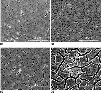
As with other dealloying methods, the precursor alloy composition must lie within a certain range in order to yield the desired nanoporous structure via vacuum thermal dealloying. The appropriate composition window for Nb/Mg precursor alloys was found to be between Nb20Mg80 and Nb30Mg70 (expressed in at. %). Four nanoporous Nb samples were fabricated by thermally dealloying various Nb/Mg precursor alloys under identical conditions. Plan view images of the resulting structures are shown in Fig. 1, corresponding to initial precursor compositions of: (a) Nb22Mg78, (b) Nb25Mg75, (c) Nb27Mg73, and (d) Nb29Mg71. The precursor alloy with a composition of Nb25Mg75 [Fig. 1(b)] generated a nanoporous structure most closely matching the desired nanostructure (i.e., that of nanoporous Au), while also minimizing micro-cracks. This precursor composition strikes a balance between providing sufficient Mg to vaporize and form the porous structure, and avoiding an overabundance of Mg that would result in widespread film cracking upon its removal. After vacuum thermal dealloying, the majority of Mg had been removed, with only 15 at.% Mg remaining in the film corresponding to the Nb25Mg75 precursor [Fig. 1(b)].
Plan view SEM micrographs of nanoporous Nb films after vacuum thermal dealloying of four different Nb/Mg precursor compositions: (a) Nb22Mg78, (b) Nb25Mg75, (c) Nb27Mg73, and (d) Nb29Mg71.

It is noted that Nb and Mg are completely immiscible in the solid state, according to the bulk equilibrium phase diagram for Nb-Mg. However, magnetron sputtering can produce thin films that do not reach the equilibrium state. A physical vapor of sputtered atoms impinges on the substrate and quickly condenses into a solid phase. This rapid quenching does not necessarily allow the deposited adatoms to fully relax into low-energy equilibrium states. Moreover, due to strain energy or surface energy, highly textured films with non-equilibrium structures are not uncommon in sputtered-deposited thin films. XRD scans of the Nb25Mg75 precursor alloy film in this study did not indicate any distinct BCC Nb peaks, but instead revealed a highly textured (basal plane orientation) HCP material that may be a Nb-Mg solid solution. In any case, there is no indication of phase separation between Nb and Mg, or the existence of additional phases.
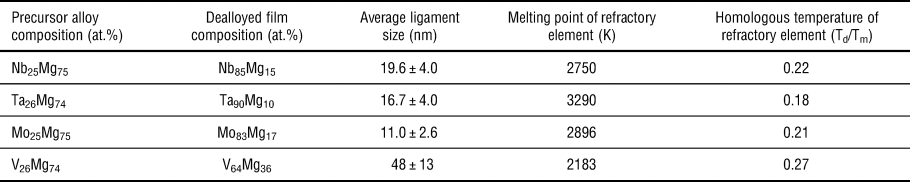
The vacuum thermal dealloying process was applied to additional alloy systems using Mg as the sacrificial element. Ta/Mg, Mo/Mg, and V/Mg alloy thin film systems were investigated. All thermally dealloyed samples contained remnant Mg levels of 17 at.% (or less), as described in Table I, except for the vanadium alloy system, which experienced higher Mg retention of 36 at.%. Precursor alloys for the other systems (Nb/Mg, Ta/Mg, and Mo/Mg) had very similar initial levels of refractory metal, and resulted in similar amounts of sublimated Mg.
Sample and processing parameters for fabrication of nanoporous refractory metals.

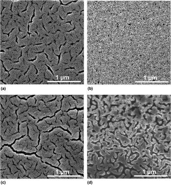
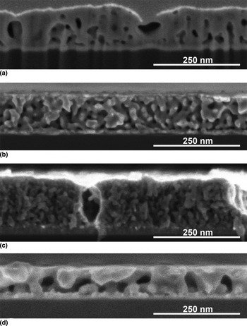
Figures 2 and 3 present plan view and cross-section micrographs showing the thermally dealloyed film structures for all four refractory alloy systems. Film cracking was observed in the Nb/Mg and Mo/Mg alloy systems, but not for the Ta/Mg or V/Mg samples investigated here. Such cracking is also sometimes observed after electrochemical dealloying of certain precursor thin films.[Reference Sun and Balk27–Reference Seker, Reed and Begley29] Removal of the sacrificial component during dealloying tends to induce a tensile stress state in a thin film.[Reference Sun and Balk27] Since the film is constrained by the Si substrate, its lateral dimensions are fixed and the film can shrink only in thickness. Thus, tensile stress can be relieved in the film plane via cracking or delamination. Further tuning of vacuum thermal dealloying parameters, such as annealing temperature (and therefore Mg removal rate), may help mitigate the cracking observed in Nb/Mg and Mo/Mg films.
Plan view SEM micrographs of nanoporous (a) Nb, (b) Ta, (c) Mo, and (d) V thin films on Si substrates, following thermal dealloying at 600 °C under vacuum.

Cross-section micrographs obtained after FIB milling of nanoporous (a) Nb, (b) Ta, (c) Mo, and (d) V thin films. Vacuum thermal dealloying at 600 °C yielded nanoporous structures with nanoscale ligaments in all films, with film cracking observed in the case of (a) Nb and (c) Mo.

Considering the observations summarized in the micrographs of Figs. 2 and 3, along with the data in Table I, it is seen that the characteristic ligament sizes in the different nanoporous alloy systems vary, despite using similar precursor compositions and dealloying conditions for all four systems. This is, however, reasonable when considering the link between temperature-dependent surface diffusion of metal adatoms and the melting point of a particular refractory metal. As discussed elsewhere,[Reference McCue, Karma and Erlebacher16] the homologous temperature (ratio of processing temperature to metal melting point, on an absolute scale) at which dealloying is performed exhibits a strong correlation with ligament size. Therefore, when dealloying is performed at a given temperature, a metal with a lower melting point will experience higher rates of surface diffusion than a metal with a higher melting point, and the former will therefore tend to develop a larger ligament size than the latter. This effect is observed for vacuum thermal dealloying as well, and explains the larger ligament size seen in nanoporous V, since V has a significantly lower melting point than the other three refractory metals studied here.
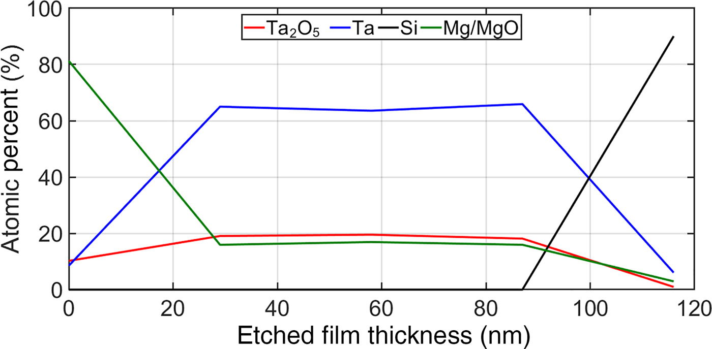
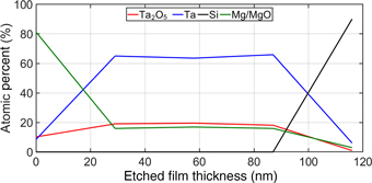
Refractory metals have a tendency to form oxides when exposed to an oxygen-containing atmosphere. However, oxidation can be prevented or mitigated when the thermal process that removes the sacrificial element (Mg) is performed in vacuum. Depth profiling analysis was performed with XPS for a sample of nanoporous Ta after vacuum thermal dealloying (see Fig. 4), and indicated that the thermally dealloyed material consisted primarily of metallic Ta, with minority amounts of oxidized Ta (Ta2O5) and remnant Mg.
XPS depth profile of nanoporous Ta, showing the variation in composition (as well as metallic versus oxidized states of Ta) as a function of thickness within the thermally dealloyed film.

In conclusion, this study focused on the fabrication of nanoporous refractory metals (Nb, Ta, Mo, and V) through thermal dealloying in vacuum. Vacuum thermal dealloying was applied to Mg-based binary alloy systems (Nb, Ta, Mo, V/Mg) to drive Mg sublimation and create refractory metals with a nanoporous structure. High-vacuum conditions with low oxygen partial pressure were essential for avoiding oxide formation, since most refractories are active metals that are prone to oxidation. The vacuum thermal dealloying technique allowed for the production of nanoporous metals that are not suitable for electrochemical techniques, and was also able to yield small ligament sizes that are difficult to achieve via dealloying in a metallic melt.
Future research will address the challenge of applying the vacuum thermal dealloying process to bulk alloy systems, where it can be difficult to alloy elements with significantly different melting points (refractory metals versus magnesium). Application of this technique to the fabrication of other nanoporous metals that are prone to oxidation (and therefore not ideal for electrochemical methods) will be studied as well.
Acknowledgments
The authors acknowledge support from the Electron Microscopy Center at the University of Kentucky, with special thanks to Nicolas J. Briot for help with FIB preparation of cross sections and EDS analysis.