Introduction
Coronary artery aneurysms caused by Kawasaki disease are likely to occur at the bifurcation of the coronary arteries or a branching site. Reference Suzuki, Kamiya and Kuwahara1–Reference Weisser, Arnold and Wallisch3 The incidence of coronary artery aneurysm at the bifurcation of the left coronary artery is exceptionally high. Its morphology varies greatly depending on the degree of distribution to peripheral arteries. Reference Tsuda, Kamiya, Ono, Kimura, Kurosaki and Echigo4 The fate of the coronary artery aneurysm at the bifurcation of the left coronary artery affects the broad myocardial wall, which is related to left ventricular ejection function and patient outcome. Reference Naoe, Shibuya, Takahashi, Wakayama, Masuda and Tanak5 Therefore, management of the aneurysms at the bifurcation of the left main trunk in this population is important. I aimed to provide an overview of the clinical course of left main trunk occlusion, which is the ultimate fate of the left coronary artery. I investigated the characteristics and clinical course in patients with left main trunk occlusion.
Methods
A total of 18 patients (12.5%) who had an occlusion of the left main trunk among 144 patients with giant aneurysms of the left coronary artery caused by Kawasaki disease between 1978 and 2022 in our hospital were reviewed based on their medical records and selective coronary angiograms (R19003-5). An occlusion of the left main trunk was confirmed with coronary angiograms. The protocol for coronary artery lesions caused by Kawasaki disease in our hospital was as follows. When coronary artery aneurysms were detected by two-dimensional echocardiography during the acute phase, the initial coronary angiogram was performed after the acute phase between 1978 and 2006. Reference Tsuda, Kamiya, Ono, Kimura and Kurosaki6 If coronary artery aneurysms were diagnosed by coronary angiograms, a follow-up coronary angiogram was performed 1 year after the initial coronary angiogram. If coronary artery aneurysms persisted, the follow-up coronary artery angiograms were repeated every 3∼5 years, depending on the degree of coronary artery lesions. From 2007, coronary computed tomography angiography was performed instead of a coronary angiogram. Reference Tsujii, Tsuda, Kanzaki and Kurosaki7 Moreover, beyond 18 years, coronary computed tomography angiography was performed every 5∼10 years depending on the degree of coronary artery lesions. When a new appearance or a progressed coronary artery lesion was suspected in coronary computed tomography angiography, a selective coronary angiogram was performed. Coronary revascularisation is indicated when myocardial ischaemia due to significant coronary stenosis is present and causing chest pain, or there is clinical evidence of ischaemia on examination. Reference Tsuda8,Reference Tsuda, Kitamura and Kimura9 As a graft, a saphenous vein was used before 1985, and an internal thoracic artery was mainly used thereafter. In patients who need multi-vessel revascularisation, the saphenous vein, radial artery, and gastroepiploic artery are also considered in each situation.
I investigated the time of the left main trunk occlusion, symptoms, coronary artery lesions, and major cardiac events, including an acute myocardial infarction and coronary artery bypass grafting, the use of devices, left ventricular ejection function, and outcome. I investigated them retrospectively based on the timing of the occlusion of the left main trunk. One was the one-time occlusion group, and the other was the two-step group. In the one-time occlusion group, the time of coronary revascularisation was defined as the time of the left main trunk occlusion. In the two-step occlusion group, the time of the left main trunk occlusion detected by selective coronary angiograms was defined as the time of the left main trunk occlusion.
Results
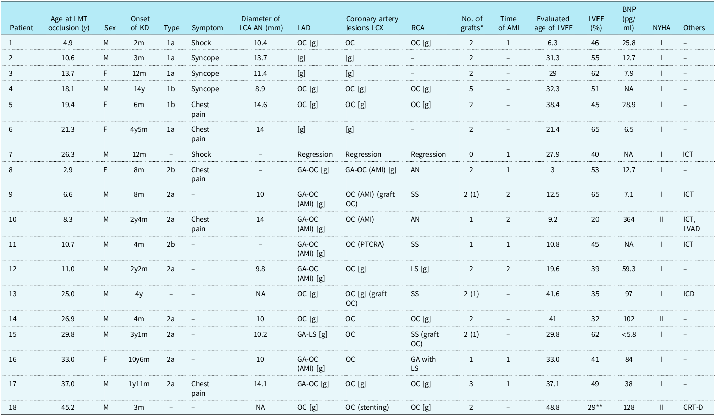
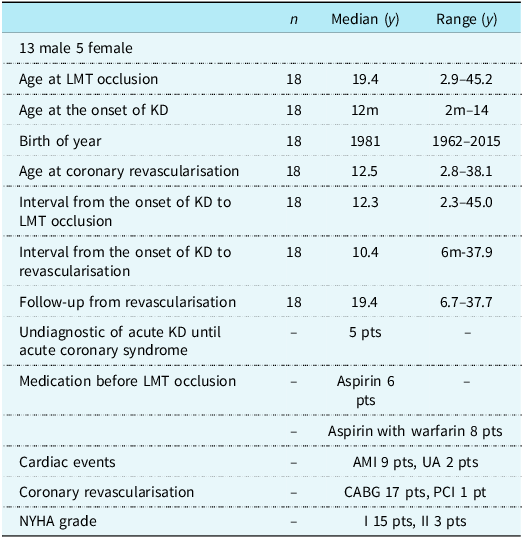
There were 13 males and 5 females (Tables 1 and 2). The age at the left main trunk occlusion ranged from 2.9 to 45.2 years, with a median of 19.4 years. The age at the onset of Kawasaki disease was 2 months to 14 years, with a median of 12 months. The interval from the onset of the Kawasaki disease to the left main trunk occlusion ranged from 2.3 to 45.0 years, with a median of 12.3 years. Their birth years ranged from 1962 to 2015, with a median of 1981. Only 3 of 18 patients received massive intravenous immunoglobulin therapy; however, they were resistant to it. In five of the 18 patients, acute Kawasaki disease was not diagnosed in the acute phase, and its diagnosis was confirmed after the occurrence of acute coronary syndrome (Patients 4, 5, 9, 16, and 18). A total of nine patients (44%) had an AMI, and three of the 9 patients had repeat myocardial infarctions. There were two adult patients who had unstable angina. For medication until the left main trunk occlusion or the initial acute myocardial infarction, eight patients took aspirin with warfarin, and 6 had aspirin. All 18 patients had multi-vessel disease. Regarding left main trunk lesions, 13 patients (72%) had a giant aneurysm at the bifurcation of the left coronary artery, and 11 of the 13 patients had an extremely giant aneurysm at the bifurcation of the left coronary artery with a maximum diameter of more than 10 mm. A total of two patients had a giant aneurysm of the proximal left anterior descending artery and left circumflex. In two adult patients, the morphology of their coronary artery lesions in the acute phase was unknown because they had acute Kawasaki disease in the 1960s. The remaining patient had a regressed aneurysm, which was a large aneurysm at the bifurcation during the acute phase. In the coronary revascularisation of the left coronary artery, 17 patients underwent a coronary artery bypass grafting, and one patient had percutaneous intracoronary thrombolysis. The age at the coronary vascularisation for the left coronary artery was 2.8 to 38.1 years, with a median of 12.5 years. The interval from the onset of Kawasaki disease to coronary revascularisation ranged from 6 months to 37.9 years, with a median of 10.4 years. The follow-up from the coronary revascularisation ranged from 6.7 to 37.7 years, with a median of 19.4 years. In 8 of the 18 patients (44%), the asymptomatic occlusion of the left main trunk was detected after coronary artery bypass grafting to the left anterior descending artery. Their left ventricular ejection fraction in the latest ranged from 20 to 65%, with a median of 46%, and their brain natriuretic peptide level varied with their left ventricular ejection fraction. For medication after coronary revascularisation, six patients took aspirin with warfarin, and 11 patients took aspirin alone. One patient had no medication. A total of fifteen patients were classified as NYHA class I, and 3 (17%) were NYHA class II. The 3 patients with NYHA class II had a left ventricular ejection fraction of < 35%. The seven patients had a preserved LVEF ≥ 50%, and eight patients had a medium LVEF ≥ 35% but <50%.
Characteristics of patients with a left main trunk occlusion

Table 1 Long description
The table presents data on 18 patients with a left main trunk occlusion, including their age at occlusion, sex, onset of Kawasaki disease, symptoms, and various medical measurements. It has 18 rows and 16 columns, with headers such as Patient, Age at LMT occlusion, Sex, Onset of KD, Type, Symptom, Diameter of LCA AN, LAD, Coronary artery lesions, No. of grafts, Time of AMI, Evaluated age of LVEF, LVEF, BNP, NYHA, and Others. Key columns include the age at occlusion ranging from 2.9 to 45.2 years, the onset of Kawasaki disease from 2 months to 14 years, and the interval from onset to occlusion ranging from 2.3 to 45.0 years. The table also details symptoms like shock and chest pain, diameters of left coronary artery aneurysms, types of coronary artery lesions, number of grafts, time of acute myocardial infarction, left ventricular ejection fraction percentages, brain natriuretic peptide levels, and NYHA classifications.
LMT= left main trunk; KD= Kawasaki disease; LCA= left coronary artery; LAD= left anterior descending; LCX= left circumflex;RCA= right coronary artery; AMI= acute myocardial infarction; LVEF= left ventricular ejection fraction; GA= giant aneuyrysm; LS= localised stenosis; OC= occlusion; AN= aneurysm; SS= segmental stenosis; ICT= intracoronary thrombolysis; LVAD= left ventricular assist device; PTCRA= percutaneous transluminal coronary rotational angioplasty; PCI= percutaneous coronary intervention; ICD= implantable cardioverter defibrillator; CRT-D= cardiac resynchronisation therapy-defibrillator; BNP, BNP= brain natriuretic peptide; [g], grafting, OC [g], graftng to an occluded vessel, ( )*, number of occluded graft, NA, not avilable; LVEF was measured on left ventriculography by cardiac catheterisation.
** The measurement was performed by 2-dimensional echocardiography.
Summary of patients with a left main trunk occlusion

Table 2 Long description
The table presents data on 18 patients with left main trunk occlusion, including 13 males and 5 females. It details their age at occlusion, age at the onset of Kawasaki disease, birth years, and intervals between key events. The table also includes information on medication, cardiac events, coronary revascularization, and NYHA grade. Notable trends include the median age at occlusion being 19.4 years, the median age at the onset of Kawasaki disease being 12 months, and the median interval from the onset of Kawasaki disease to occlusion being 12.3 years. Most patients underwent coronary artery bypass grafting, and the follow-up period ranged from 6.7 to 37.7 years.
LMT = left main truncus; KD = Kawasaki disease; ACS = acute coronary syndrome; AMI = acute myocardial infarction; UA = unstable angina; CABG = coronary artery bypass grafting; PCI = percutaneous coronary intervention; NYHA = New York Heart Association.
One-time occlusion group
Patients with coronary revascularisation of the left coronary artery immediately after an occlusion of the left main trunk
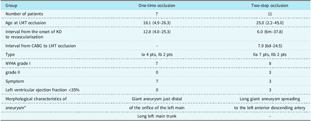
There were four male and three female patients (Table 3). The age at the left main trunk occlusion ranged from 4.9 to 26.3 years, with a median of 18.1 years. The interval from the onset of Kawasaki disease to coronary revascularisation ranged from 4.0 to 25.3 years, with a median of 12.8 years. Regarding coronary artery lesions, all 7 patients had giant aneurysms at the bifurcation of the left coronary artery. A total of four of the 7 patients had an inlet stenosis of the giant ball-shaped aneurysm of the left main trunk, where the distal portion of the orifice of the left coronary artery was also (Ia) (Figure 1). They had had a longer left main trunk. The two adult patients had an obstruction of the left anterior descending artery and left circumflex in just the distal portions of the giant aneurysm at the bifurcation of the left coronary artery (Ib). The symptoms were as follows: cardiogenic shock, 2 patients; syncope, 3 patients; and chest pain, 2 patients.
Morphology of the left coronary aneurysm of each left main trunk occlusion. Ia, A giant aneurysm at the bifurcation of the left coronary artery with inlet stenosis. There is a severe stenosis of just the proximal portion of the giant aneurysm, where just the distal portion of the orifice of the left coronary artery is located. Ib, A giant aneurysm at the bifurcation with occlusion of the left anterior descending artery and the left circumflex artery. This condition can be found in patients with undiagnosed coronary artery lesions caused by Kawasaki disease. IIa, A giant aneurysm at the bifurcation, which spread into the left anterior descending artery, creating a huge cylinder. IIb. Giant aneurysms of proximal lesions of the left anterior descending artery and the left circumflex artery. This type does not involve the bifurcation of the left coronary artery.

Figure 1 Long description
The image consists of four panels labeled Ia, Ib, IIa, and IIb, each depicting different types of left coronary artery aneurysms. Panel Ia shows a giant aneurysm at the bifurcation of the left coronary artery with inlet stenosis, where the proximal portion of the aneurysm is severely narrowed. Panel Ib illustrates a giant aneurysm at the bifurcation with occlusion of the left anterior descending artery and the left circumflex artery. Panel IIa displays a giant aneurysm at the bifurcation that extends into the left anterior descending artery, forming a large cylindrical shape. Panel IIb shows giant aneurysms in the proximal lesions of the left anterior descending artery and the left circumflex artery, without involving the bifurcation of the left coronary artery. Arrows in the images indicate specific areas of interest or abnormalities.
Characteristics of the one-time occlusion group and the second-step occlusion group

Table 3 Long description
The table compares characteristics of one-time occlusion and two-step occlusion groups in patients with Kawasaki disease. It has two main columns: One-time occlusion and Two-step occlusion, each with several rows detailing specific characteristics. The table includes data on the number of patients, age at left main trunk occlusion, interval from the onset of Kawasaki disease to revascularisation, interval from coronary artery bypass grafting to left main trunk occlusion, type of lesion, New York Heart Association grade, symptoms, left ventricular ejection fraction, and morphological characteristics of aneurysms. Notable trends include differences in age at occlusion, intervals to revascularisation, types of lesions, and symptoms between the two groups.
* It is not always possible to divide it closely.
LMT = left main truncus; KD = Kawasaki disease; NYHA = New York Heart Association.
A total of two patients had cardiogenic shock due to the left main trunk occlusion. A 4-year-old boy underwent emergency coronary artery bypass grafting for his cardiogenic shock (Patient 1). Reference Tamaki, Tsuda, Nakajima, Kobayashi and Shiono10 He was going to undergo a coronary artery bypass grafting to the left anterior descending artery and left circumflex with internal thoracic arteries on the next day because of a severe stenosis at just the distal portion of the orifice of the left coronary artery (Figure 2, left). He had grafting to the left anterior descending artery and right coronary artery with the internal thoracic arteries immediately after the cardiogenic shock. Although bilateral internal thoracic artery grafts were patent 14 years after the operation, a hypoperfusion of the lateral wall of the left ventricle was detected in exercise 99mTc-myocardial perfusion imaging (Figure 2, right). A 25-year-old male patient had sudden cardiogenic shock, followed by chest pain in the middle of the night after drinking (Patient 7). He was a smoker. Previously, he had a regression of large aneurysms of the left and right coronary arteries 1 year after acute Kawasaki disease. His follow-up was stopped at the age of 7 years. He had an acute myocardial infarction due to thrombotic occlusion of the left main trunk. He underwent emergency percutaneous coronary intracoronary thrombolysis. Reference Tsuda, Hanatani, Kurosaki, Naito and Echigo12 He had no stenosis in the left main trunk. These two patients with rapid coronary revascularisation recovered to NYHA class I; however, their left ventricular ejection fraction decreased to the 40% level.
Coronary angiogram in a patient with an inlet stenosis of a giant aneurysm in the left main trunk and 99mTc myocardial perfusion images (Patient 1) (Left) coronary angiogram showed a severe inlet stenosis of a giant aneurysm in the left main trunk before the operation. He underwent coronary artery bypass grafting immediately after an acute myocardial infarction at the age of 4 years. The left internal thoracic artery was anastomosed to the left anterior descending artery. The right internal thoracic artery was anastomosed to the right coronary artery. Bilateral grafts were patent. (Right) 99mTc myocardial perfusion images 14 years after operation. A hypoperfusion of the lateral wall of the left ventricle was detected.

Figure 2 Long description
The image contains three elements: one coronary angiogram and two myocardial perfusion images. The angiogram on the left shows a severe inlet stenosis of a giant aneurysm in the left main trunk before surgery. The two images on the right are 99mTc myocardial perfusion images taken 14 years after the operation. The top image shows stress (exercise) conditions with a hypoperfusion in the lateral wall of the left ventricle, while the bottom image shows rest conditions. The images are side-by-side, with the angiogram on the left and the perfusion images on the right.
In total, three patients had a severe stenosis of just the proximal portion of the giant aneurysm at the bifurcation of the left coronary artery. In their right coronary artery, there was no stenosis. Two patients had syncope after exercise at their school (Patients 2 and 3), and one patient had chest pain with ST-T changes on her electrocardiogram after exercise (Patient 6) (Figure 1, Ia). They underwent coronary artery bypass grafting to the left anterior descending artery and left circumflex. Their left ventricular ejection function is preserved. The two adult patients were referred to our hospital because of unstable angina. A 19-year-old female patient with hemiparesis due to a cerebral infarction at the age of 2 years complained of chest pain. She had acute Kawasaki disease at the age of 7 months (Patient 5). A 19-year-old male patient had syncope and dyspnoea on effort (Patient 4) Reference Wakisaka, Tsuda and Asakura11 (Figure 1, Ib). Their coronary angiograms revealed giant aneurysms at the bifurcation of the left coronary artery and an occlusion of the left anterior descending artery, left circumflex, and right coronary artery. Subsequently, they underwent coronary artery bypass grafting to multiple vessels. The male patient was diagnosed as having incomplete Kawasaki disease after the operation. He had had a history of “hepatitis due to a viral infection” at the age of 14 years old. He had had a fever of unknown origin for a month with a conjunctival injection.
Two-step occlusion group
Patients with an occlusion of the left main trunk after an occlusion of the left anterior descending artery
There were nine male and two female patients; six patients had giant aneurysms of the left coronary artery, which spread into the left anterior descending artery, creating a huge cylinder (IIa, LCA type) (Figure 1, IIa) (Figure 3). A 6-year-old boy also developed a giant aneurysm of the left main trunk that spread into the left anterior descending artery and left circumflex (Patient 9). The two patients had giant aneurysms of proximal lesions of the left anterior descending artery and left circumflex (IIb, branch type) (Patients 8, 11) (Figure 1, IIb). The detailed morphology of the coronary aneurysm in the acute phase was unknown in two adult patients because their coronary artery lesions were diagnosed many years after the onset (Patients 13 and 18). All patients had had an occlusion of the left anterior descending artery before the left main trunk occlusion. A total of five patients had a symptomatic acute myocardial infarction, and five had an asymptomatic occlusion of the left anterior descending artery, and one patient underwent grafting to the left anterior descending artery because of a severe localised stenosis just at the outlet of the giant aneurysm at the bifurcation (Patient 15) (Figure 3). For the right coronary artery, 10 of the 11 patients had stenotic lesions, and four underwent a coronary artery bypass grafting. All eleven patients had three-vessel disease. The age at the left main trunk occlusion ranged from 2.2 to 45.0 years, with a median of 25.0 years. All 11 patients had coronary artery bypass grafting (one graft, 4 patients; two grafts, 6 patients; three grafts, 1 patient). All 11 patients underwent a coronary artery bypass grafting to the left anterior descending artery. The ten patients had an internal thoracic artery graft, and one patient had a saphenous vein graft. The interval from the onset of Kawasaki disease to coronary artery bypass grafting ranged from 6 months to 37.9 years, with a median of 6.0 years (n = 11). The interval from the coronary artery bypass grafting to the left main trunk occlusion ranged from 6 days to 24.5 years, with a median of 7.9 years (n = 11).
Left coronary angiograms in a patient with a two-step occlusion of the left main trunk and 99mTc myocardial perfusion images (Patient 15). A 14-year-old male had had coronary artery bypass grafting with bilateral internal thoracic arteries to the left anterior descending artery and right coronary artery. The right internal thoracic artery graft to the right coronary artery was occluded immediately after coronary artery bypass grafting, and there was a left internal thoracic artery graft stenosis in the anastomosis of the left anterior descending artery 1 month after operation. The left anterior descending artery was occluded 11 months after operation. He had undergone percutaneous balloon angioplasty for anastomotic stenosis of the left internal thoracic artery 3 months after operation. ( a ) Left coronary angiogram 1 month after coronary artery bypass grafting (15 years 1 month). There was a localised stenosis of the left anterior descending artery. ( b ) Left coronary angiogram 11 months after coronary artery bypass grafting (15 years 11 months). The left anterior descending artery was occluded 11 months after operation. ( c ) Left coronary angiogram at the age of 29 years. The left main trunk was asymptomatically occluded 14 years after operation. ( d ) Left internal thoracic artery angiogram at the age of 29 years. The left internal thoracic artery was patent. ( e ) Right coronary angiogram at the age of 29 years. The collateral arteries from the right coronary artery to the circumflex artery. ( f ) 99mTc myocardial perfusion images. A slight hypoperfusion at the inferior wall of the left ventricle was detected.

Figure 3 Long description
The composite image consists of six elements: four angiograms and two myocardial perfusion images. The first angiogram (a) shows a localized stenosis of the left anterior descending artery one month after coronary artery bypass grafting. The second angiogram (b) depicts the occlusion of the left anterior descending artery eleven months after the operation. The third angiogram (c) illustrates the asymptomatic occlusion of the left main trunk at the age of twenty-nine years. The fourth angiogram (d) shows a patent left internal thoracic artery at the age of twenty-nine years. The fifth angiogram (e) displays collateral arteries from the right coronary artery to the circumflex artery at the age of twenty-nine years. The sixth element (f) presents 99mTc myocardial perfusion images, indicating slight hypoperfusion at the inferior wall of the left ventricle.
In 8 of the 11 patients, a left main trunk occlusion was first recognised in the follow-up coronary angiograms, and they were asymptomatic, even if a left main trunk occlusion was detected. Collateral arteries from the left anterior descending artery to the internal thoracic artery graft or the right coronary artery to the left circumflex had developed (Patients 9, 15, and 16) (Figures 3–5). A 6-year-old boy with sausage-like giant aneurysms of the 3 vessels, including the left main trunk, had a repeat acute myocardial infarction immediately after the acute phase, and he underwent repeat intracoronary thrombolysis (Patient 9). He had a complicated cerebral infarction with an acute myocardial infarction. He underwent coronary artery bypass grafting to the left anterior descending artery and obtuse marginal branch with bilateral internal thoracic arteries. The graft to the left anterior descending artery was patent 4 years after the operation, but the graft to the obtuse marginal branch was occluded. Although hypoperfusion of the left ventricle was detected in 99mTc myocardial perfusion imaging, his left ventricular ejection fraction was preserved (Figure 4).
Coronary angiograms 4 years after CABG and 99mTc myocardial perfusion imaging (Patient 9). ( a ) The left main trunk was occluded. ( b ) The right internal thoracic artery graft to the left anterior descending artery was patent. ( c ) Collateral arteries from the RCA to the left circumflex are well developed. ( d ) A slight hypoperfusion of the anterior wall of the left ventricle was detected in myocardial perfusion imaging.

Figure 4 Long description
The image consists of four panels showing different views of coronary angiograms and a myocardial perfusion scan. Panel (a) shows an occlusion in the left main trunk of the coronary artery. Panel (b) displays a patent right internal thoracic artery graft to the left anterior descending artery. Panel (c) illustrates well-developed collateral arteries from the right coronary artery to the left circumflex artery. Panel (d) presents a myocardial perfusion scan indicating slight hypoperfusion of the anterior wall of the left ventricle.
Coronary angiograms in a patient with a two-step occlusion of the left main trunk (Patient 16). (A1, A2) Left coronary angiograms at the age of 10 years. (A1) Right anterior oblique (RAO) 30 (A2) Left anterior oblique (LAO) 60. She had had an acute myocardial infarction at the age of 10 years. After the acute myocardial infarction, she was diagnosed with Kawasaki disease and bilateral giant aneurysms caused by Kawasaki disease. (B1, B2) Left coronary angiograms at the age of 29 years before operation. (B1) RAO 30 caudal 25. (B2) LAO 60 cranial 25. She underwent a CABG to the left anterior descending artery with the left ITA at the age of 29 years old. (C1, C2) Angiograms at the age of 33 years. (C1) Left internal thoracic angiogram. (C2) Left coronary angiogram. Four years after CABG, the left main trunk was asymptomatically occluded. Collateral arteries from the left anterior descending artery to the left circumflex artery had developed. (D) 99mTc myocardial perfusion images at the age of 33 years. Severe hypoperfusion at the anteroseptal wall was detected. Slight hypoperfusion at the lateral wall of the left ventricle was detected in the stressed myocardial perfusion imaging.

Figure 5 Long description
The composite image consists of six angiograms and one set of myocardial perfusion images. The angiograms are presented in pairs, showing the left coronary artery at different ages and angles. The first pair (A1, A2) shows the left coronary angiograms at the age of 10 years, with views in right anterior oblique (RAO) 30 and left anterior oblique (LAO) 60. The second pair (B1, B2) displays the left coronary angiograms at the age of 29 years before operation, with views in RAO 30 caudal 25 and LAO 60 cranial 25. The third pair (C1, C2) presents angiograms at the age of 33 years, including a left internal thoracic angiogram and a left coronary angiogram. The final image (D) shows 99mTc myocardial perfusion images at the age of 33 years, highlighting severe hypoperfusion at the anteroseptal wall and slight hypoperfusion at the lateral wall of the left ventricle during stress.
In the remaining three patients, a left main trunk occlusion was recognised when they had an acute myocardial infarction due to an occlusion of the left circumflex. They had chest pain, and two of the 3 underwent emergency coronary thrombolysis. A 4-year-old girl with a previous asymptomatic occlusion of the left anterior descending artery had heart failure and an acute myocardial infarction due to an occlusion of the left circumflex (Patient 8). She underwent coronary artery bypass grafting to the left anterior descending artery and left circumflex with bilateral internal thoracic arteries immediately after the acute myocardial infarction. A 9-year-old boy had an acute myocardial infarction due to an occlusion of the left circumflex 6 days after the coronary artery bypass grafting to the left anterior descending artery with the internal thoracic artery (Patient 10). Although he underwent coronary thrombolysis intravenously, he had cardiogenic shock, and his left ventricular ejection function decreased to 20%. His heart failure slightly improved with carvedilol and enalapril over 7 years. However, he underwent a left ventricular assist device implantation at the age of 17 years because his heart failure worsened with his somatic growth. A 35-year-old male patient had a coronary artery bypass grafting with three-vessel disease at the age of 11 years (Patient 17). Both the right coronary artery and the left anterior descending artery had complete occlusions. Bilateral internal thoracic arteries were used for left anterior descending and right coronary artery grafts, and the left circumflex artery was grafted with the saphenous vein. Follow-up was stopped a few years after the post-operative coronary angiograms, and he had no medications. His acute myocardial infarction was caused by thrombosis due to a thrombus in the saphenous vein graft at the age of 35 years. Reference Fujimoto, Tsuda, Kurosaki, Shiraishi, Kitamura and Masuda13 The left main trunk occlusion had supposedly occurred many years before the acute myocardial infarction.
Regarding the coronary artery lesions, seven patients had giant aneurysms at the bifurcation of the left coronary artery. Giant aneurysms with a thrombus on the coronary arterial wall existed at the bifurcation after an occlusion of the left anterior descending artery. A total of ten of the 11 patients had undergone a coronary artery bypass grafting after an occlusion of the left anterior descending artery. In most of those patients, their left ventricular ejection fraction had reduced at the time of left anterior descending artery occlusion (Table 1). After the left main trunk occlusion, the changes of the left ventricular ejection fraction were as follows: no changes in 5 patients, reduced in 3, and unknown in 3. Two adult patients with a low left ventricular ejection fraction failed because of ventricular fibrillation. One patient had an implantable cardioverter defibrillator (Patient 13), and another patient had a cardiac resynchronisation therapy-defibrillator device (Patient 18).
Discussion
There were two timings in the cases with left main trunk occlusions. One was a one-time occlusion, and the other was a second-step occlusion after an occlusion of the left anterior descending artery. Furthermore, there were two types of one-time occlusion, depending on the occlusion site. One was an inlet occlusion in a giant aneurysm, which meant it was just near the orifice of the left coronary artery. An occlusion or severe stenosis of this type induces cardiogenic shock or syncope after exercise, which can lead to a fatal cardiac event. This type can occur in an extremely large aneurysm involving the left main trunk with a diameter of more than 10 mm. To prevent a fatal cardiac event, a coronary artery bypass graft to the left anterior descending artery and the left circumflex artery is needed at an optimal time. When myocardial ischaemia is detected with a drug or exercise loading examination in patients with severe stenosis, coronary artery bypass grafting is recommended. If coronary artery bypass grafting were successful, the left ventricular ejection function would be preserved, and the prognosis would be good. However, the grafts would be occluded in some cases where the native flow is preserved, because of competition with the native flow. Reference Tsuda8,Reference Tsuda, Kitamura and Kimura9,Reference Miyazaki, Tsuda, Miyazaki, Kitamura, Tomita and Echigo14–Reference Kitamura, Tsuda and Kobayashi17 The decision regarding the optimal time for coronary revascularisation is very critical. In another one-time occlusion type, patients already had asymptomatic occlusions of the left anterior descending and left circumflex arteries. They did not have a diagnosis of acute Kawasaki disease or coronary artery lesions until the occurrence of acute coronary syndrome. Coronary artery bypass grafting is recommended in such cases with multiple-vessel occlusion. Reference Tsuda8 A total of one adult patient with a regressed large aneurysm had an acute myocardial infarction; however, its incidence is infrequent. Percutaneous coronary intervention may be considered in patients with thrombotic occlusions without severe coronary stenosis due to endothelial dysfunction in regressed large aneurysms; however, its selection is also critical. Medication for patients with regression of large aneurysms might be considered.
In patients of the second-step occlusion group, there were also two morphological types of coronary artery lesions of the left coronary artery. One was a giant aneurysm at the bifurcation of the left coronary artery. A giant aneurysm at the bifurcation of the left coronary artery spreads into the left anterior descending artery, creating a huge cylinder (IIa, LCA type). Another is giant aneurysms of the left anterior descending artery and left circumflex without giant aneurysms of the bifurcation of the left coronary artery (IIb, branch type). The incidence of coronary artery aneurysms in the left anterior descending artery was significantly higher than in the left circumflex. Reference Suzuki, Kamiya and Kuwahara1,Reference Tsuda, Tsujii, Kimura and Suzuki2 The left anterior descending artery is more likely to occlude than the left circumflex artery. Reference Barca, Menendes and Palacios15 The former had a persistent aneurysm at the bifurcation of the left coronary artery after a previous thrombotic occlusion of the proximal left anterior descending artery. Later, they often developed an asymptomatic left main trunk occlusion. Because those patients had a graft to the left anterior descending artery, they remained asymptomatic even if the left main trunk was occluded. In patients with asymptomatic occlusions of the left main trunk, collateral arteries also developed, and left ventricular ejection fraction did not change before the occlusion in some patients. On the other hand, the left ventricular ejection fraction was further reduced in some patients. Patients with a low left ventricular ejection function can experience fatal ventricular arrhythmias in adults. An implantable cardioverter defibrillator or cardiac resynchronisation therapy-defibrillator device would be required in such patients. Reference Tsuda and Yamada18
Once an acute myocardial infarction occurs, the left ventricular ejection function would decrease depending on the degree of myocardial involvement. Reference Tsuda, Hirata, Matsuo, Abe, Sugiyama and Yamada19–Reference Tsuda, Miyake and Iwasa21 Timely coronary artery bypass grafting is needed to prevent myocardial involvement; however, the decision on the optimal time is not easy. Grafting to the left anterior descending artery after a previous myocardial infarction not only improves myocardial ischaemia in the infarcted area but also is helpful as the root to another vessel through the collateral arteries (Patient 16) (Figure 5). It can prevent a fatal event due to a left main trunk occlusion. Even if a patient has a three-vessel occlusion in the late period, a graft to the left anterior descending artery can supply three vessels through collateral arteries. If a coronary artery bypass grafting is successful, the left ventricular ejection function may be preserved in some cases.
Anticoagulation should be given to patients with giant aneurysms of the left coronary artery, and thrombotic occlusion within 2 years should be noted. After that, pay attention to the progression of stenotic lesions. If there is a three-branch lesion with left anterior descending artery occlusion, the following approach is recommended: 1. If cardiac function decreases, a coronary artery bypass grafting to the left anterior descending artery will be performed. 2. If there is no decrease in cardiac function, considering the progression of stenotic lesions in other branches, careful follow-up is performed. If there is occlusion or significant stenosis of other branches, multi-vessel bypass may be considered.
The outcomes of patients with giant aneurysms at the bifurcation of the left coronary artery varied depending on the morphological development of the left anterior descending artery and the left circumflex. Each giant aneurysm morphology may depend on patient-specific factors, such as the length of the left main trunk and the branching of vessels. Classification of left main trunk occlusions in this report was considered from the retrospective course. Therefore, the pattern of left main trunk occlusion cannot necessarily be predicted from the morphology of giant aneurysms in the acute phase. However, careful and continuous follow-up may be helpful in the management of this population. I must manage such patients to learn the characteristics of the coronary artery lesions caused by Kawasaki disease.
Limitation
The treatment for acute Kawasaki disease during the last half-century has remarkably changed. With the development of equipment and technology, the treatment of accidents has improved. However, the underlying responses for cardiac events are common at some points. I thought that it was important to present the changes of the left main coronary artery in the long-term clinical course. In the two-step occlusion group, the time when the left main trunk occlusion was detected by selective coronary angiograms was defined as the time of the left main trunk occlusion. In the asymptomatic patients in the second-step occlusion group, the interval from the previous coronary angiogram to the time of the first detected occlusion ranged from 0.8 to 14 years, with a median of 5.8 years (n = 7). Therefore, I could not compare the time of the left main trunk occlusion between the one-step occlusion and the second-step occlusion, because the time of the left main trunk occlusion in the second-step occlusion group included unclear values.
Conclusions
There were two types of left main trunk occlusion in the timing. One was a severe stenosis just proximal to an extremely giant aneurysm of the left coronary artery, which led to a fatal cardiac event. The other was an asymptomatic occlusion of a persistent aneurysm of the left main trunk after a graft to the left anterior descending artery. Grafting to the left anterior descending artery was needed at an optimal time in patients with an extremely giant aneurysm of the left main trunk complicating a stenotic lesion of the left anterior descending artery. Even if a left main trunk occlusion occurs, it would not become fatal.
Acknowledgements
I greatly appreciate all the surgeons and the paediatricians who have been working at the National Cardiovascular Center (Suita, Osaka, Japan). I thank Mr John Martin for his kind English language consultation.
Financial support
This report received no specific grant from any funding agency, commercial, or not-for-profit sectors.
Competing interests
The author states that I have no conflict of interest.
Ethical standard
All procedures performed in studies involving human participants were in accordance with the ethical standards of the institutional committee, the 1964 Helsinki Declaration and its later amendments or comparable ethical standards.