Nijhawan presents a compelling argument in support of the existence of delay compensation in visual processing. Specifically, he draws a distinction between “discrete” and “continuous” stimuli and focuses on the flash-lag effect as a useful illustration of differential processing between the two. Our laboratory investigates an important class of stimuli that extends the discrete-continuous continuum. We use sequential stimuli, consisting of consecutive stimulation separated by brief blank periods. This kind of visual stimulation is frequently encountered by the retina.
Humans make rapid eye movements, or saccades, approximately three times per second. These movements shift the position of the world on the retina. Thus, the seemingly simple act of looking at an object, such as a face, involves the successive repositioning of its image features on disparate locations of the retina. Importantly, we do not perceive these shifts of the visual world or the period of lost time during a saccade, because of a general phenomenon known as saccadic suppression. Natural vision is therefore largely comprised of stable visual input that is frequently interrupted by brief periods of saccadic suppression (although continuous stimulation resulting from object tracking is possible in the form of “smooth pursuit” movements; for review, see Krauzlis Reference Krauzlis2004). Saccadic suppression adds appreciable “dead time” to the normal delays that Nijhawan reviews; yet we fail to notice all of the lost perceptual time just as we fail to be disturbed by the afferent delays. Instead, we perceive a world that is spatially and temporally stable.
How do we maintain this two-dimensional visual stability in the face of the brain's physical limitations? Nijhawan discusses feedforward signaling as one way of coping with visual delays, and our earlier work demonstrated the existence of such signaling in a pathway from the midbrain to prefrontal cortex (Sommer & Wurtz Reference Sommer and Wurtz2002; Reference Sommer and Wurtz2006). This pathway seems to help keep the brain and retinotopic visual world in spatial register before and after a saccade. Our recent work has focused on how the brain maintains temporal stability (Mayo & Sommer, submitted). We sequentially present two visual probes in the receptive field of neurons in awake, fixating monkeys. We systematically vary the amount of time between probe presentations (interstimulus interval, or ISI). The purpose is to mimic the fixation-saccade-fixation timing of natural vision. We have recorded neuronal activity in two brain areas: the retinally recipient superficial layers of the superior colliculus (supSC) and the frontal eye field (FEF), a “higher” visuomotor area in prefrontal cortex.
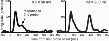
Sequential stimulation elicited sequential visual responses in neurons, with each response separated by a period of baseline activity equivalent to the ISI. Therefore, the timing of neuronal responses accurately encodes the timing of real-world visual stimulation. However, the relative magnitude of the neuronal responses also varied significantly with ISI (Fig. 1). Second visual responses were only a fraction of the first; this is a well-known phenomenon called neuronal adaptation.
Example visual responses from a small group of single neurons in superior colliculus. The results of using two probes presented at interstimulus intervals of 50 msec (left) and 200 msec (right) are shown.

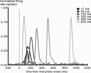
We suggest that neuronal adaptation may be useful for timing, because it carries information about the ISI during sequential visual stimulation. Extremely short ISIs (<50 msec) lead to negligible second neuronal responses (Fig. 1, left), whereas longer ISIs lead to monotonically increasing responses (Fig. 1, right) until ISI≈400 msec, where second responses attain normal magnitudes (Fig. 2). We found that adaptation followed the same time-course in FEF and supSC, but was stronger overall in FEF. As a point of reference with previous studies, our supSC data matched those reported by other laboratories (Robinson & Kertzman Reference Robinson and Kertzman1995; Wurtz et al. Reference Wurtz, Richmond and Judge1980).
Normalized visual responses in the superior colliculus population. Vertical dotted line indicates time of first probe onset. Grayscale lines depict mean responses to the second probe after sequential, paired stimulation, at various interstimulus intervals. Dashed line represents typical response to first probe.

In light of the apparent need for a compensatory physiological mechanism as described by Nijhawan, we posit that neuronal adaptation may act as an alternative source of latency encoding. As the target article explains, the exact timing of neuronal responses in identical conditions can vary by tens of milliseconds from trial to trial. Neurons downstream of these time-varying sensory responses therefore receive an imprecise measurement of stimulus onset. However, for sequential stimuli presented within 400 msec of each other, downstream neurons could corroborate the raw timing of the neuronal responses with relative magnitude comparisons of the incoming neuronal responses. Neuronal adaptation therefore may function as a complementary latency code. The presence of this code in ecologically relevant scenarios – in response to sequential stimuli, and at presentation times that mimic typical periods of fixation and saccades – lends further support to our hypothesis.
Using the relative magnitudes of sequential visual responses as a clue to the timing of visual stimuli requires an assumption that the sequential visual stimuli themselves are nearly identical. Accepting this assumption means that differences in visual responses that occur within 400 msec of each other must be due to timing. The assumption seems valid for many real-world situations, such as making saccades across continuously textured images or when an object under inspection is briefly occluded. In the latter case, neuronal adaptation would provide a robust encoding of the duration of occlusion.
Although it is still unclear how exactly neuronal adaptation could account for delay compensation, it has two important traits that are central to Nijhawan's proposal. First, neuronal adaptation is a sensory process that occurs only one synapse beyond the retina in cats and monkeys (Coenen & Eijkman Reference Coenen and Eijkman1972; Mayo & Sommer, submitted) and has been reported within the retina in lower species (Schwartz et al. Reference Schwartz, Harris, Shrom and Berry2007a). Thus, neuronal adaptation seems to be an immediate, automatic sensory mechanism. Second, neuronal adaptation is pervasive, seen at all stages of striate and extrastriate visual processing. This substantial concordance between adaptation and Nijhawan's proposal calls for additional research and computational modeling. An improved understanding of the neurophysiology of timing would provide an important complement to Nijhawan's thoughtful analysis.
ACKNOWLEDGMENTS
This research was supported by the Alfred P. Sloan foundation and NIH R01-EY017592.