Introduction
Critical CHD is the most common group of congenital malformations, affecting close to 1% of live births, and remains a leading contributor to neonatal morbidity and mortality.1–Reference Liu, Chen and Zühlke3 Yet it is no longer a disease defined solely by the malformed heart. Although advances in prenatal diagnosis, prostaglandin-based ductal management and neonatal cardiac surgery have transformed survival over recent decades, the neonate who reaches the operating room has, in many cases, already sustained injury to organ systems far beyond the heart.Reference Peyvandi, Latal, Miller and McQuillen4 The same haemodynamic derangements that define complex CHD—parallel rather than series circulation, ductal dependence, systemic-to-pulmonary steal, cyanosis, and low cardiac output—propagate to the lungs, the splanchnic bed, and the developing brain during the fragile preoperative interval. Critical CHD is therefore best conceptualised not as an isolated cardiac lesion but as a multisystem disorder in which the heart is the origin and other organs are the silent recipients of disturbed perfusion and oxygen delivery. This reframing has direct clinical consequences: preoperative stabilisation, the timing of surgery, and the anticipation of complications all depend on an accurate, organ-by-organ appraisal of the neonate as a whole, rather than on cardiac anatomy alone.
The magnitude of this extracardiac burden is now well documented across each of the three organ systems most exposed to abnormal CHD physiology. In the brain, preoperative injury—predominantly white matter injury and arterial ischaemic stroke—is demonstrable on MRI in approximately 26–43% of neonates with complex CHD, frequently in the absence of overt clinical signs, and is a recognised antecedent of adverse neurodevelopmental outcome.Reference Peyvandi, Latal, Miller and McQuillen4–Reference McQuillen, Barkovich and Hamrick7 In the gut, chronically reduced and diastolically reversed mesenteric flow predisposes to a distinct entity termed cardiac necrotising enterocolitis (NEC), which, unlike classical NEC, affects predominantly term infants and clusters in lesions with the most severe systemic hypoperfusion, such as hypoplastic left heart syndrome and truncus arteriosus, carrying considerable mortality.Reference Kelleher, McMahon and James8–Reference Spinner, Morris and Nandi10 In the lungs, pulmonary overcirculation, interstitial oedema, and effusion are near-ubiquitous accompaniments of left-to-right shunting and elevated pulmonary blood flow, and they directly govern oxygenation, ventilatory requirements, and fluid balance.Reference Liang, Liang and Chen11,Reference Rodríguez-Fanjul, Llop, Balaguer, Bautista-Rodriguez, Hernando and Jordan12 Crucially, each of these processes is dynamic and potentially modifiable in the hours and days before surgery—provided it is detected.
It is precisely this requirement for timely, repeatable, organ-level information at the bedside that has propelled the rise of point-of-care ultrasound (POCUS). Since the seminal demonstration by Lichtenstein and colleagues that clinician-performed ultrasound could alter therapeutic decisions in roughly a quarter of critically ill patients, POCUS has evolved from a radiology-bound diagnostic test into a dynamic extension of the physical examination.Reference Singh13–Reference Lichtenstein and Axler16 Its diffusion through neonatal and paediatric critical care has been rapid and, tellingly, has been led largely by neonatologists, paediatric intensivists, and emergency physicians rather than by imaging specialists; international evidence-based guidance issued by the European Society of Paediatric and Neonatal Intensive Care (ESPNIC) and endorsement of paediatric POCUS by the American Academy of Pediatrics reflect this clinician-driven trajectory.Reference Singh13,Reference Singh, Tissot and Fraga Mía14,Reference Marin and Lewiss17,Reference Miller, Stoller and Fraga18 Importantly, the value of POCUS lies not merely in detection but in its documented capacity to change management: implementation studies in the neonatal ICU report that bedside ultrasound directly influences therapeutic and procedural decisions, and this yield is plausibly greater when multiple organ systems are interrogated within a single integrated examination rather than one in isolation.Reference Pai, Dasani and Vallandingham19
Within this clinician-performed paradigm, each constituent modality has reached a degree of evidentiary maturity. Lung ultrasound discriminates the common neonatal respiratory phenotypes and detects pneumothorax and pleural effusion with accuracy at least equivalent to chest radiography while avoiding ionising radiation; in the specific context of CHD, it can quantify pulmonary overcirculation and, postoperatively, has been shown to predict extubation success and to guide diuretic and respiratory therapy.Reference Liang, Liang and Chen11,Reference Rodríguez-Fanjul, Llop, Balaguer, Bautista-Rodriguez, Hernando and Jordan12,Reference Corsini, Parri, Ficial and Dani20,Reference Georgiev, Haase and Neunhoeffer21 Abdominal ultrasound enables early recognition of NEC and other intra-abdominal pathology, and cranial ultrasound remains the first-line bedside modality for intraventricular haemorrhage, ventriculomegaly, and white matter injury.Reference Stewart, Elsayed and Fraga Mía15,Reference Dudink, Steggerda and Horsch22 Indeed, a recent expert opinion from the Association for European Paediatric and Congenital Cardiology argued that there is now sufficient evidence to support the systematic use of lung ultrasound in neonates and children with cardiac disease, yet noted that its uptake in paediatric cardiology remains limited and that the available experience is centred on the post-surgical period.Reference Cantinotti, Di Salvo and Voges23 The preoperative window—when end-organ injury is accruing and when management decisions are arguably most consequential—has received comparatively little attention.
Against this background, a structured, multimodal, preoperative ultrasound assessment—and its concrete impact on clinical management—has not been systematically characterised in neonates with CHD. We hypothesised that a single, integrated preoperative POCUS protocol encompassing the lung, abdomen, and brain would frequently reveal clinically significant pathology and would meaningfully inform their management. Accordingly, the present prospective study was designed to evaluate the feasibility of such a multimodal preoperative protocol and, in particular, to quantify the extent to which its findings support and modify clinical decision-making in neonates with CHD awaiting surgery.
Methods
Study design and setting
This was a prospective, single-centre observational study conducted in the neonatal ICU of Başakşehir Çam and Sakura City Hospital, İstanbul, Türkiye. The study protocol was approved by the institutional Clinical Research Ethics Committee (approval no. 2025/462; 26 November 2025), and the study was conducted in accordance with the principles of the Declaration of Helsinki. Written informed consent was obtained from the parents or legal guardians of all participating neonates. Patient enrollment took place between December 2025 and March 2026.
Study population
Neonates with a confirmed diagnosis of CHD who were admitted to the neonatal ICU and considered candidates for cardiac surgery were eligible for inclusion. Neonates were excluded if they had major noncardiac congenital anomalies that would independently dictate management, if adequate sonographic windows could not be obtained, or if parental consent was not provided. CHD was diagnosed and classified by transthoracic echocardiography performed by the paediatric cardiology team. All eligible neonates meeting the inclusion criteria during the study period were enrolled consecutively, yielding a total of 60 neonates. All included neonates underwent a cardiac surgical or interventional procedure during the neonatal admission. For lesions typically requiring intervention in the neonatal period (e.g., hypoplastic left heart syndrome, d-transposition of the great arteries, aortic coarctation/interrupted aortic arch, truncus arteriosus, pulmonary atresia, obstructive total anomalous pulmonary venous drainage, and other single-ventricle physiology), neonates underwent primary corrective or first-stage palliative surgery according to the underlying anatomy. For lesions in which complete repair is usually deferred beyond the neonatal period (e.g., tetralogy of Fallot, ventricular septal defect/atrioventricular septal defect, and nonobstructive total anomalous pulmonary venous drainage), only neonates who were symptomatic during the neonatal period—presenting with cyanotic spells, congestive heart failure, or failure to thrive—were included; these patients underwent an early palliative procedure (a modified Blalock–Taussig–Thomas shunt or pulmonary artery banding, according to the underlying physiology) rather than deferred complete repair. No neonate awaiting elective repair scheduled beyond the neonatal period was enrolled. The date of the surgical or interventional procedure was determined in advance by the multidisciplinary cardiology–cardiovascular surgery team once the neonate had been sufficiently stabilised and medically optimised. The preoperative multimodal POCUS examination was performed as a dedicated preoperative assessment 24–48 hours before the scheduled procedure. The median postnatal age at POCUS (10 days; IQR 6–14) reflects our institutional preoperative pathway, in which a considerable proportion of neonates were transferred from external centres and required a period of preoperative stabilisation, medical optimisation, completion of pre-surgical work-up, and multidisciplinary and parental counselling before elective neonatal cardiac surgery. Before the preoperative POCUS assessment, a standardised baseline clinical evaluation was performed for every neonate in the cohort. This comprised a formal transthoracic echocardiogram performed by the paediatric cardiology team (on which the CHD diagnosis and haemodynamic characterisation were based), a routine preoperative anteroposterior chest radiograph, arterial or capillary blood gas analysis, a structured clinical examination including detailed cardiovascular, respiratory and abdominal assessment, and a standard preoperative laboratory panel (complete blood count, C-reactive protein, urea and creatinine, electrolytes, and coagulation profile). This baseline evaluation was uniformly applied across all 60 neonates and did not vary between patients, ensuring that the comparison between the pre-POCUS and post-POCUS management plans was made against a common informational baseline.
POCUS protocol
All POCUS examinations were performed in the preoperative period, during intensive care admission, as a single multimodal assessment prior to cardiac surgery, using a Hitachi Arietta 65 ultrasound system (Fujifilm Healthcare, Tokyo, Japan). A modality-specific transducer was used for each examination: a high-frequency linear-array probe (approximately 5–18 MHz; L64) for lung ultrasound, a convex-array probe (approximately 4–9 MHz; C41) for abdominal ultrasound, and a microconvex/sector probe (approximately 5–8 MHz; C251) applied through the anterior fontanelle for cerebral ultrasound. Examinations were performed by a radiology associate professor with substantial experience in paediatric cardiac imaging; in selected patients, the examinations were performed by a paediatric cardiology specialist under his direct supervision. The operator was blinded to the patient’s current treatment plan and management intentions at the time of image acquisition and recorded only the sonographic findings. The three modalities—lung, abdominal, and cerebral ultrasound—were performed sequentially at the bedside without interrupting ongoing intensive care.
Lung ultrasound was performed with the neonate in the supine position, with lateral repositioning as needed to access the posterior regions. Each hemithorax was systematically scanned in three regions (anterior, lateral, and posterior), for a total of six lung regions,Reference Brat, Yousef, Klifa, Reynaud, Shankar Aguilera and De Luca24 assessing B-line pattern, pulmonary oedema, consolidation (differentiated as atelectasis with a static air bronchogram versus pneumonia with a dynamic air bronchogram), pleural effusion, pleural line characteristics, and pneumothorax (including evaluation of lung sliding and lung point) (Figure 1a). Abdominal ultrasound was performed systematically, assessing the liver, spleen, kidneys, urinary tract, gallbladder, and bowel. Free fluid/ascites was evaluated in the perihepatic (hepatorenal recess), perisplenic, and pelvic regions. Hydronephrosis was assessed by the anteroposterior diameter of the renal pelvis and graded as mild, moderate, or severe. Bowel was evaluated for wall thickness and other sonographic signs of NEC (bowel wall thickening, reduced peristalsis, pneumatosis intestinalis, portal venous gas, free air), and suspected NEC was staged according to the modified Bell criteria,Reference Walsh and Kliegman25 with sonographic signs interpreted in line with current bowel ultrasound evidenceReference Bhattacharjee, Dolinger, Singh and Singh26 (Figure 1c). Cerebral ultrasound was performed primarily through the anterior fontanelle in standard coronal and sagittal/parasagittal planes, with the mastoid fontanelle used additionally to assess the posterior fossa and cerebellum. The examination assessed intracranial haemorrhage (staged by the modified Papile classification),Reference Papile, Burstein, Burstein and Koffler27 parenchymal injury, lateral ventricular dimensions, and sonographic signs of cerebral oedema (Figure 1b).
Representative multimodal point-of-care ultrasound (POCUS) images obtained during preoperative assessment of neonates with CHD. (a) Lung POCUS showing the pleural line and adjacent rib shadows. (b) Cerebral POCUS through the anterior fontanelle demonstrating parenchymal and ventricular anatomy. (c) Abdominal POCUS view of the liver and adjacent structures. All images were acquired with a convex transducer (Hitachi Arietta 65; Fujifilm Healthcare, Tokyo, Japan).

Definition of pathological findings
For each modality, the presence of a clinically significant pathological finding was pre-defined as the presence of at least one of a specified set of abnormalities, while nonspecific or mild findings commonly observed in CHD were excluded. For lung POCUS, a pathological finding comprised multifocal or bilateral diffuse B-line pattern, sonographic pulmonary oedema, consolidation, pleural effusion or pneumothorax; focal B-line pattern and pleural line irregularity were not counted. For abdominal POCUS, a pathological finding comprised suspected NEC, ascites/free fluid, hydronephrosis, bowel wall thickening, pneumatosis intestinalis, or obstructive uropathy; isolated hepatomegaly, splenomegaly, and gallbladder wall thickening—findings generally reflecting systemic venous congestion in CHD—were not counted in isolation. For cerebral POCUS, a pathological finding comprised germinal matrix–intraventricular haemorrhage, ventriculomegaly, posthemorrhagic hydrocephalus, parenchymal infarct, cerebellar haemorrhage, cystic periventricular leukomalacia, cerebral oedema (loss of grey–white matter differentiation), or midline shift ≥5 mm; isolated mild periventricular echogenicity was not counted. These criteria were specified a priori.
Outcome measures
The primary outcome was a change in clinical management attributable to the POCUS findings. To minimise attribution bias, the sonographic findings and the management decision were assigned by different parties: the operator (blinded to the treatment plan) reported only the findings, whereas the decision on whether a management change was warranted and attributable to those findings was made prospectively by the treating paediatric cardiology and cardiovascular surgery team, who did not perform the examination. For each modality, the team recorded whether the examination prompted any change in management, and the specific change(s) were documented (e.g., diuretic adjustment, fluid restriction, initiation of antibiotics, withholding of feeds, respiratory support adjustment, additional imaging, or specialist consultation). A multimodal management change was defined as a change prompted by at least one of the three modalities. As a secondary, subjective measure, the treating team recorded whether they judged that a different clinical decision would have been made had POCUS not been performed. This counterfactual assessment was conducted as a “hot recall”: the treating team was asked to make this judgement immediately after the POCUS examination and the resulting management decision, in order to minimise recall bias.
Statistical analysis
Continuous variables are presented as median (interquartile range, IQR) and categorical variables as frequency (percentage). Birth anthropometric measurements were converted to sex-specific percentiles using the Fenton 2013 growth charts.Reference Fenton and Kim28 The association between the presence of a pathological finding and a management change was assessed using the Fisher exact test, with odds ratios (OR) and 95% confidence intervals (CI) calculated using the Haldane-Anscombe correction. The relationship between the number of modalities showing a pathological finding and the probability of a management change was evaluated using the Cochran-Armitage test for trend. A two-sided p value < 0.05 was considered statistically significant; for the principal comparisons, a Bonferroni-corrected threshold of p < 0.0125 was additionally applied. Analyses were performed using IBM SPSS Statistics version 29.0 (IBM Corp., Armonk, NY, USA).
Results
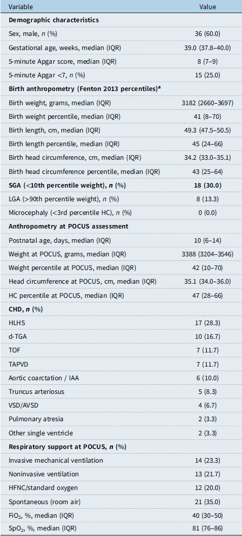
During the study period, a total of 60 neonates with CHD underwent multimodal POCUS assessment and were included in the analysis. The median gestational age was 39.0 weeks (IQR 37.8–40.0), and 36 infants (60.0%) were male. The median 5-minute Apgar score was 8 (IQR 7–9), with a score below 7 recorded in 15 neonates (25.0%). Birth anthropometry, expressed as sex-specific Fenton 2013 percentiles, showed a median birth-weight percentile of 41 (IQR 8–70); 18 neonates (30.0%) were small for gestational age (<10th percentile) and 8 (13.3%) were large for gestational age. No infant had microcephaly. At the time of POCUS assessment, the median postnatal age was 10 days (IQR 6–14). The most frequent diagnoses were hypoplastic left heart syndrome (28.3%), dextro-transposition of the great arteries (16.7%), tetralogy of Fallot (11.7%), and total anomalous pulmonary venous drainage (11.7%). At assessment, 14 neonates (23.3%) were receiving invasive mechanical ventilation and 21 (35.0%) were breathing spontaneously on room air. The detailed baseline characteristics are presented in Table 1.
Patient demographics and anthropometric characteristics

Table 1. Long description
A table detailing patient demographics and anthropometric characteristics. The table has 25 rows and 2 columns. Column headers are Variable and Value. Row 1: Sex, male, n (percent), 36 (60.0). Row 2: Gestational age, weeks, median (IQR), 39.0 (37.8-40.0). Row 3: 5-minute Apgar score, median (IQR), 8 (7-9). Row 4: 5-minute Apgar score <7, n (percent), 15 (25.0). Row 5: Birth weight, grams, median (IQR), 3182 (2660-3697). Row 6: Birth weight percentile, median (IQR), 41 (8-70). Row 7: Birth length, cm, median (IQR), 49.3 (47.5-50.5). Row 8: Birth length percentile, median (IQR), 45 (24-66). Row 9: Birth head circumference, cm, median (IQR), 34.2 (33.0-35.1). Row 10: Birth head circumference percentile, median (IQR), 43 (25-64). Row 11: SGA (<10th percentile weight), n (percent), 18 (30.0). Row 12: LGA (>90th percentile weight), n (percent), 8 (13.3). Row 13: Microcephaly (<3rd percentile HC), n (percent), 0 (0.0). Row 14: Postnatal age, days, median (IQR), 10 (6-14). Row 15: Weight at POCUS, grams, median (IQR), 3388 (3204-3546). Row 16: Weight percentile at POCUS, median (IQR), 42 (10-70). Row 17: Head circumference at POCUS, cm, median (IQR), 35.1 (34.0-36.0). Row 18: HC percentile at POCUS, median (IQR), 47 (28-66). Row 19: CHD, n (percent), HLHS, 17 (28.3). Row 20: CHD, n (percent), d-TGA, 10 (16.7). Row 21: CHD, n (percent), TOF, 7 (11.7). Row 22: CHD, n (percent), TAPVD, 7 (11.7). Row 23: CHD, n (percent), Aortic coarctation / IAA, 6 (10.0). Row 24: CHD, n (percent), Truncus arteriosus, 5 (8.3). Row 25: CHD, n (percent), VSD/AVSD, 4 (6.7). Row 26: CHD, n (percent), Pulmonary atresia, 2 (3.3). Row 27: CHD, n (percent), Other single ventricle, 2 (3.3). Row 28: Respiratory support at POCUS, n (percent), Invasive mechanical ventilation, 14 (23.3). Row 29: Respiratory support at POCUS, n (percent), Noninvasive ventilation, 13 (21.7). Row 30: Respiratory support at POCUS, n (percent), HFNC/standard oxygen, 12 (20.0). Row 31: Respiratory support at POCUS, n (percent), Spontaneous (room air), 21 (35.0). Row 32: FiO2, percent, median (IQR), 40 (30-50). Row 33: SpO2, percent, median (IQR), 81 (76-86).
a Percentiles calculated according to sex-specific Fenton 2013 growth charts. IQR = interquartile range; SGA = small for gestational age; LGA = large for gestational age; HC = head circumference; HLHS = hypoplastic left heart syndrome; d-TGA = dextro-transposition of the great arteries; TOF = tetralogy of Fallot; TAPVD = total anomalous pulmonary venous drainage; IAA = interrupted aortic arch; VSD = ventricular septal defect; AVSD = atrioventricular septal defect; HFNC = high-flow nasal cannula.
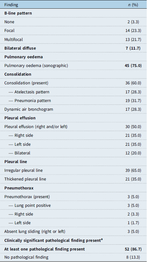
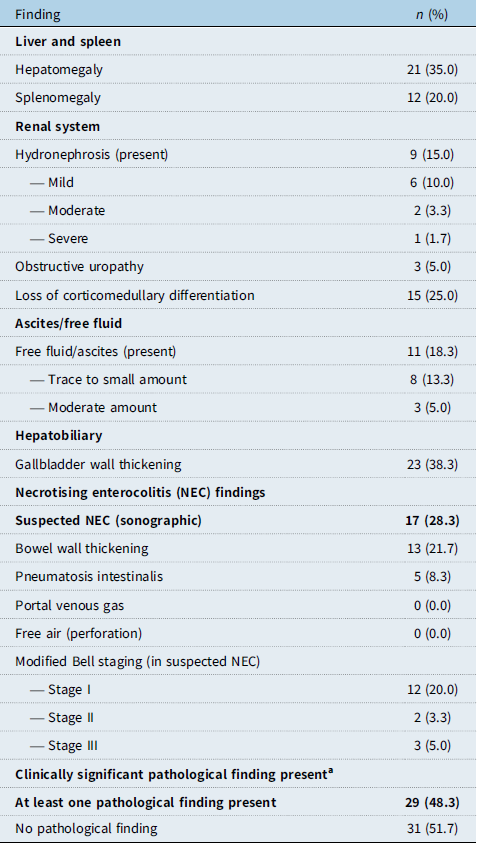
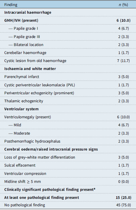
Lung POCUS revealed a high burden of abnormalities: sonographic pulmonary oedema was the most common finding (75.0%), followed by consolidation (60.0%) and pleural effusion (50.0%). A clinically significant pathological lung finding was present in 52 of 60 neonates (86.7%) (Table 2). On abdominal POCUS, suspected NEC was identified in 17 neonates (28.3%), and a clinically significant pathological abdominal finding was present in 29 (48.3%) (Table 3). Cerebral POCUS was within normal limits in the majority of patients; germinal matrix–intraventricular haemorrhage was detected in 6 neonates (10.0%), and a clinically significant pathological cerebral finding was present in 15 (25.0%) (Table 4). Overall, at least one modality showed a clinically significant pathological finding in 54 of 60 neonates (90.0%).
Lung POCUS findings

Table 2. Long description
A table with 15 rows and 2 columns. The columns are labeled ‘Finding’ and ‘n (%)’. The table lists various lung POCUS findings and their respective counts and percentages. Row 1: B-line pattern, None, 2 (3.3). Row 2: B-line pattern, Focal, 14 (23.3). Row 3: B-line pattern, Multifocal, 13 (21.7). Row 4: B-line pattern, Bilateral diffuse, 7 (11.7). Row 5: Pulmonary oedema, Pulmonary oedema (sonographic), 45 (75.0). Row 6: Consolidation, Consolidation (present), 36 (60.0). Row 7: Consolidation, Atelectasis pattern, 17 (28.3). Row 8: Consolidation, Pneumonia pattern, 19 (31.7). Row 9: Consolidation, Dynamic air bronchogram, 17 (28.3). Row 10: Pleural effusion, Pleural effusion (right and/or left), 30 (50.0). Row 11: Pleural effusion, Right side, 21 (35.0). Row 12: Pleural effusion, Left side, 21 (35.0). Row 13: Pleural effusion, Bilateral, 12 (20.0). Row 14: Pleural line, Irregular pleural line, 39 (65.0). Row 15: Pleural line, Thickened pleural line, 21 (35.0). Row 16: Pneumothorax, Pneumothorax (present), 3 (5.0). Row 17: Pneumothorax, Lung point positive, 3 (5.0). Row 18: Pneumothorax, Right side, 2 (3.3). Row 19: Pneumothorax, Left side, 1 (1.7). Row 20: Pneumothorax, Absent lung sliding (right or left), 3 (5.0). Row 21: Clinically significant pathological finding present, At least one pathological finding present, 52 (86.7). Row 22: Clinically significant pathological finding present, No pathological finding, 8 (13.3).
a A clinically significant pathological lung POCUS finding was pre-defined as the presence of at least one of the following: (i) multifocal or bilateral diffuse B-line pattern; (ii) sonographic pulmonary oedema; (iii) consolidation; (iv) right- or left-sided pleural effusion; and (v) pneumothorax. Non-specific or mild findings such as focal B-line pattern and pleural line irregularity were excluded from this category.
Sonographic findings were classified according to standard criteria: pulmonary oedema (multiple B-lines), consolidation (subpleural hypoechoic area), atelectasis (with static air bronchogram), and pneumonia (with dynamic air bronchogram).
Abdominal POCUS findings

Table 3. Long description
A table with 16 rows and 2 columns. The columns are labeled Finding and n (%). The table lists various abdominal POCUS findings and their corresponding percentages. Row 1: Liver and spleen, Hepatomegaly, 21 (35.0). Row 2: Liver and spleen, Splenomegaly, 12 (20.0). Row 3: Renal system, Hydronephrosis (present), 9 (15.0). Row 4: Renal system, Mild, 6 (10.0). Row 5: Renal system, Moderate, 2 (3.3). Row 6: Renal system, Severe, 1 (1.7). Row 7: Renal system, Obstructive uropathy, 3 (5.0). Row 8: Renal system, Loss of corticomedullary differentiation, 15 (25.0). Row 9: Ascites/free fluid, Free fluid/ascites (present), 11 (18.3). Row 10: Ascites/free fluid, Trace to small amount, 8 (13.3). Row 11: Ascites/free fluid, Moderate amount, 3 (5.0). Row 12: Hepatobiliary, Gallbladder wall thickening, 23 (38.3). Row 13: Necrotising enterocolitis (NEC) findings, Suspected NEC (sonographic), 17 (28.3). Row 14: Necrotising enterocolitis (NEC) findings, Bowel wall thickening, 13 (21.7). Row 15: Necrotising enterocolitis (NEC) findings, Pneumatosis intestinalis, 5 (8.3). Row 16: Clinically significant pathological finding present, At least one pathological finding present, 29 (48.3).
a A clinically significant pathological abdominal POCUS finding was pre-defined as the presence of at least one of the following: (i) suspected NEC; (ii) ascites/free fluid; (iii) hydronephrosis; (iv) bowel wall thickening; (v) pneumatosis intestinalis; (vi) obstructive uropathy. Isolated hepatomegaly, splenomegaly and gallbladder wall thickening, findings generally reflecting systemic venous congestion in CHD, were not counted in isolation.
Cerebral POCUS findings

Table 4. Long description
A table summarizing cerebral POCUS findings in neonates. The table has 15 rows and 2 columns. The columns are labeled ‘Finding’ and ‘n (%)’. Row 1: Intracranial haemorrhage. Row 2: GMH/IVH (present), 6 (10.0). Row 3: Papile grade I, 4 (6.7). Row 4: Papile grade III, 2 (3.3). Row 5: Bilateral location, 2 (3.3). Row 6: Cerebellar haemorrhage, 1 (1.7). Row 7: Cystic lesion from old haemorrhage, 7 (11.7). Row 8: Ischaemia and white matter. Row 9: Parenchymal infarct, 3 (5.0). Row 10: Cystic periventricular leukomalacia (PVL), 1 (1.7). Row 11: Periventricular echogenicity (prominent), 3 (5.0). Row 12: Thalamic echogenicity, 2 (3.3). Row 13: Ventricular system. Row 14: Ventriculomegaly (present), 6 (10.0). Row 15: Mild, 4 (6.7). Row 16: Moderate, 2 (3.3). Row 17: Posthemorrhagic hydrocephalus, 2 (3.3). Row 18: Cerebral oedema/raised intracranial pressure signs. Row 19: Loss of grey-white matter differentiation, 3 (5.0). Row 20: Sulcal effacement, 1 (1.7). Row 21: Ventricular compression, 1 (1.7). Row 22: Midline shift ≥ 5 mm, 0 (0.0). Row 23: Clinically significant pathological finding present. Row 24: At least one pathological finding present, 15 (25.0). Row 25: No pathological finding, 45 (75.0).
a A clinically significant pathological cerebral POCUS finding was pre-defined as the presence of at least one of the following: (i) GMH/IVH; (ii) ventriculomegaly; (iii) posthemorrhagic hydrocephalus; (iv) parenchymal infarct; (v) cerebellar haemorrhage; (vi) cystic PVL; (vii) cerebral oedema (loss of grey–white matter differentiation); and (viii) midline shift ≥ 5 mm. Non-specific findings such as isolated mild periventricular echogenicity were excluded from this category.
GMH = germinal matrix haemorrhage; IVH = intraventricular haemorrhage; PVL = periventricular leukomalacia. Intraventricular haemorrhage was staged using the modified Papile classification. In this population, the majority of cerebral POCUS findings were within normal limits (75%).
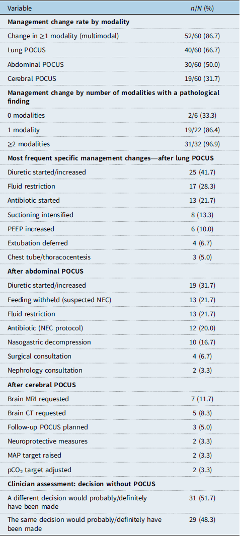
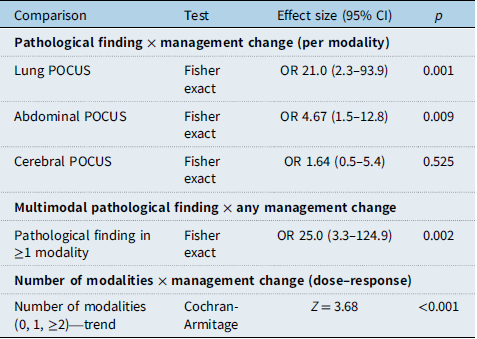
POCUS assessment was associated with a change in clinical management in 52 of 60 neonates (86.7%) in at least one modality. Management changes were most frequent following lung POCUS (66.7%), followed by abdominal (50.0%) and cerebral (31.7%) POCUS (Table 5; Figure 2). The presence of a clinically significant pathological finding was strongly associated with a subsequent management change for lung POCUS (75.0% versus 12.5%; OR 21.0, 95% CI 2.3–93.9; p = 0.001) and abdominal POCUS (69.0% versus 32.3%; OR 4.67, 95% CI 1.5–12.8; p = 0.009). In contrast, no significant association was observed for cerebral POCUS (40.0% versus 28.9%; OR 1.64, 95% CI 0.5–5.4; p = 0.525), suggesting that cerebral POCUS was used predominantly for prognostic rather than immediate decision-making purposes in this population. The presence of a pathological finding in at least one modality markedly increased the odds of a management change (OR 25.0, 95% CI 3.3–124.9; p = 0.002). These associations are summarised in Table 6 (Figure 3).
Clinical management changes following POCUS

Table 5. Long description
The table presents data on management change rates by modality and specific management changes after POCUS. It has 32 rows and 2 columns. The columns are labeled ‘Variable’ and ‘n/N (%)’. The table is divided into sections: Management change rate by modality, Management change by number of modalities with a pathological finding, Most frequent specific management changes after lung POCUS, After abdominal POCUS, After cerebral POCUS, and Clinician assessment: decision without POCUS. Each section lists variables and their corresponding values in terms of n/N (%). For example, under Management change rate by modality, Change in ≥1 modality (multimodal) has a value of 52/60 (86.7%), Lung POCUS has 40/60 (66.7%), Abdominal POCUS has 30/60 (50.0%), and Cerebral POCUS has 19/60 (31.7%). The table also lists specific management changes and their frequencies after each type of POCUS.
POCUS = point-of-care ultrasound; NEC = necrotising enterocolitis; PEEP = positive end-expiratory pressure; MAP = mean arterial pressure. Percentages are of the total cohort (n = 60). Statistical analyses of these associations are presented in Table 6.
Clinical management change rates following POCUS, by modality. Odds ratios (OR) and p-values refer to the association between pathological finding presence and management change (Fisher exact test).

Statistical analysis of the association between POCUS findings and clinical management change

Table 6. Long description
The table presents data on clinical management changes following POCUS, focusing on different modalities and their impact. It includes three main sections: pathological finding by management change per modality, multimodal pathological finding by any management change, and the number of modalities by management change (dose-response). The table has 11 rows and 4 columns. Column headers are Comparison, Test, Effect size (95% CI), and p. Row labels include Lung POCUS, Abdominal POCUS, Cerebral POCUS, Pathological finding in ≥1 modality, and Number of modalities. Notable trends include significant effect sizes for Lung POCUS and Abdominal POCUS, with p-values indicating statistical significance. The table also shows the most frequent specific management changes after each type of POCUS and clinician assessments on decision changes.
Dose–response relationship between the number of modalities showing a pathological finding and the probability of a management change (Cochran-Armitage trend test, Z = 3.68; p < 0.001).

A dose–response relationship was observed between the number of modalities showing a pathological finding and the probability of a management change, which rose from 33.3% when no modality was abnormal to 86.4% with one abnormal modality and 96.9% with two or more abnormal modalities (Cochran-Armitage trend test, Z = 3.68; p < 0.001; Table 6). The corresponding management-change rates by number of affected modalities are presented in Table 5. The most frequent specific management changes were initiation or escalation of diuretics (41.7% after lung POCUS) and fluid restriction (28.3%); after abdominal POCUS, diuretic adjustment (31.7%) and withholding of feeds for suspected NEC (21.7%) predominated. After cerebral POCUS, the majority of changes consisted of requests for additional imaging rather than direct therapeutic interventions. The full list of specific management changes is provided in Table 5. On the clinician self-assessment, in 31 of 60 cases (51.7%) the attending team judged that a different decision would probably or definitely have been made had POCUS not been performed (Table 5). After Bonferroni correction for multiple comparisons (threshold p < 0.0125), the lung, abdominal and multimodal associations, and the dose–response trend remained statistically significant (Table 6).
Discussion
In this prospective study of 60 neonates with CHD awaiting cardiac surgery, a structured preoperative multimodal POCUS protocol encompassing the lung, abdomen, and brain revealed a clinically significant pathological finding in at least one organ system in 90.0% of patients and was associated with a change in clinical management in 86.7%. To our knowledge, this is among the first studies to evaluate an integrated, multi-organ POCUS assessment performed specifically in the preoperative period in neonates with CHD and quantify its influence on bedside decision-making.
The overall rate of POCUS-associated management change observed here is consistent with, and at the upper end of, the existing literature. Since the seminal report by Lichtenstein and Axler that clinician-performed ultrasound altered treatment in approximately a quarter of critically ill patients, implementation studies have reported substantial management change rates, reaching 62% for a dedicated neonatal lung ultrasound service.Reference Elsayed, Narvey, Lashin, Alammary, Gigolyk and Louis29 When examined modality by modality, our findings align closely with the largest neonatal implementation series to date: in the programme reported by Pai and colleagues, POCUS influenced management in 14% of lung, 53% of abdominal, and 43% of cranial examinations, figures broadly comparable to the present cohort (66.7%, 50.0%, and 31.7%, respectively).Reference Pai, Dasani and Vallandingham19 The higher overall and lung-specific impact in our study likely reflects the deliberately selected, high-risk population of surgical CHD candidates, in whom pulmonary overcirculation, oedema, and effusion are particularly prevalent and, importantly, directly amenable to bedside therapeutic adjustment. Two further points warrant emphasis. First, a high diagnostic yield does not equate to a high decision-making impact: although a pathological finding was identified in 90.0% of neonates, not every finding mandated a change in management, and the gap between these figures reflects the appropriate clinical judgement that some abnormalities were either already known or did not in themselves require a new intervention. Second, the multimodal design itself contributes to the high overall rate, since interrogating three organ systems inevitably increases the probability that at least one will inform a decision—a structural feature that distinguishes an integrated protocol from any single-organ assessment and that we examine further below.
The strong association between a pathological finding and a subsequent management change for lung (OR 21.0) and abdominal (OR 4.67) POCUS is clinically coherent. Pulmonary findings such as oedema and effusion are readily actionable because they reflect a haemodynamic state that can be modified within hours; in our cohort, they most often led to the initiation or escalation of diuretics and to fluid restriction. Abdominal findings, dominated by the sonographic suspicion of NEC, prompted decisions to withhold feeds and to adjust antibiotic and fluid therapy, consistent with the recognised vulnerability of the CHD gut to a distinct “cardiac NEC” driven by mesenteric hypoperfusion. The choice of a six-region lung protocol is itself supported by evidence that 6- and 12-region acquisitions provide comparable predictive value in neonates, favouring a pragmatic approach without loss of information.
In contrast, the presence of a pathological cerebral finding was not significantly associated with an immediate change in management (OR 1.64; p = 0.525). Rather than diminishing the value of cranial POCUS, this finding clarifies its role in this setting. The majority of cerebral examinations that did influence management did so by prompting further imaging—most often brain MRI—rather than by triggering an immediate therapeutic intervention. This pattern is consistent with the nature of the lesions detected: established intracranial haemorrhage, white matter injury and ventricular changes are generally not acutely reversible, and although cranial ultrasound reliably detects the larger haemorrhagic lesions while being less sensitive than MRI for subtle white matter injury,Reference Benders, Kersbergen and de Vries30 their identification carries substantial prognostic weight, informs neurodevelopmental surveillance and supports family counselling, given that preoperative brain injury is demonstrable in a substantial proportion of these neonates and predicts later outcome. Cranial POCUS in the preoperative CHD neonate may therefore be best regarded as a prognostic and triage tool rather than an instrument of immediate therapeutic change.
A central observation of this study is the graded relationship between the number of organ systems showing a pathological finding and the probability of a management change, which rose from 33.3% in neonates with no pathological finding to 96.9% in those with two or more affected systems (trend p < 0.001). This dose–response pattern provides internal support for the multimodal approach itself: whereas previous work has largely interrogated organ systems in isolation, our data suggest that the diagnostic and decision-making yield is cumulative when the lung, abdomen, and brain are assessed within a single integrated examination. In a population in which multiple organ systems are simultaneously exposed to abnormal circulatory physiology, a protocol that mirrors this multisystem involvement appears better suited to inform care than any single-organ assessment alone.
The timing of the preoperative POCUS assessment in our study deserves particular consideration when interpreting our findings. In this cohort, POCUS was performed 24–48 hours prior to the scheduled surgical or interventional procedure, at a median postnatal age of 10 days (IQR 6–14). This reflects our institutional preoperative pathway rather than a delay in surgical planning; nevertheless, this preoperative window is longer than that reported by centres that undertake surgical intervention for critical CHD within the first 3–5 postnatal days. This longer window is likely to have contributed to the burden of pathological findings observed in some modalities. However, this effect is probably not uniform across organ systems. Pulmonary and abdominal pathology in this population is largely a dynamic consequence of ongoing abnormal circulatory physiology—pulmonary overcirculation with progressive interstitial oedema and effusion, and low-cardiac-output-related mesenteric hypoperfusion contributing to the “cardiac NEC” pattern—and would therefore be expected to accrue with time. In contrast, the majority of clinically significant cerebral findings in preoperative CHD neonates (established intraventricular haemorrhage, ventriculomegaly, and pre-existing white matter injury) represent largely fixed structural or antenatally acquired lesions rather than pathologies that evolve within a few postnatal days; this is consistent with our finding that cerebral POCUS was not significantly associated with an immediate management change and functioned primarily as a prognostic tool. Our management-change estimates for lung and abdominal POCUS should therefore be interpreted in the context of our centre’s preoperative timeline and may not directly generalise to centres in which surgery is performed within the first 3–5 postnatal days; the cerebral estimates, in contrast, are less likely to be time-sensitive.
Limitations
Several methodological features strengthen these findings. Patients were enrolled consecutively, reducing selection bias; the operator was blinded to the prevailing treatment plan and recorded only the sonographic findings; and the determination of whether a management change was attributable to POCUS was made by the treating cardiology and cardiovascular surgery team rather than by the operator, thereby separating the detection of findings from the attribution of decisions and mitigating attribution bias. Pathological findings were defined a priori, avoiding subjective severity grading.
Several limitations should nevertheless be acknowledged. This was a single-centre study with a modest sample size, which limits generalisability. All examinations were performed by a single experienced operator, so inter-observer reproducibility could not be assessed. Furthermore, the operator in this study was a radiology associate professor with dedicated neonatal ultrasound expertise, whereas in many centres bedside POCUS is performed by neonatal or paediatric intensivists themselves. While the involvement of a single trained expert operator standardised image acquisition across all three organ systems and helped separate image acquisition from clinical decision-making, it may limit direct generalisation of our management-change estimates to settings in which POCUS is performed by the treating intensivist team. Confirmation of our findings in multicentre studies encompassing different operator profiles is therefore warranted. The secondary measure of whether a different decision would have been made without POCUS is inherently subjective and susceptible to recall and hypothetical bias. While this measure was collected as an immediate (“hot”) recall to minimise this bias, it remains a clinician-reported estimate of perceived added value and does not on its own establish a causal added value of POCUS beyond the standardised baseline clinical assessment. Accordingly, our primary evidence for the decision-making impact of POCUS rests on the formal statistical associations between pathological POCUS findings and objectively recorded management changes (ORs and trend statistics, Table 6), rather than on the clinician-reported counterfactual measure alone. Finally, the present analysis was confined to immediate decision-making and did not address whether POCUS-guided changes translate into improved clinical outcomes.
Conclusion
A structured preoperative multimodal POCUS protocol, encompassing the lung, abdomen and brain, frequently identified clinically significant pathology and was associated with a meaningful change in clinical management in neonates with CHD awaiting surgery. The decision-making impact was strongest for lung and abdominal assessment, whereas cerebral POCUS appeared to serve a predominantly prognostic role. The probability of a management change increased with the number of organ systems affected, supporting the value of an integrated multi-organ approach. Prospective, multicentre, and outcome-oriented studies are now warranted to determine whether this decision-level benefit translates into improved patient outcomes.
Data availability statement
The data that support the findings of this study are available from the corresponding author upon reasonable request.
Financial support
This research received no external funding.
Competing interests
The authors declare no conflicts of interest.
Institutional review board statement
This study was conducted in accordance with the Declaration of Helsinki and approved by the Ethics Committee of Istanbul Başakşehir Çam and Sakura City Hospital, University of Health Sciences of Turkey (Protocol Code: 2025/462, date: 26 November 2025).
Informed consent statement
Written informed consent for participation was obtained from the legal guardians (parents) of all neonates included in the study prior to inclusion.
Use of artificial intelligence
During the preparation of this work, the authors used Claude (Anthropic) to assist with manuscript drafting, refine sentence structure, and improve readability. After using this tool, the authors reviewed and edited the content as needed and take full responsibility for the content of the published article.