Introduction
The origin of some or all lissamphibians is hypothesized to fall among small upper Carboniferous temnospondyls, the amphibamiform dissorophoids (Bolt, Reference Bolt1969; Anderson et al., Reference Anderson, Reisz, Scott, Fröbisch and Sumida2008a; Maddin et al., Reference Maddin, Jenkins and Anderson2012; Pardo et al., Reference Pardo, Small and Huttenlocker2017; Schoch, Reference Schoch2019). This became increasingly clear after the discovery of pedicellate teeth in Doleserpeton annectens Bolt, Reference Bolt1969 and Amphibamus grandiceps Cope, Reference Cope1865 (Bolt, Reference Bolt1969, Reference Bolt and Nitecki1979), the identification of cylindrical centra in some amphibamiforms (Bolt, Reference Bolt1969), and the description of the very lissamphibian-like Gerobatrachus hottoni Anderson et al., Reference Anderson, Reisz, Scott, Fröbisch and Sumida2008a, which has a partially open cheek and shortened trunk.
Yet the origin and diversification of amphibamiforms within the Dissorophoidea remain obscure. For instance, most taxa (sensu Schoch, Reference Schoch2019) are of early Permian age and reveal a remarkable diversity that suggests a much earlier diversification sometime in the Carboniferous (Schoch and Milner, Reference Schoch, Milner and Sues2014). Upper Carboniferous deposits in the Czech Republic (Nýřany, Broumov), Germany (Stützerbach in the Thuringian forest), France (Montceau-les-Mines in Sâone et Loire), and the United States (Linton and Five Points in Ohio, Mazon Creek in Illinois, Hamilton Quarry and Garnett in Kansas) have yielded a diverse record of dissorophoid material that documents the presence of most major clades within the group: the micromelerpetids Limnogyrinus elegans Fritsch, Reference Fritsch1881 and Nyranerpeton amilneri Werneburg, Reference Werneburg2012 (Werneburg, Reference Werneburg1994, Reference Werneburg2012; Schoch and Witzmann, Reference Schoch and Witzmann2018); the branchiosaurids Branchiosaurus and Apateon (Boy, Reference Boy1987; Werneburg, Reference Werneburg2012); the trematopids Actiobates peabodyi Eaton, Reference Eaton1973, Anconastes vesperus Berman et al., Reference Berman, Reisz and Eberth1987, Fedexia striegeli Berman et al., Reference Berman, Henrici, Brezinski and Kollar2010, Mordex calliprepes Steen, Reference Steen1938, and Mattauschia laticeps (Fritsch, Reference Fritsch1881) (Berman et al., Reference Berman, Reisz and Eberth1987, 2010; Werneburg, Reference Werneburg2012; Milner, Reference Milner2019; Gee and Reisz, Reference Gee and Reisz2020a); and the amphibamiforms Platyrhinops lyelli (Wyman, Reference Wyman1858) (Hook and Baird, Reference Hook and Baird1984; Clack and Milner, Reference Clack and Milner2010), Amphibamus grandiceps (Watson, Reference Watson1940; Milner, Reference Milner1982), and Eoscopus lockardi Daly, Reference Daly1994.
As recently outlined (Schoch, Reference Schoch2019), major phylogenetic questions remain regarding the Dissorophoidea that range from their early diversification, to the position of branchiosaurids and lissamphibians, to the early evolution of olsoniform and amphibamiform branches (Boy, Reference Boy1981; Anderson et al., Reference Anderson, Reisz, Scott, Fröbisch and Sumida2008a, Reference Anderson, Henrici, Sumida, Martens and Bermanb; Schoch, Reference Schoch2012, Reference Schoch2013; Maddin et al., Reference Maddin, Fröbisch, Evans and Milner2013). It is clear that in addition to applying new preparation techniques to existing material, the discovery of new specimens will play an important role in resolving these uncertainties.
Such a discovery is the basis of this study. The previously unreported specimen we describe was found in 1994 at Five Points, Ohio. Because all the coal mines in the Five Points area were reclaimed by the middle 1990s, no additional collecting is possible today. The deposit is somewhat older than the classic Diamond Mine locality at Linton, which is ~42 km to the south in Jefferson County, Ohio, but both originated as sapropelic fillings of deep, abandoned channels (Hook and Ferm, Reference Hook and Ferm1988; Hook and Hower, Reference Hook and Hower1988; Hook and Baird, Reference Hook and Baird1994). Not surprisingly, aquatic taxa dominate both assemblages, yet comparatively small numbers of amphibious to terrestrial forms also occur. Among these rarest forms is a single outstanding specimen from Five Points that represents the distinctively new dissorophoid genus reported here.
Materials and methods
Material examined
A single specimen (CM [Carnegie Museum] 34608) forms the basis of the present study. It consists of two pieces that are counterpart blocks. After the blocks were etched in dilute HCl, latex casts of the natural molds were made. These and the original material were studied under a WILD M3Z binocular microscope. Illustrations are based on photographs taken with a Panasonic Lumix DMC-TZ22. For Figure 5, the latex cast was dusted with ammonium chloride before images were taken with a Canon EOS 20D.
In preparing the description, we referred to the following works for comparative studies: Actiobates peabodyi (Gee and Reisz, Reference Gee and Reisz2020a), Amphibamus grandiceps (Milner, Reference Milner1982), Branchiosauridae (Boy, Reference Boy1987), Cacops morrisi Reisz, Schoch, and Anderson, Reference Reisz, Schoch and Anderson2009 (Reisz et al., Reference Reisz, Schoch and Anderson2009; Gee and Reisz, Reference Gee and Reisz2018), Eoscopus lockardi (Daly, Reference Daly1994), Micromelerpetidae (Werneburg, Reference Werneburg1991; Boy, Reference Boy1995; Schoch and Witzmann, Reference Schoch and Witzmann2018), Nanobamus macrorhinus Schoch and Milner, Reference Schoch, Milner and Sues2014 (Gee and Reisz, Reference Gee and Reisz2020b), Pasawioops mayi Fröbisch and Reisz, Reference Fröbisch and Reisz2008, Platyrhinops lyelli (Clack and Milner, Reference Clack and Milner2010), Scapanops neglectus Schoch and Sues, Reference Schoch and Sues2013, and Tersomius texensis Case, Reference Case1910 (Carroll, Reference Carroll1964; Maddin et al., Reference Maddin, Fröbisch, Evans and Milner2013). In addition, we have studied nearly all of the published specimens of dissorophoids first-hand and have assessed a sizeable number of unreported specimens from Linton and Five Points.
Maturity of specimen
Some features of the type and only specimen of P. bairdi n. gen. n. sp. indicate that it was not fully mature. These are, (1) the poorly established ornament in the snout; (2) the existence of a nasolacrimal groove rather than a closed channel; and (3) the absence of any ossifications in the sphenethmoid, otic, and exoccipital regions. On the other hand, this specimen is post-larval because of the ossified quadrate, the lack of lateral line sulci, and the absence of any branchial arch elements or branchial denticles. We conclude that the specimen was a late juvenile.
Repository and institutional abbreviation
The type and only specimen is housed in the Carnegie Museum of Natural History (CM), Pittsburgh, USA.
Systematic paleontology
Temnospondyli Zittel, Reference Zittel1888
Rhachitomi Watson, Reference Watson1919 sensu Schoch, Reference Schoch2013
Dissorophoidea Bolt, Reference Bolt1969
Palodromeus new genus
Type species
Palodromeus bairdi n. gen. n. sp. (by monotypy).
Diagnosis
As for the type species by monotypy.
Etymology
Greek palos = swamp, dromeus = walker.
Remarks
None.
Palodromeus bairdi new species
Figures 1–7
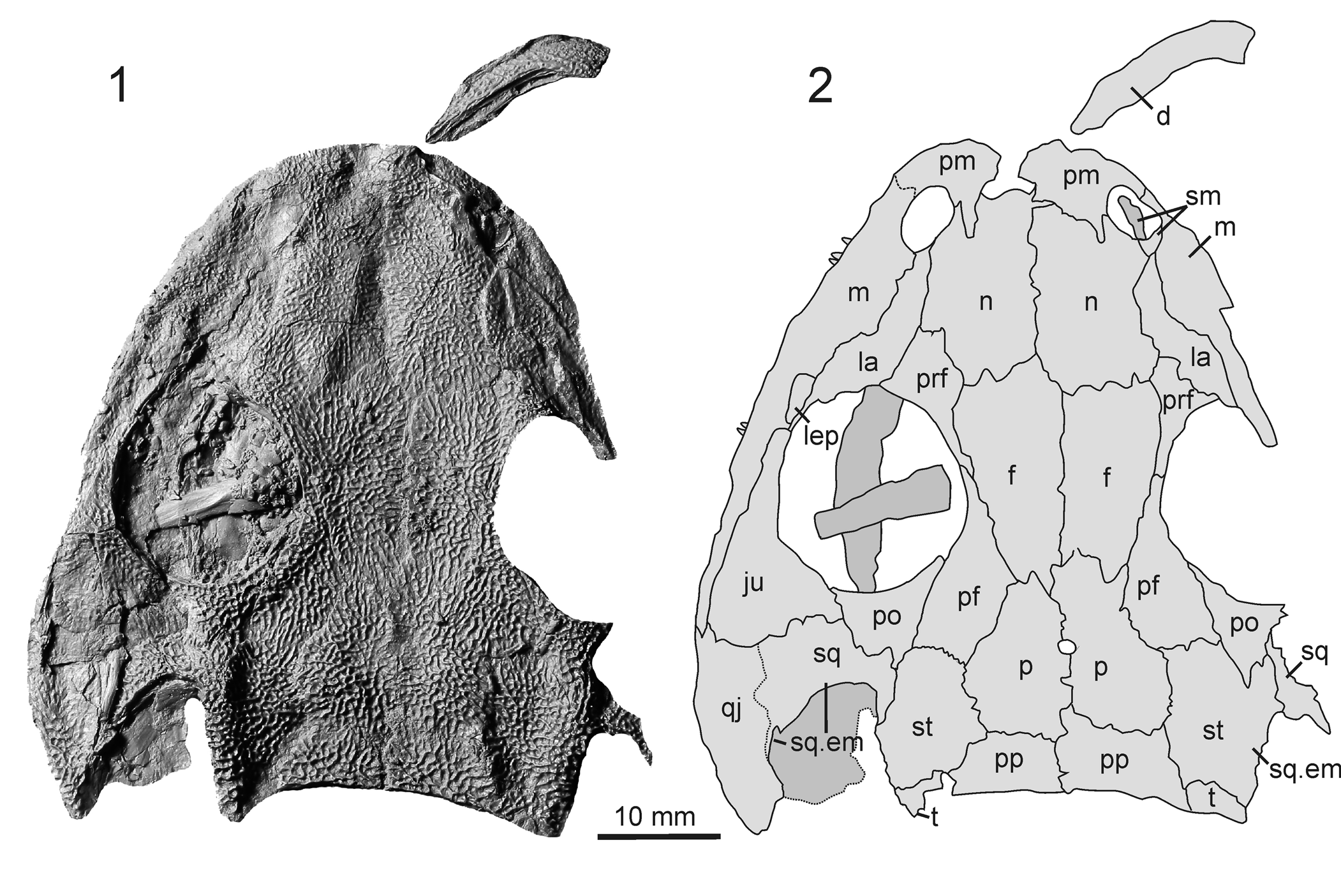
Palodromeus bairdi n. gen. n. sp. Skull roof in dorsal view (CM 34608). (1) Photograph of latex peel; (2) drawing. d = dentary; f = frontal; ju = jugal; la = lacrimal; lep = laterally exposed palatine; m = maxilla; n = nasal; p = parietal; pf = postfrontal; pm = premaxilla; po = postorbital; pp = postparietal = prf, prefrontal; qj = quadratojugal; sm = septomaxilla; sq = squamosal; sq.em = squamosal embayment; st = supratemporal; t = tabular.

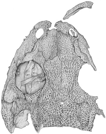
Palodromeus bairdi n. gen. n. sp. Skull roof in dorsal view (CM 34608). See Figure 1 for scale.

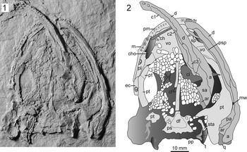
Palodromeus bairdi n. gen. n. sp. Palate, skull roof and mandible in ventral view (CM 34608). (1) Photograph of peel; (2) drawing. a = angular; ar = articular; c1 = anterior coronoid; c2 = middle coronoid; c3 = posterior coronoid; cf = cultriform process; ch = ceratohyal; cho = choana; cl = clavicle; d = dentary; df = denticle field; ec = ectopterygoid; m = maxilla; mw = Meckelian window; os = dentigerous palatal plates; pa = prearticular; pl = palatine; pm = premaxilla; pp = postparietal; ps = parasphenoid; psp = postsplenial; pt = pterygoid; q = quadrate; sa = surangular; sp, splenial; sta = stapes; t = tabular; vo = vomer.

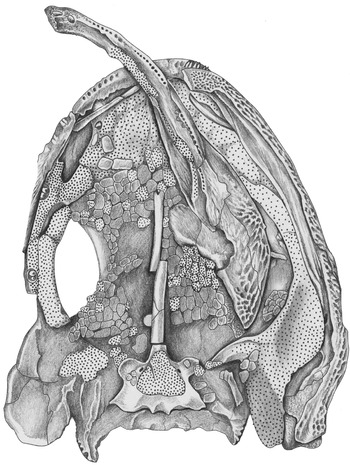
Palodromeus bairdi n. gen. n. sp. Palate, skull roof and mandible in ventral view (CM 34608). See Figure 3 for scale.

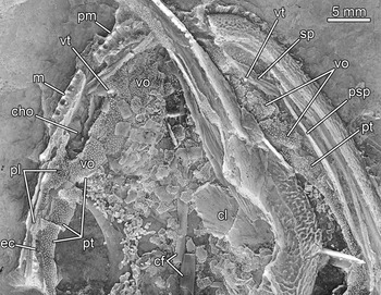
Palodromeus bairdi n. gen. n. sp. Close-up of anterior palate and mandible of CM 34608. See Figure 3 caption for abbreviations. vt = vomerine tusk.

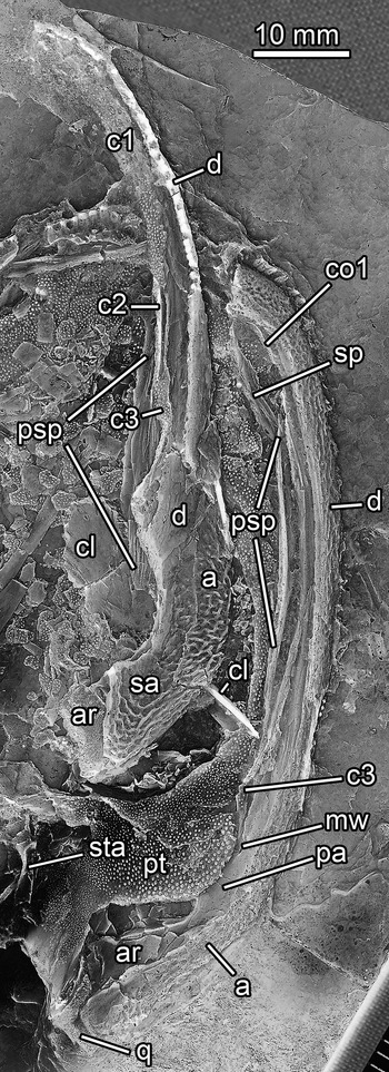
Palodromeus bairdi n. gen. n. sp. Close-up of mandible halves of CM 34608. See Figure 3 caption for abbreviations. co1 = coronoid 1.

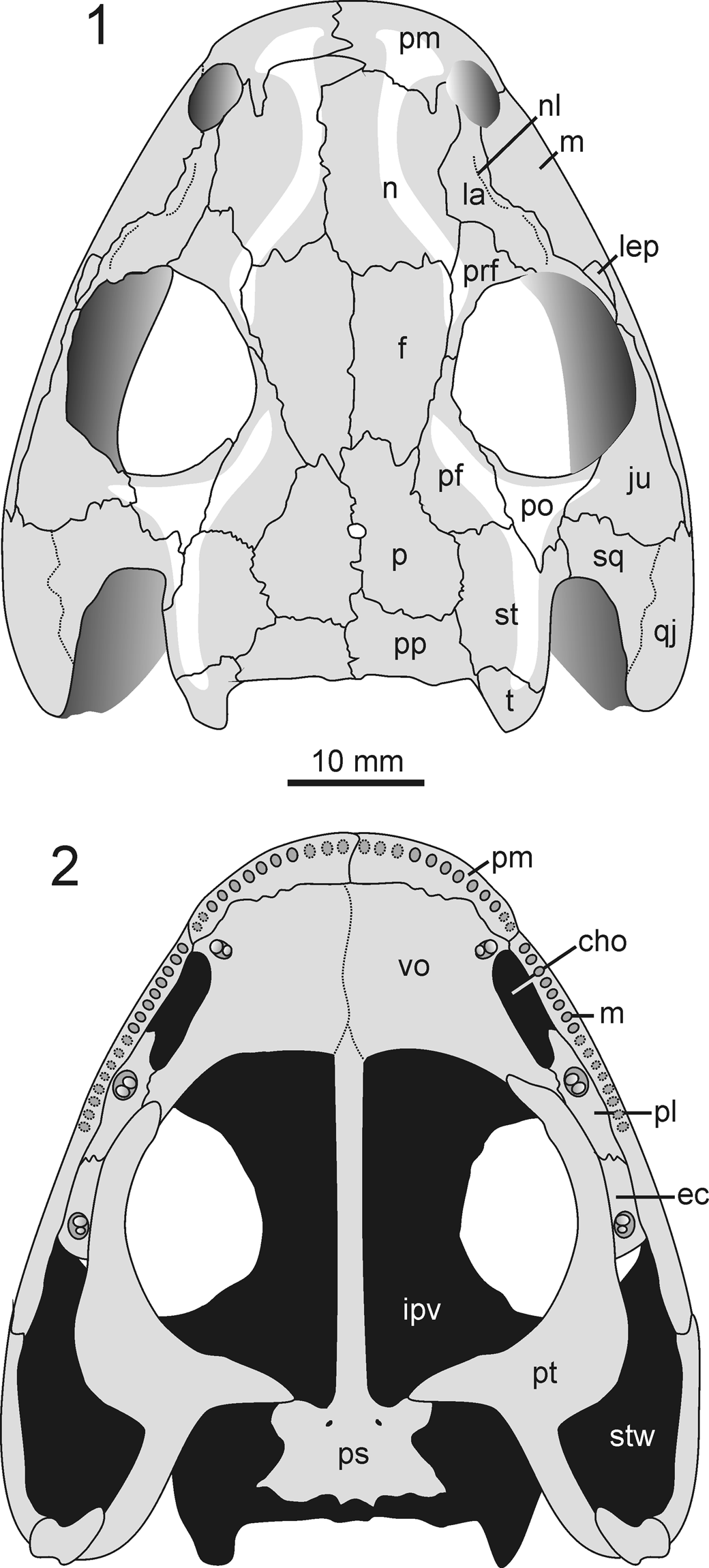
Palodromeus bairdi n. gen. n. sp. Reconstruction of the skull based on CM 34608. (1) dorsal view; (2) ventral view. cho = choana; ec = ectopterygoid; f, frontal; ipv = interpterygoid vacuity; ju = jugal; la = lacrimal; lep = laterally exposed palatine; m = maxilla; n = nasal; nl = nasolacrimal canal; p = parietal; pf = postfrontal; pl = palatine; pm = premaxilla; po = postorbital; pp = postparietal; prf = prefrontal; ps = parasphenoid; pt = pterygoid; qj = quadratojugal; sq = squamosal; st = supratemporal; stw = subtemporal fossa; t = tabular; vo = vomer.

Holotype
CM 34608, a complete skull with mandible (49 mm skull midline length, premaxilla-postparietal), preserved as natural molds in counterpart blocks, in dorsal and ventral views.
Diagnosis
Autapomorphies: (1) interorbital distance relative to skull length moderately wide (IOW:SL = 0.326); (2) postfrontal, postorbital, and supratemporal with pronounced ridge; (3) parietal small, approximately two-thirds the length of the frontal; and (4) lacrimal laterally constricted by wide maxilla shelf, narrower than alary process of the premaxilla.
Plesiomorphies (contrasting the condition in Platyrhinops lyelli and Amphibamus grandiceps): quadrate and occipital condyles level in dorsal perspective, lateral margin of supratemporal straight, postorbital slender and posteriorly pointed, parasphenoid with a small triangular denticle field, leaving most of the cultriform process edentulous. Features shared with P. lyelli and A. grandiceps: prefrontal and postfrontal sutured, no lateral line sulci.
Occurrence
Five Points Cannel Mine of Ted Mesmer and Sons, Inc., 1.3 km northeast of North Lima, Beaver Township, Mahoning County, Ohio (USA); cannel coal below the Lower Kittanning coal, Allegheny Group, upper Carboniferous. This exact same deposit was reported incorrectly by Clack (Reference Clack2011, p. 1150) as the “Canfield Cannel Formation.”
Description
The skull of Palodromeus bairdi n. gen. n. sp. is relatively wide, with the greatest width at the level of the pineal foramen (Figs. 1–5). In dorsal view, it is a parabola attenuated slightly in the region of the suspensorium, which is consistent with many dissorophoids. The preorbital region of P. bairdi n. gen. n. sp. is unusually wide, with nares set well posterior to the anterior snout margin. The length of the nearly circular orbits measures one-third that of the skull, and preorbital and postorbital regions are nearly equal in length. Although postmortem flattening disrupted the snout margin on the right side, the less-disturbed left side indicates an internarial width greater than the interorbital measure. There is no unequivocal evidence for an interpremaxillary fontanelle. Whereas the anterior margin of the snout is blunt and similar to Platyrhinops lyelli, the shape of the nares is markedly different: in P. bairdi n. gen. n. sp., the opening is ovoid and lengthened parallel to the lateral margins of the premaxilla and maxilla; in Platyrhinops lyelli, the external naris is a compressed ellipse whose long axis runs perpendicular to the midline of the skull. In dorsal perspective, the posterior outline of the skull differs from most dissorophoids in having the inferred occipital and quadrate condyles in near alignment.
The skull roof has ridges that are most pronounced in the postorbital skull table, where a longitudinal ridge runs anteriorly from the tabular horn to along the lateral margin of the supratemporal. At the supratemporal-postorbital junction, the ridge bifurcates into a lateral branch that continues toward the jugal, where it terminates rather abruptly, and a medial ramus that forms the posteromedial orbital margin on the postfrontal. This system of ridges is fairly consistent with that of dissorophids, notably Cacops spp., Conjunctio multidens Carroll, Reference Carroll1964, Dissorophus multicinctus Cope, Reference Cope1895, and Broiliellus spp. (Williston, Reference Williston1910; Schoch, Reference Schoch2012). The ridge that runs from the tabular to the postorbital, however, is broader than in dissorophids, expands over most of the postorbital, and it does not continue onto the jugal. Other ridges typical of dissorophids, such as along the occipital margin of the skull table, at mid-level of the frontals, or posterior to the pineal foramen, are absent in Palodromeus bairdi n. gen. n. sp.
In P. bairdi n. gen. n. sp., dermal ornament is well developed on the skull and mandibles and consists mostly of pits and short grooves framed by thin ridges. On the snout, sculpture is somewhat fainter than in the posterior skull table, a condition typical of immature temnospondyls (Bystrow and Efremov, Reference Bystrow and Efremov1940; Boy, Reference Boy1988). Likewise, the lateral flank of the jugal is only faintly ornamented. In the cheek region, an area shared by the posterior jugal, anterior squamosal, and medial quadratojugal, only subdued ridge-and-furrow ornament, not the finely reticulated pattern present on most of the skull, is present. Lateral line sulci are absent throughout the dermal bones.
Compared to most other dissorophoids, the premaxilla forms a major portion of the snout. It is a robust element best studied from the left premaxilla as exposed in dorsal view because its counterpart on the right side is less complete and disrupted by compaction. Both can be observed only partially in palatal view, in which they are overlain by the left mandible. The midline junction between the premaxillae is not exposed in either perspective. Although the most medial portion of the left premaxilla has a modest embayment confluent with a similar feature on the left nasal, there is no unequivocal evidence from the right side for an interpremaxillary fontanelle. A posteriorly directed, straight alary process intrudes the nasal. In palatal view, the right premaxilla holds at least 10 closely placed tooth positions, seven of which are occupied by short (longest tooth ~1.7 mm as preserved), peg-like, distally incomplete teeth. The left premaxilla bears six fairly well-preserved teeth. The teeth are conical, not recurved, and taper to a pointed crown. The tooth bases are not exposed, and a pedicel is not present in the preserved portion of the teeth. There is a very modest size increase in basal tooth diameter anteriorly. Otherwise, no details of tooth morphology can be rendered.
The anteromedial part of the maxilla is wide and has a stepped suture with the lacrimal. It is excluded from the orbital margin by a thin posterior projection of the lacrimal and the thicker suborbital ramus of the jugal and first contacts the quadratojugal at the level of the posterior edge of the orbit as seen on the left side. Although an exact maxillary tooth count is impossible, there are 20 closely set tooth spaces, nine of which are occupied by remnants of teeth, on the right side to the level of the back of the choana. Posterior to the choana, tooth size decreases uniformly. The broken and incomplete right maxilla in this region preserves teeth or sockets, but an exact tooth count cannot be given. The teeth of the right maxilla and premaxilla are all preserved without crowns. This resembles a common preservation of pedicellate teeth, in which the crowns had fallen off prior to burial (Bolt, Reference Bolt1977). However, in P. bairdi n. gen. n. sp., the tooth bases appear to be broken off at various levels, rather than at equal height, which does not agree with the morphology of tooth bases in Doleserpeton annectens. In addition, the preserved premaxilla teeth on the left side are monocuspid, unlike the bicuspid crowns of Doleserpeton annectens and Amphibamus grandiceps.
The septomaxilla is exposed in the right naris. As in other temnospondyls, it has a semilunar portion, which is sutured to the nasal, and an anterior process, which points into the naris. The semilunar portion houses a deep transverse groove. It is unclear if the septomaxilla excludes the lacrimal from the margin of the naris.
The nasal is shorter and only slightly wider than the frontal. Laterally, it has a long suture with the lacrimal. On both sides of the skull, this contact has been disrupted by postmortem crushing. The nasal forms the medial margin of the narial opening and narrows gently towards the suture with the premaxilla. The elongate frontals are excluded from the orbital margin by a thin suture between prefrontal and postfrontal, and the frontals reach the parietals along a short, highly interdigitated suture. As in many dissorophoids, there is a conspicuous asymmetry of these sutures on the left and right sides.
Perhaps the most distinctive feature of the skull roof is the short and wide parietals, which are approximately two-thirds the length of the frontals and approximately twice the length of the postparietals. Among xerodromes (Schoch and Milner, Reference Schoch, Milner and Sues2014), only Scapanops neglectus has comparably small parietals, but the postorbital skull table and cheek as a whole are miniaturized in comparison to the orbits and snout in that taxon. The pineal foramen is situated posterior to the frontal-parietal suture, just short of half the length of the common parietal junction. The postparietals are much larger than those of either Amphibamus grandiceps, Platyrhinops lyelli, or Georgenthalia clavinasica Anderson et al., Reference Anderson, Henrici, Sumida, Martens and Berman2008b.
The circumorbital series is consistent with Platyrhinops lyelli and consists of moderately large lacrimal, prefrontal, postfrontal, postortbital, and jugal. A small dorsal portion of the palatine (LEP) is exposed between the lacrimal, maxilla, and possibly the jugal. It appears to be excluded from the orbital margin by a thin posterior process of the lacimal that contacts the jugal. The lacrimal is elongate and has a semilunar outline. It is very slender, more so even than in Cacops morrisi, being constricted by a substantially wider alary process of the premaxilla and a wide nasal. It is about equal to the nasal in length. At the margin of the orbit, the lacrimal wedges broadly between the prefrontal and the laterally exposed palatine. A narrow but well-defined, dorsally open nasolacrimal canal traverses the anterior half of the lacrimal. This condition is known from immature eryopiforms (Onchiodon labyrinthicus Geinitz, Reference Geinitz1862: Boy, Reference Boy1990; Sclerocephalus haeuseri Goldfuss, Reference Goldfuss1847; Schoch, Reference Schoch2003) and branchiosaurid amphibamiforms (Schoch, Reference Schoch1992). The prefrontal makes a narrow sutural contact with the postfrontal at roughly the midlevel of the orbit, a contact preserved on both sides of the skull. The prefrontal is anteriorly blunt with a slender lateral wing, a convex medial margin, and an elongate but thin posterior or postfrontal process. The postfrontal is large and has a wide and gently curved posteromedial portion, and by area almost equaling that of the parietal. Unlike many amphibamiforms, the postfrontal does not contribute greatly to the orbital margin. Instead it makes only a small contribution to the margin and extends far posteriorly, much as in eryopiforms. In contrast to amphibamiforms, the postorbital is rather narrow, truncated laterally, and extends well posteriorly to terminate in a pointed end on the right side; a more complicated serrated suture with the squamosal is present on the left side. The length and proportional size of the postorbital are consistent with basal dissorophoids, such as Limnogyrinus elegans, eryopiforms, and edopoids.
The jugal is the largest bone of the circumorbital series. It reaches farther anteriorly than in amphibamiforms and has an elongate and rather broad suborbital ramus that is well sculptured. The morphology of the suborbital ramus is most consistent with dissorophids such as Cacops morrisi. The posterior half, in dorsal aspect, of the jugal is broadened medially and is widest at the level where it meets the postorbital. There, it forms a serrated suture with the squamosal.
The supratemporal is subrectangular, almost twice as long as wide, and equal in length to the parietal. Unlike Amphibamus grandiceps and Platyrhinops lyelli, it is not constricted laterally and has a rather straight, aligned lateral margin. Its entire lateral portion is markedly raised, forming part of a ridge that runs from the tabular to the orbit margin. In dorsolateral view, a semilunar flange is exposed in which the supratemporal forms a ventral flange that participates in the medial bordering of the otic notch.
The squamosal has a rather short, dorsally exposed suture with the supratemporal because the postorbital wedges deeply between the two elements, and the squamosal embayment reaches relatively far anteriorly. The squamosal covers a relatively small area, but its lateral extension is unclear. At any rate, the quadratojugal is much longer than the squamosal and forms a relatively large part of the cheek.
The tabular is narrower and smaller than the postparietal. There is a distinct tabular horn that is well exposed in ventral view (Fig. 4). Its medial margin is markedly ornamented and bears a ventral crest. The morphology of the tabular horn resembles that of branchiosaurids and Gerobatrachus hottoni (Schoch, Reference Schoch2019). With the exception of the tabulars, the posterior margin of the skull table is straight and lacks an occipital flange. As in many xerodromes, the otic notch is deep and very wide.
Palate
On CM 34608, the palate is well preserved, although partially concealed by the right mandible (Figs. 3–5). The pterygoids and parasphenoid are slightly dislocated from each other. As in many temnospondyls, the interpterygoid vacuities were filled by a mosaic of small dentigerous plates that are partially disarticulated in the holotype specimen. These vary considerably in size and shape, and their minute teeth agree in size and density with the denticles that mantle the oral surfaces of the vomer, palatine, ectopterygoid, and pterygoid, as well as part of the parasphenoid. Shelves on the palatine or pterygoid, such as reported from Cacops morrisi, are not exposed because the bone margins are covered by plates. In life, these plates formed a continuous dentigerous sheath under parts of the palatine and vomer, along the lateral margin of the parasphenoid, and posterolaterally to the general area of the basal articulation.
The general proportions of bones are similar to those of dissorophids, with moderately large oval interpterygoid vacuities, slender palatine and ectopterygoid, but a robust and elongate palatal ramus of the pterygoid. The vomer forms a large quadrangular plate that is fairly completely exposed on the right side. Because its posteromedial portion is covered by palatal plates, a posteromedial process cannot be confirmed. Two vomerine tusks and accompanying replacement pits are located about mid-level on the bone, near the anteromedial margin of the choana. The tusk pair is relatively small compared to those of other dissorophoids. The posterolateral corner tapers to a blunt point and is wedged between the palatine and pterygoid, which is a feature consistent with many trematopids, as discussed in the last section. Based on evidence from the right side, the palatine is excluded from the interpterygoid vacuity, and the vomer forms all of the medial margin of the choana. The latter forms a long, slit-like opening about as long as the palatine, which completely frames it posteriorly. The lateral margin of the palatine is gently widened anterior to the maxilla-palatine suture. The elongate and slender shape and marginal position of the choana are most consistent with that of dissorophids.
The palatine and ectopterygoid, exposed only on the right side of the palate, are not as narrow as in most amphibamiforms (except Platyrhinops lyelli), but are somewhat more slender than in all olsoniforms. However, they appear slender only because of their narrow ventral exposure; that is, the medial, smooth margins of these elements were covered by the palatal ramus of the pterygoid. As in Platyrhinops lyelli and Eoscopus lockardi, the ventrally exposed areas of the palatine and ectopterygoid are completely covered by denticles, and both bones bear tusk pairs. Whereas the palatine tusks are located at the lateral margin and removed from the posterior end of the choana, the ectopterygoid tusks are situated posterior to the midlevel of the element. The ectopterygoid forms a rectangular strip of bone that appears to be shorter than the palatine and is somewhat narrower than the adjacent palatal ramus of the pterygoid.
In contrast to most other dissorophoids, the pterygoid extends anteriorly well past the midlevel of the interpterygoid vacuity and approaches the posterior limit of the choana. The anteriormost part of the right pterygoid is broken and slightly displaced, but a non-denticulated flange on the medial margin of the vomer indicates clearly the extent and nature of the junction. There is some asymmetry in the topology of sutures, with the anatomical left pterygoid (ventral view) having a stepped lateral suture and reaching farther anterior than the right (Fig. 4). There is no transverse flange, and the bone broadens markedly near the base of the palatal ramus. The interpterygoid vacuities are narrower than in amphibamiforms—a feature that is almost certainly plesiomorphic. Another primitive aspect of the pterygoid is its almost complete covering of denticles, a feature closely resembling the condition in colosteids and edopoids.
The cultriform process of the parasphenoid is narrow and ventrally flat with straight margins and a slightly raised area at about midlevel between the orbits; its anterior contact with the vomers is not exposed. Unlike many amphibamiforms, the cultriform process is entirely edentulous and smooth. A well-defined, triangular area on the basal plate of the parasphenoid, however, is heavily denticulated in the manner of the pterygoids and other bones of the palate. The basal plate is transversely rectangular, about two-thirds as long as wide. The basipterygoid joint is well offset and separated from the main plate by a deep groove that borders the slightly raised, rounded-triangular denticle field. The groove originates near the base of the cultriform process, where a tiny foramen of the internal carotid artery is located on both sides; this foramen is much smaller than in most dissorophoids. The basipterygoid region of the basal plate is large and the facets face anterolaterally. The posterior portion of the basal plate is smooth, but contains five (two pairs and one medial) faint longitudinal depressions arranged transversely along the posterior margin.
The quadrate is well ossified, and its posterior and ventromedial portions are exposed on the right side in palatal view. The enlarged medial portion is well developed, and a marked central trochlea is present, consistent with other temnospondyls. A dorsal process is not exposed.
Mandible
Both mandibular rami are preserved and allow a fairly complete description of the lower jaw. Whereas the left ramus is essentially in place and exposed in ventral aspect, the right is detached and twisted ~90° near the posterior end of the dentary such that the posterior one-third of the jaw is seen in lateral view, and the remainder is exposed in dorsal or occlusal view (Figs. 3, 5, 6). The anterior end of this displaced ramus extends beyond the snout and is visible in ventral perspective on the counterpart. A small portion of the same ramus is seen in medial view within the left orbit (Fig. 1).
The mandible conforms to a plesiomorphic dissorophoid design, such as known from Tersomius texensis (Carroll, Reference Carroll1964) and Platyrhinops lyelli (Clack and Milner, Reference Clack and Milner2010). It is constructed more lightly than in eryopiforms and enlarged only modestly in the adductor fossa area. Dermal sculpture is present on the lateral surface and ventral edge of the dentary and angular, the ventral face of the postsplenial, and the lateral face of the surangular. No lateral line sulci are present.
The dentary is a fairly gracile element that forms, in conjunction with the coronoid series, splenial, and postsplenial on the medial and ventral surfaces, the anterior two-thirds of the mandible. At the jaw symphysis, the dentary becomes more robust and supports marginal teeth of modestly increased size similar to those of the opposing premaxilla. Because the symphyseal region is not well preserved, it cannot be determined if the splenial contributed to the symphysis or if an adsymphyseal ossification was present. Although imperfectly shown in the peels, two large alveoli for two symphyseal tusks are present on the lingual side of the marginal tooth row of the right dentary. The right dentary has exposed the labial surface of the lower jaw to about the posterior end of the orbit, where it tapers posteriorly above the angular.
The surangular is exposed in lateral aspect in the right mandible, and it is missing its dorsal portion near the level of the ventral edge of the adductor fossa. Because of this, the development of the coronoid process cannot be determined. The prearticular forms the ventral border of the adductor fossa and extends anteriorly to the level of the postsplenial, as seen in the left mandible. A tiny Meckelian window pierces the prearticular near the midpoint of its exposed length and has an elongate oval shape. The articular is exposed on both sides. Corresponding with the above described quadrate trochlea, the articular condyle is saddle shaped, and the medial face of the articular overlaps the posterior end of the prearticular.
The coronoid series is better preserved in the right mandible, where it runs from the symphysis to the adductor fossa and is covered in minute denticles resembling those of the palate. As in Platyrhinops lyelli, all three elements are covered by a shagreen of denticles. The anterior end of coronoid 1, the longest of the series, is indistinct, but an oblique suture between it and coronoid 2 is preserved. Posterior to this suture, the coronoids are twisted such that the anterior part of coronoid 2 and the posterior end of coronoid 3 have a vertical orientation. The ventral edge of the anterior end of coronoid 1 is preserved in the left mandible, based on the presence of a shagreen of teeth preserved on its lingual face. The bone becomes indistinct posteriorly.
A bone that is most likely the splenial is preserved in association with the anterior end of the left mandible. It is displaced laterally and has rotated so that the lingual surface is facing ventrally. Whether or not it contributed to the symphysis cannot be determined.
The postsplenial is preserved in both mandibles. Whereas the right is preserved in labial aspect in which thin striations of the suture scar are preserved, the left has fallen out of articulation of and lies medially adjacent to the mandible. A suture scar for the posterior end of the postsplenial, which also bears thin striations, is preserved on the prearticular, adjacent to the overlap of the dentary and angular in the left mandible. The postsplenial is about three times the length of the presumed splenial. The two bones would join near the level of the posterior part of the choana, and the postsplenial extends to about the posterior third of the orbit.
Visceral skeleton
The stapes is exposed on the left side (Figs. 3, 4). It has a widened proximal portion and a shaft about twice as long as the head region. The head preserves an elongate, thin, and pointed ventral process. Near the ventral margin of the head region, a large oval depression is present that probably housed a stapedial foramen. The shaft is relatively long and straight and is more consistent with that of Micromelerpeton credneri Bulman and Whittard, Reference Bulman and Whittard1926 (Boy, Reference Boy1995) than amphibamiforms (Bolt and Lombard, Reference Bolt and Lombard1985). A slender rod-like element is preserved on the right side of the anterior palate. It is very similar to a paired element in Platyrhinops lyelli that was hypothesized to be ceratohyals by Clack and Milner (Reference Clack and Milner2010).
Postcranial skeleton
The only preserved postcranial bone lies between the palate and right mandible. The element consists of a rod-like portion that is visible within the left orbit (Figs. 1, 2) and a blade-like component that is largely obscured by the right mandible (Figs. 3, 4). This is a clavicle that is indistinguishable from that of Platyrhinops lyelli (Clack and Milner, Reference Clack and Milner2010, fig. 2g).
Etymology
The species name honors the late Dr. Donald Baird, whose nearly half century of work contributed profoundly to our understanding of late Paleozoic and Mesozoic vertebrate deposits and early tetrapod evolution (Sues et al., Reference Sues, Hook and Olsen2013). Following the discovery of vertebrate fossils at Five Points by G.A. McComas in 1983, Don initiated a collecting program in 1988 that continued intermittently for 10 years (Hook and Baird, Reference Hook and Baird1994). He found and prepared (acid etching and latex microcasting; see Baird, Reference Baird1955) the holotypic specimen in 1994.
Remarks
None.
Results
Data set
We conducted a phylogenetic analysis based upon the dissorophoid data set recently composed by Schoch (Reference Schoch2019). The characters were renumbered and grouped according to morphological regions (see Appendix in Supplemental Data). The character definitions and character-taxon matrix are provided in the Appendix (in Supplemental Data). All multistate characters were ordered. When these characters were left unordered, the topology of the strict consensus tree remained unchanged. Dendrysekos (=Dendrerpeton of many former authors who referred to Holmes et al., Reference Holmes, Carroll and Reisz1998) has served as outgroup taxon. The non-dissorophoid ingroup taxa are Trimerorhachis (for Dvinosauria), Sclerocephalus (for Eryopiformes), and Acanthostomatops (for Zatracheidae). Five additional taxa were added: Actiobates peabodyi (Gee and Reisz, Reference Gee and Reisz2020a), Mattauschia laticeps and Mordex calliprepes (Milner, Reference Milner2019), Nanobamus macrorhinus (Gee and Reisz, Reference Gee and Reisz2020b), and Palodromeus bairdi n. gen. n. sp. Seven new characters were added (14, 16, 28, 42, 60, 63, and 72) to the existing data set, and one character (former 30) was omitted because it overlapped with former character 17 (new number 30) (see Appendix in Supplemental Data for definitions).
Analysis
The cladistic analysis was performed with TNT 1.5 (available at https://cladistics.org/tnt/), under New Technology Search option, which found five equally most parsimonious trees (tree length [TL] = 304 steps; consistency index [CI] = 0.4013; retention index [RI] = 0.698). A variant analysis omitting Mattauschia laticeps and Mordex calliprepes gave almost the same topology, but six most parsimonious trees (see below).
Phylogenetic position of Palodromeus n. gen
The resulting five most parsimonious trees contain four polytomies in the strict consensus (see Fig. 8 and below). Homoplasies are common, with raised levels within the Amphibamiformes. The topology is consistent with that of Schoch (Reference Schoch2019), especially in the higher amphibamiform part of the tree, with the exception that Platyrhinops lyelli is nested now more robustly with Amphibamus grandiceps and Doleserpeton annectens, and Nanobamus forming the basalmost taxon of the post-micropholid amphibamiforms.
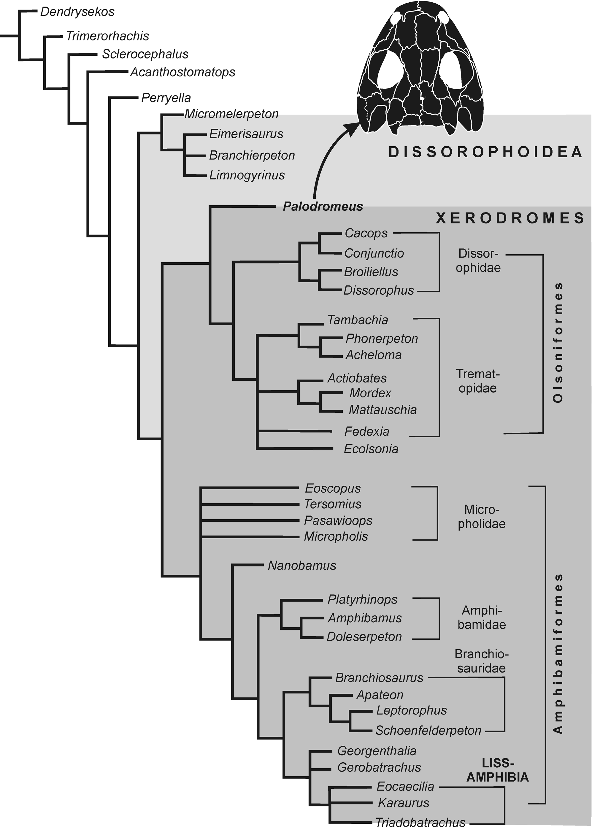
Hypothesis of relationships of dissorophoid temnospondyls, with particular emphasis on the position of Palodromeus bairdi n. gen. n. sp.
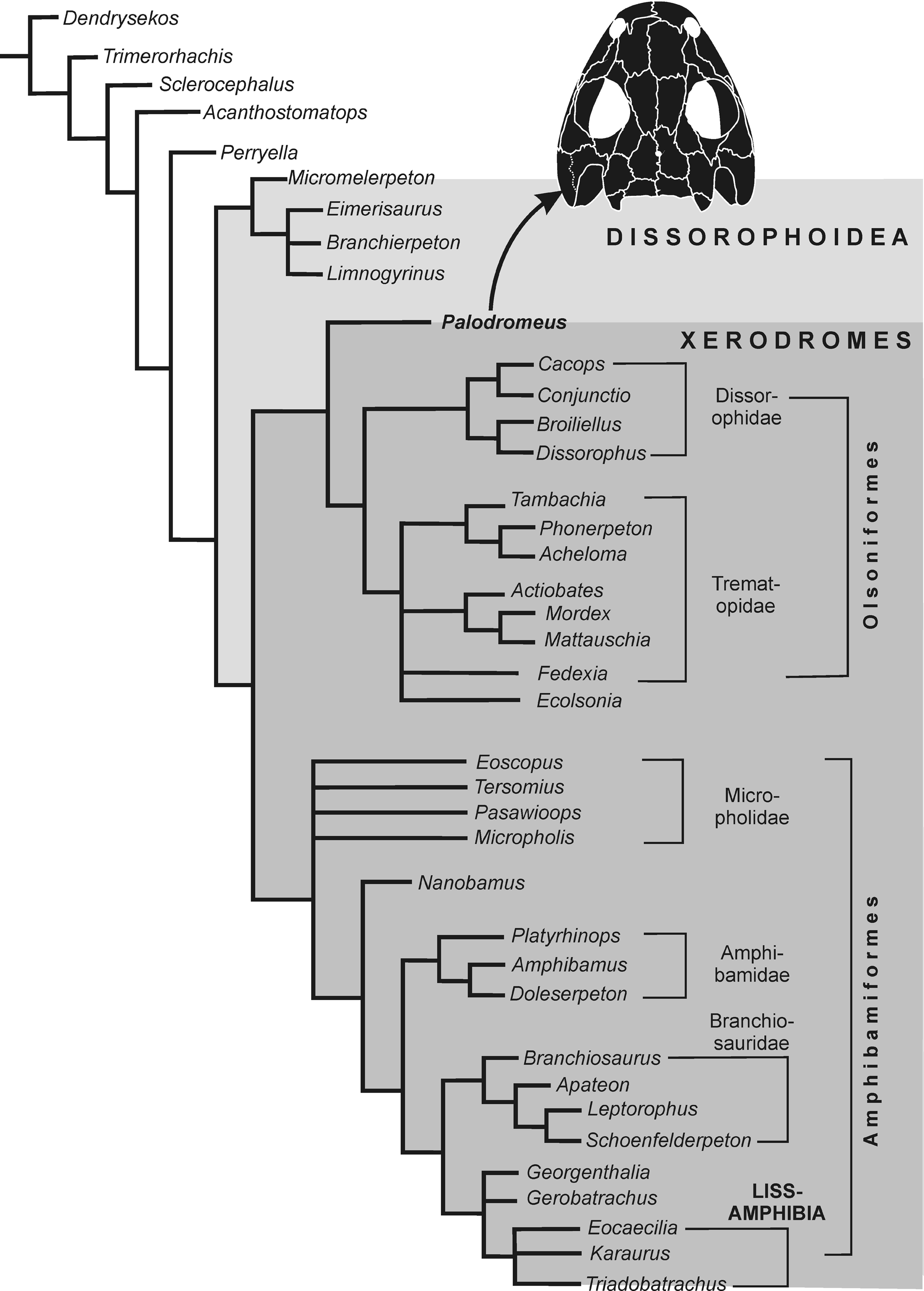
Perryella olsoni Carlson, Reference Carlson1987 is found as the sister taxon to the Dissorophoidea, the Micromelerpetidae form its basalmost clade, and the Olsoniformes plus Amphibamiformes together comprise the monophyletic Xerodromes (Schoch and Milner, Reference Schoch, Milner and Sues2014). Amphibamiformes fall into three assemblages, only two of which are well supported: (1) the basal (here polytomic) Micropholidae (Eoscopus lockardi, Tersomius texensis, Pasawioops mayi, and Micropholis stowi Huxley, Reference Huxley1859), (2) the monophyletic Amphibamidae (Platyrhinops lyelli, Amphibamus grandiceps, and Doleserpeton annectens), and (3) an unnamed group containing Georgenthalia clavinasica (basalmost taxon), the Branchiosauridae (Branchiosaurus salamandroides Fritsch, Reference Fritsch1876, Apateon spp., Schoenfelderpeton prescheri Boy, Reference Boy1986, and Leptorophus tener [Schönfeld, Reference Schönfeld1911]), Gerobatrachus hottoni, and Lissamphibia (Eocaecilia micropodia Jenkins and Walsh, Reference Jenkins and Walsh1993, Karaurus sharovi Ivakhnenko, Reference Ivakhnenko1978, and Triadobatrachus massinoti Piveteau, Reference Piveteau1936).
In this framework, Palodromeus bairdi n. gen. n. sp. nests at the base of the Olsoniformes (Dissorophidae plus Trematopidae). All other olsoniforms form a clade with a (1) monophyletic Dissorophidae (Dissorophus multicinctus + Broiliellus spp., Cacops spp. + Conjunctio multidens); (2) a small clade encompassing Tambachia trogallas Sumida, Berman, and Martens, Reference Sumida, Berman and Martens1998, Phonerpeton pricei (Olson, Reference Olson1941), and Acheloma cumminsi Cope, Reference Cope1882; and (3) the late Carboniferous taxa Actiobates peabodyi, Mattauschia laticeps, and Mordex calliprepes from the Czech Republic.
In a variant analysis, the poorly known Czech genera Mattauschia and Mordex were omitted, which resulted in the same topology except for the base of the Olsoniformes being somewhat better resolved. Here, Palodromeus bairdi n. gen. n. sp. and Actiobates peabodyi form successive sister taxa of all other olsoniforms, with Fedexia striegeli and Ecolsonia cutlerensis Vaughn, Reference Vaughn1969 falling within a polytomy with Trematopidae and Dissorophidae, and Eoscopus lockardi is found to nest at the base of the Amphibamiformes.
Discussion
Palodromeus bairdi n. gen. n. sp. bridges the morphological gap between the two major, widely disparate clades of xerodromes, the olsoniforms and amphibamiforms (see Schoch, Reference Schoch2019 for the phylogenetic definition of these clades). The new genus shares with olsoniforms, particularly dissorophids: (1) the morphology and alignment of some ridges on the skull roof, and (2) an elongated and uniformly narrow choana. In contrast, there are no characters that suggest an amphibamiform placement, even though the skull roof of P. bairdi n. gen. n. sp. is generally consistent with some less-derived taxa within that clade, notably Eoscopus lockardi and Micropholis stowi. Major differences between P. bairdi n. gen. n. sp. and all amphibamiforms are: (1) the anterior extension of the jugal (longer and broader in P. bairdi n. gen. n. sp.), (2) the relatively smaller orbits and interpterygoid vacuities, and (3) the morphology of the palate. In particular, the far anterior extension of the palatal ramus of the pterygoid never occurs in amphibamiforms, and the palatine and ectopterygoid are substantially broader than in most amphibamiforms (except Platyrhinops lyelli and Eoscopus lockardi). Basal amphibamiform taxa (Micropholidae) retain an elongated postorbital skull table, with configurations of the supratemporal, parietal, and squamosal much like those in the currently recognized basalmost dissorophoids, the Micromelerpetidae (Boy, Reference Boy1972; Schoch, Reference Schoch2019).
The short parietal and the laterally constricted, lunate lacrimal probably are autapomorphies of P. bairdi n. gen. n. sp. because to that extreme extent they do not occur in either olsoniforms or amphibamiforms except the highly derived Scapanops. In P. bairdi n. gen. n. sp., the broadening of the skull evidently was reached by a widening of the posterior lacrimal and the medial process of the jugal. This differs from the amphibamiforms Platyrhinops lyelli and Amphibamus grandiceps, in which medial skull roof elements broaden and increase skull width.
Amphibamiform interrelationships are still controversial, especially with Platyrhinops lyelli and Amphibamus grandiceps being so similar in many features (including a presacral vertebral count of 24, contra Clack and Milner, Reference Clack and Milner1993), that the question needs to be addressed if they are sister taxa. This cannot be attempted here, however, because a significant amount of Ohio material requires further assessment and reexamination.
Palodromeus bairdi n. gen. n. sp. also retains various plesiomorphic features that are found in basal taxa of both the olsoniforms and amphibamiforms. Among these, the completely dentigerous vomer, palatine, ectopterygoid, and pterygoid stand out, shared with some trematopids (Phonerpeton pricei, Acheloma cumminsi, and Mordex calliprepes), micropholids (Eoscopus lockardi), and amphibamids (Platyrhinops lyelli and Amphibamus). The retention of dentigerous palatal plates adds to this; such plates appear to have been absent in Doleserpeton annectens, branchiosaurids, Gerobatrachus hottoni, and Lissamphibia (see Gee et al., Reference Gee, Haridy and Reisz2017, for literature survey). The narrow contact between vomer and pterygoid, as interpreted here, is consistent with the condition in Acheloma cumminsi, Phonerpeton pricei, Ecolsonia cutlerensis, Fedexia striegeli, and Mordex calliprepes. Like Mordex calliprepes and Mattauschia laticeps, P. bairdi n. gen. n. sp. also retains a prefrontal-postfrontal suture, a feature not found in any dissorophid, but shared with the late Carboniferous amphibamids Platyrhinops lyelli and Amphibamus grandiceps. These plesiomorphic features support P. bairdi n. gen. n. sp. as a basal olsoniform that combined various cranial plesiomorphies, a pattern consistent with its relatively old (late Carboniferous) age.
The shared synapomorphies of P. bairdi n. gen. n. sp. and the dissorophids are further consistent with the short posterior skull table formed by abbreviated parietals and supratemporals (except the probably derived Broiliellus texensis Williston, Reference Williston1914 and Platyhistrix rugosus Case, Reference Case1910), whereas the anterior snout region is substantially wider than in any dissorophid, being most consistent with Mordex calliprepes among olsoniforms. The overall broad skull is further consistent with other Late Carboniferous xerodromes, notably Mordex calliprepes, Mattauschia laticeps, Eoscopus lockardi, Platyrhinops lyelli, and Amphibamus grandiceps. Moreover, the consistent system of ridges on the skull roof especially prompts us to think of P. bairdi n. gen. n. sp. as the most basal dissorophid that retained various basal olsoniform or even primitive xerodrome features.
The Olsoniformes were first found to be monophyletic and defined by Anderson et al. (Reference Anderson, Reisz, Scott, Fröbisch and Sumida2008a), which was later corroborated by Schoch (Reference Schoch2012), Schoch and Sues (Reference Schoch and Sues2013), and Maddin et al. (Reference Maddin, Fröbisch, Evans and Milner2013). The placement of P. bairdi n. gen. n. sp. in a polytomy at the base of this clade suggests that the early evolution of this group started with small forms that lacked specializations in the skull, such as the modified nares of trematopids or the spectrum of otic notch morphologies found in dissorophids and trematopids. In fact, the otic region of P. bairdi n. gen. n. sp. is relatively plesiomorphic, and the shortened distance between the squamosal embayment and orbit appears to be a shared xerodrome character (Schoch and Milner, Reference Schoch, Milner and Sues2014). That said, the apparent immaturity of the P. bairdi n. gen. n. sp. holotype indicates that adults would have been larger, which renders the small-size hypothesis questionable.
In conclusion, Palodromeus bairdi n. gen. n. sp. from Five Points, Ohio, adds to the picture of early dissorophoid and xerodrome diversification and relationships. As the oldest known non-trematopid olsoniform, it clearly indicates that the diversification of dissorophoids must have started earlier than the upper Carboniferous and that a substantial ghost lineage awaits discovery.
Acknowledgments
The Five Points collecting program was supported by a National Geographic Society grant to D.S Berman, Carnegie Museum of Natural History. For nearly a decade, the East Fairfield Coal Company accommodated collecting activities by providing a secure storage location for the fossiliferous stockpiles. A. Kollar, Carnegie Museum of Natural History, kindly provided assistance with taking the photographs in Figures 5 and 6, and A. McAfee conducted the labelling. We are grateful to N. Fröbisch and B. Gee for their very helpful and thoughtful reviews.
Accessibility of supplemental data
Data available from the Dryad Digital Repository: https://doi.org/10.5061/dryad.nk98sf7rr.