

INTRODUCTION
Microbial diversity in oceans is rich (Strom, Reference Strom2008). Microorganisms reside in every available space of the oceans; including open water, sediment, bodies of marine macro- and microorganisms, and hydrothermal vents. This microbial community is dominated by bacteria, showing a similar diversity to that of terrestrial soil (Santelli et al., Reference Santelli, Orcutt, Banning, Bach, Moyer, Sogin, Staudigel and Edwards2008). Thus, it is clear that prokaryotes are by far the most abundant and phylogenetically diverse group inhabiting our planet (Oren, Reference Oren2004).
Several pathogen species have been commonly found in zooplankton populations (Decaestecker et al., Reference Decaestecker, De Meester and Ebert2002). Among such a plethora of pathogenic organisms, bacterial species such as Vibrio cholerae (Tamplin et al., Reference Tamplin, Gauzens, Huq, Sack and Colwell1990; Heidelberg et al., Reference Heidelberg, Heidelberg and Colwell2002; Binsztein et al., Reference Binsztein, Costagliola, Pichel, Jurquiza, Ramírez, Akselman, Vacchino, Huq and Colwell2004), Vibrio alginolyticus (Carli et al., Reference Carli, Pane, Casareto, Bertone and Pruzzo1993), Campylobacter lari, Arcobacter butzleri, Aeromonas spp., Escherichia coli (Jones, Reference Jones2001; Asmat & Gires, Reference Asmat and Gires2002; Maugeri et al., Reference Maugeri, Carbone, Fera, Irrera and Gugliandolo2004) and Helicobacter pylori (Cellini et al., Reference Cellini, Vecchio, Candia, Campli, Favaro and Donelli2004) appear as some of the most conspicuous members of the domain Bacteria colonizing zooplankton communities.
Necrotizing hepatopancreatitis (NHP) is a disease affecting several species of penaeid shrimp, which has been putatively attributed to an unclassified Gram-negative, pleomorphic, obligately intracellular α-proteobacterium residing almost permanently in the cytoplasm of tubular epithelial cells of the hepatopancreas of infected shrimp (Lightner et al., Reference Lightner, Redman and Bonami1992; Loy et al., Reference Loy, Dewhirst, Weber, Frelier, Garbar, Tasca and Templeton1996; Bradley-Dunlop et al., Reference Bradley-Dunlop, Pantoja and Lightner2004; Vincent & Lotz, Reference Vincent and Lotz2007). Reported host species include Penaeus vannamei, P. setiferus, P. stylirostris, P. aztecus and P. californiensis (Vincent & Lotz, Reference Vincent and Lotz2007) and experimental infection indicates that American lobster Homarus americanus is susceptible to this pathogen (Avila-Villa et al., Reference Avila-Villa, Gollas-Galván, Martínez-Porchas, Mendoza-Cano and Hernández-López2012). Since its identification in 1985 as a disease causative agent (Johnson, Reference Johnson and Johnson1989; Frelier et al., Reference Frelier, Sis, Bell and Lewis1992) NHP has been recognized as one of the most important bacterial diseases for marine shrimp, causing up to 95% mortality in populations not treated with antibiotics.
Studies concerning potential vectors for transmission of this disease are particularly important to devise appropriate control measures to allow the reduction or avoidance of the deleterious impact of this infectious agent on marine shrimp populations. Besides, the impact of this disease, if any, in other crustacean species, deserves to be investigated in detail. Thus, the aim of this study was to investigate the occurrence of NHP associated with zooplankton in the Gulf of California.
MATERIALS AND METHODS
Sampling
A zooplankton sample was monthly collected during February, March, May and June 2008 on six different stations located on the eastern shore of the Gulf of California, an area geographically adjacent to the coast of Sonora, Mexico (Figure 1) (Bahía de Kino, Bahía el Choyudo, Bahía de Guásimas, Bahía de Lobos, Bahía de Yavaros, and Bahía de Agiabampo). Samples (N = 24) were collected by using a small boat, slowly towing horizontally a 30 cm diameter mouth, 200 µm mesh plankton net for 10 min, and were immediately fixed with 70% ethanol.
Location map of the zooplankton sampling stations in the Gulf of California, México. 1, Bahía de Kino; 2, Bahía el Choyudo; 3, Bahía de Guásimas; 4, Bahía de Lobos; 5, Bahía de Yavaros; and 6, Bahía de Agiabampo. Locations of NHP-B positive detection sites are indicated by arrows.

Genomic DNA extraction
Zooplankton samples were allowed to settle by gravity overnight, after which the supernatant was removed and discarded. The samples were transferred into a 1.7 ml Eppendorf tube and were centrifuged at 8000 × g for 10 min, and supernatant removed by pipetting. The cells were suspended in 150 µl of lysis buffer (100 mM NaCl, 50 mM Tris pH 8.0, 100 mM EDTA pH 8.0, 1% SDS) and homogenized with a Teflon pestle (one stroke, 30 seconds). Samples were centrifuged at 8000 × g for 10 min and 50 µl of cell lysate were transferred into a new 1.7 ml Eppendorf tube containing 150 µl of GeneClean® Spin Glass Milk® (MP Biomedicals, Inc). Samples were gently stirred and incubated at room temperature for 5 min, after which they were centrifuged at 6000 × g for 5 min. Supernatant was decanted and the Glass Milk/DNA complex was gently resuspended in 150 µl of a mixture of 50% TBE buffer (130 mMTris, 45 mM boric acid, and 2.5 mM EDTA, pH 8.0), 50% ethanol. Samples were centrifuged at 6000 × g for 5 min, and supernatant was removed by decantation. The washing process was repeated once more. After last decantation, tubes were inverted and samples were air-dried at room temperature for 15 min. Finally, DNA was eluted from Glass Milk matrix adding 30 µl of water of molecular biology grade.
NHP-B by qPCR analysis
DNA extracted from zooplankton samples was used as template to detect NHP-B. Amplification was performed by real time PCR (Rotor-Gene thermocycler, Corbett Research), the reaction mixture contained iQTM SYBR® Green Supermix (BIORAD) and the primers previously described by Nunan et al. (Reference Nunan, Pantoja and Lightner2008) (NHPF2: 5′-CGT-TGG-AGG-TTC-GTC-CTT-CAG-T-3′, and NHPR2: 5′-GCC-ATG-AGG-ACC-TGA-CAT-CAT-C-3′), with an initial denaturation step at 95°C for 5 min and 35 cycles of 95°C for 30 s, 60°C for 30 s, 72°C for 10 s (acquiring), and final extension step at 72°C for 5 min. Fluorescence graphic and melting curve analysis was performed to detect NHP-B.
Further the PCR products (10 µl) were loaded on a 2% agarose gel stained with SYBR SafeTM (E-gel) (Invitrogen) and then electrophoresed in an E-Gel PowerBaseTM v.4 for 15 min, and then visualized under ultraviolet light and digitally photographed by a Gel Logic 100 imaging system (Kodak). The primers used for this work were those reported by Nunan et al. (Reference Nunan, Pantoja and Lightner2008) and have been used by Gracia-Valenzuela et al. (Reference Gracia-Valenzuela, Avila-Villa, Yepiz-Plascencia, Hernández-López, Mendoza-Cano, Garcia-Sánchez and Gollas-Galvan2011) and Avila-Villa et al. (Reference Avila-Villa, Gollas-Galván, Martínez-Porchas, Mendoza-Cano and Hernández-López2012) for detection of NHP-B in crustacean tissues. Those primers amplify a 379 base pairs (bp) fragment from the 16S ribosomal RNA gene.
DNA sequencing and sequence analysis
DNA sequencing was performed using a Taq FS Dye Terminator Cycle Sequencing kit in an ABI 3730 automated DNA sequencer (Applied Biosystems) at the oligonucleotide synthesis and sequencing facilities of the Instituto de Biotecnología of the Universidad Nacional Autónoma de México (UNAM). Chromatogram sequencing files were inspected with Chromas 2.33 (Technelysium Pty Ltd) and the obtained sequences were submitted for homology searching in non-redundant databases using Basic Local Alignment Search Tool for DNA (BLASTN) alignment program (Altschul et al., Reference Altschul, Gish, Miller, Myers and Lipman1990) from the EMBL-EBI website (http://www.ebi.ac.uk/Tools/blast2/nucleotide.html).
RESULTS
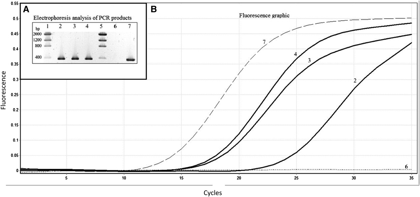
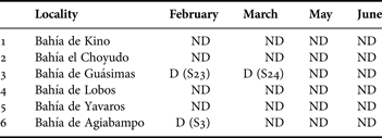
Necrotizing hepatopancreatitis was detected in three different zooplankton samples by qPCR (~13.0%) of the total number of analysed samples (24), NHP was positively detected in samples S3 (collected in February 2008), S23 (collected in February 2008) and S24 (collected in March 2008) (Table 1). These samples are geographically located in Bahía de Agiabampo (S3) and Bahía de Guasimas (S23 and S24), and these sampling sites are distributed along the length of the coast in the vicinity of a shrimp farm area (Figure 1). In accord with Nunan et al. (Reference Nunan, Pantoja and Lightner2008), a single PCR fragment (379 bp) was obtained from samples S3, S23 and S24. These results were checked using a qPCR assay with SYBR Green Supermix (Figure 2).
Necrotizing hepatopancreatitis bacterium (NHP-B) in zooplankton samples. Agarose gel electrophoresis analysis of PCR products and fluorescence graphic analysis from zooplankton samples using specific primers for amplification of the 16S rRNA gene of the NHP-B (379 bp). Lane 1: low DNA mass ladder; lane 2: sample 3; lane 3: sample 23; lane 4: sample 24; lane 5: low DNA mass ladder; lane 6: no template negative control; and lane 7: NHP-positive control. The PCR products were loaded on a 2% agarose gel stained with SYBR SafeTM (E-gel) (Invitrogen).

Occurrence of NHP-B in zooplankton samples collected in different locations in the Gulf of California as determined by PCR.

ND, not detected; D, detected.
One sample from each geographical site with NHP detection was sequenced and analysis of the partial sequence by BLASTN showed a high degree of identity (97–98%) with a previously published sequence of the 16S ribosomal RNA gene of an α-proteobacterium (GenBank Accession no. U65509) identified as NHP (Loy et al., Reference Loy, Dewhirst, Weber, Frelier, Garbar, Tasca and Templeton1996) (Figure 3).
Partial base sequence of NHP-B associated with the zooplankton samples from the Gulf of California (S3 and S24) and comparison with the 16S ribosomal RNA gene sequence from NHP-B (U65509). Dots in the sequences represent identical bases to sequences. Space represents any changes to sequences.

DISCUSSION
The colonization of zooplankton by potentially pathogenic bacteria has long been recognized (Nagasawa, Reference Nagasawa1988; Nagasawa & Nemoto, Reference Nagasawa and Nemoto1988; Plante et al., Reference Plante, Jumars and Baross1989; Gowing & Wishner, Reference Gowing and Wishner1992; Jones, Reference Jones2001; Asmat & Gires, Reference Asmat and Gires2002; Heidelberg et al., Reference Heidelberg, Heidelberg and Colwell2002; Binsztein et al., Reference Binsztein, Costagliola, Pichel, Jurquiza, Ramírez, Akselman, Vacchino, Huq and Colwell2004; Cellini et al., Reference Cellini, Vecchio, Candia, Campli, Favaro and Donelli2004; Maugeri et al., Reference Maugeri, Carbone, Fera, Irrera and Gugliandolo2004; Møller et al., Reference Møller, Riemann and Søndergaard2007; Rodrigues et al., Reference Rodrigues, Duffy, Tessier, Ebert, Mouton and Schmidt2008). However, NHPB, an intracellular bacterium associated with a severe disease affecting the marine shrimp (Lightner et al., Reference Lightner, Redman and Bonami1992; Loy et al., Reference Loy, Dewhirst, Weber, Frelier, Garbar, Tasca and Templeton1996; Vincent & Lotz, Reference Vincent and Lotz2007) and other crustaceans (Avila-Villa et al., Reference Avila-Villa, Gollas-Galván, Martínez-Porchas, Mendoza-Cano and Hernández-López2012) has never been found in a different organism than shrimp in a natural shape. In this study, the presence of NHP on samples of zooplankton from different locations of the Gulf of California was searched. As previously mentioned NHPB positively diagnosed samples were collected near the vicinity of a shrimp farming area. High prevalence of NHPB has been detected in these areas, and it is well known that the water used in shrimp farming ponds must be released and replaced periodically. In this way, NHP may be dispersed in to the natural environment throughout the release of water from these ponds.
The present results demonstrate the occurrence of the recognized aetiological agent of the shrimp necrotizing hepatopancreatitis disease associated with zooplankton samples from the Gulf of California. This finding may have significant implications on the biological fitness of this fragile bacterium, which loses its viability rapidly once released into the extracellular environment. Thus, the ability to colonize zooplankton surfaces may be an important strategy of this bacterium to survive adverse conditions. It has been well documented that certain pathogenic bacteria survive and persist in water by their ability to adhere to chitin-containing surfaces, such as those of zooplanktonic organisms (Kaneko & Colwell, Reference Kaneko and Colwell1973, Reference Kaneko and Colwell1975; Huq et al., Reference Huq, Small, West, Huq, Rahman and Colwell1983; Pruzzo et al., Reference Pruzzo, Tarsi, Lleó, Signoretto, Zampini, Pane, Corwell and Canepari2003; Signoretto et al., Reference Signoretto, Burlacchini, Lleó, Pruzzo, Zampini, Pane, Franzini and Canepari2004, Reference Signoretto, Burlacchini, Pruzzo and Canepari2005; Kirn et al., Reference Kirn, Jude and Taylor2005; Zampini et al., Reference Zampini, Pruzzo, Bondre, Tarsi, Cosmo, Bacciaglia, Chhabra, Srivastava and Srivastava2005; Rawlings et al., Reference Rawlings, Ruiz and Colwell2007; Vezzulli et al., Reference Vezzulli, Pezzati, Repetto, Stauder, Giusto and Pruzzo2008). Avila-Villa et al. (Reference Avila-Villa, Martínez-Porchas, Gollas-Galván, López-Elías, Mercado, Murguia-López, Mendoza-Cano and Hernández-López2011) recently demonstrated the possibility that the benthic microalgae Navicula sp., and the crustacean Artemia sp., may serve as vectors of necrotizing hepatopancreatitis bacteria.
Furthermore, it is relevant to mention that this finding does not necessarily imply that NHP exists on zooplankton in a pathogenic state. Merely finding the organism does not imply that it is causing a disease. There is a clear possibility of detection by PCR of inactivated but structurally intact NHP bacteria. If the NHP causing bacteria is able to colonize the guts or mid-gut gland of zooplankton composing species or if it is associated to chitin-containing surfaces and uses such sophisticated strategy to survive still remains to be elucidated.
Although NHP-B has been reported in several penaeid species, this is the first report, as far as we know, that sheds light on the presence of this shrimp pathogenic bacteria associated or colonizing zooplankton caught in the field which may be considered as a vector of potential importance in the spread of this disease. It remains to be elucidated if NHP-B shows a broader host range among marine organisms considering that it is well recognized that zooplankton may serve as vector of several pathogens, including viruses (Kitamura et al., Reference Kitamura, Kamata, Nakano and Suzuki2003), bacteria (Nese & Enger, Reference Nese and Enger1993), fungi (Czeczuga et al., Reference Czeczuga, Kozlowzka, Godlewska and Velu2008), nematodes (Austin & Welch, Reference Austin and Welch1972) and others (Carrasco et al., Reference Carrasco, López-Flores, Alcaraz, Furones, Berthe and Arzul2007).
The results obtained in this work give rise to several questions about the NHP-B/zooplankton association. Ongoing studies in our laboratory are aimed to define if this bacterium is internally distributed in the mid-gut gland of zooplankton or if it is attached to the chitinaceous exoskeleton. In case the NHP-B is found in the mid-gut gland of zooplankton, it must be demonstrated if it is a transient non-pathogenic or a resident infective bacteria. Colonizing bacteria should grow and proliferate in the mid-gut gland at a rate that equals or exceeds their rate of elimination from the gut. As long as these issues have not been solved, the complex life cycle of this bacterium will remain unclear.
ACKNOWLEDGEMENTS
We express our gratitude to the technical staff of the Laboratorio Especializado de Pesquerías (CIBNOR, Campus Guaymas), for kindly providing the experimental zooplankton samples. This work was supported by Centro de Investigaciones Biologicas del Noroeste, S.C.