Introduction
Despite major advances in both surgical and transcatheter repair of coarctation of the aorta, recurrent narrowing remains a frequent and challenging issue during follow-up. Reference Forbes, Kim and Du1–Reference Lala, Scali and Feezor4 In larger children and adults, endovascular approaches are generally preferred over redo surgery, providing a minimally invasive solution with faster recovery and lower early morbidity. Reference Forbes, Kim and Du1–Reference Lala, Scali and Feezor4 With the evolution of techniques and the expansion of device options, tailored strategies are becoming increasingly feasible, thereby reducing the need for unnecessary technical or procedural risks. Reference Forbes, Kim and Du1–Reference Lala, Scali and Feezor4 Here, we report a hybrid repair of complex re-coarctation of the aorta using a custom thoracic endograft reinforced by a balloon-expandable stent.
Case description
We report a 40-year-old man (167 cm, 69 kg) with a history of a bicuspid aortic valve, coarctation of the aorta, and ventricular septal defect. He underwent neonatal aortic arch repair with reversed subclavian flaps and pulmonary artery banding, followed at 13 months by pulmonary debanding, ventricular septal defect closure, and resection of infundibular muscular obstruction. At 33 years, he developed severe aortic regurgitation with a 55-mm ascending aortic aneurysm, for which he underwent a Bentall procedure with mechanical aortic valve implantation and a 25-mm prosthetic conduit.
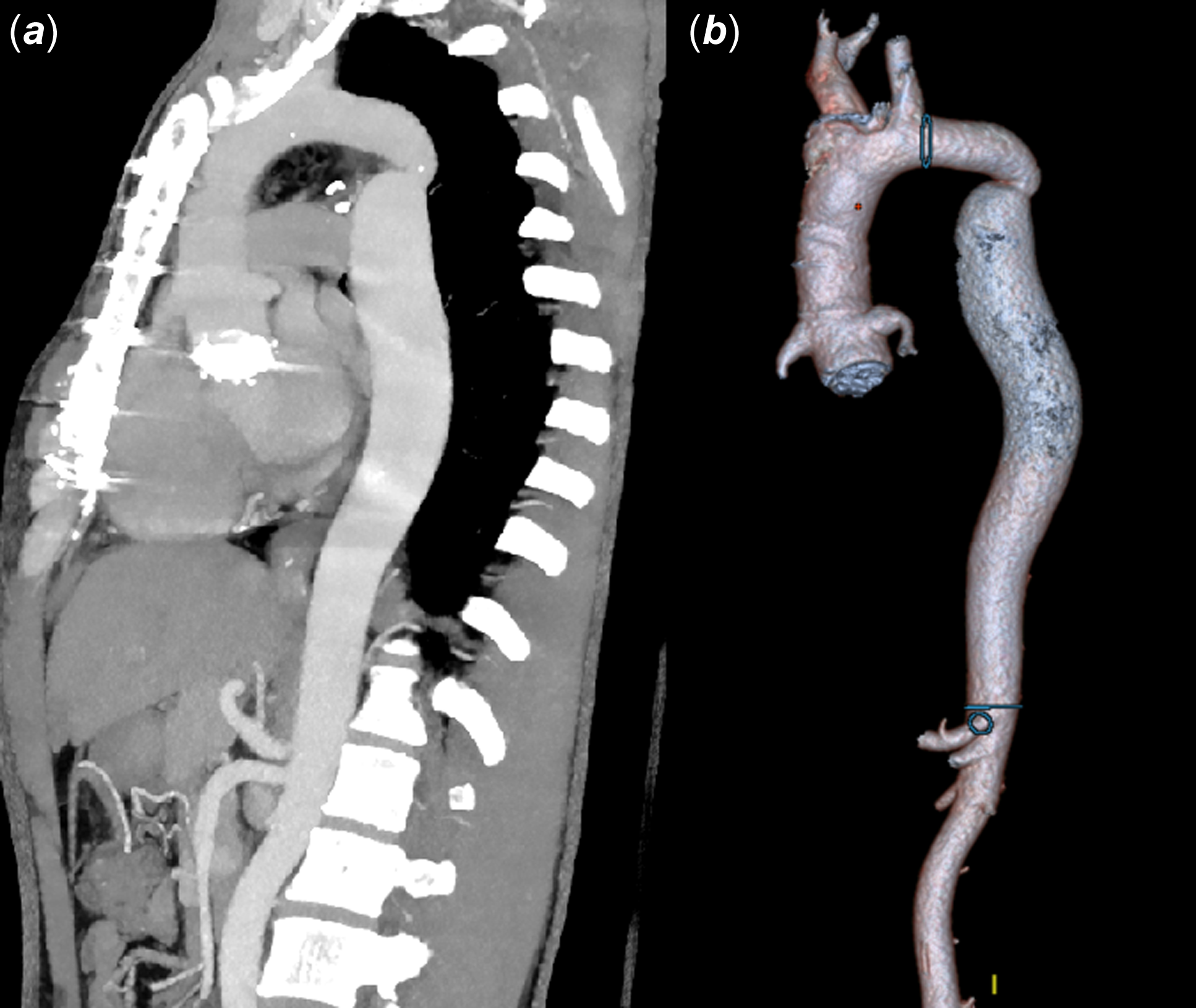
At presentation, he had arterial hypertension requiring dual therapy and a 45 mmHg right upper–lower limb gradient. Left ventricular function was preserved, the aortic valve function was normal, and the ascending aortic conduit was nondilated at 34 mm. CT angiography revealed recurrent coarctation of the aorta with an aortic arch of 27 mm, a focal isthmic narrowing of 19 × 19 mm (area 2.9 cm2), and a post-stenotic dilation of the proximal descending aorta to 36 × 32 mm (Figure 1). The 17 mm diameter mismatch posed a major challenge: a standard endograft risked poor distal sealing, while longer coverage increased the danger of spinal cord ischaemia. Balloon-expandable stenting alone carries high risks of dissection or perforation, given the abrupt calibre change and unfavourable arch geometry. The patient declined surgery, so the heart team opted for a tailored hybrid solution—a custom thoracic endograft (Cook Medical, USA) to bridge the diameter transition, reinforced with a balloon-expandable stent to optimise luminal gain and long-term stability.
Preprocedural CT angiogram showing recurrent coarctation of the aorta.

Figure 1. Long description
The image consists of two parts. On the left, a CT angiogram in a sagittal view displays the recurrent coarctation of the aorta. The aorta is highlighted, showing areas of narrowing. The spine and surrounding anatomical structures are visible, with the aorta appearing as a prominent, contrast-filled tube. On the right, a 3D reconstruction of the aorta is shown, illustrating the complex anatomy and areas of narrowing marked by blue circles. The image provides a detailed view of the aorta’s structure and the specific locations of recurrent narrowing, which are critical for planning a hybrid repair procedure.
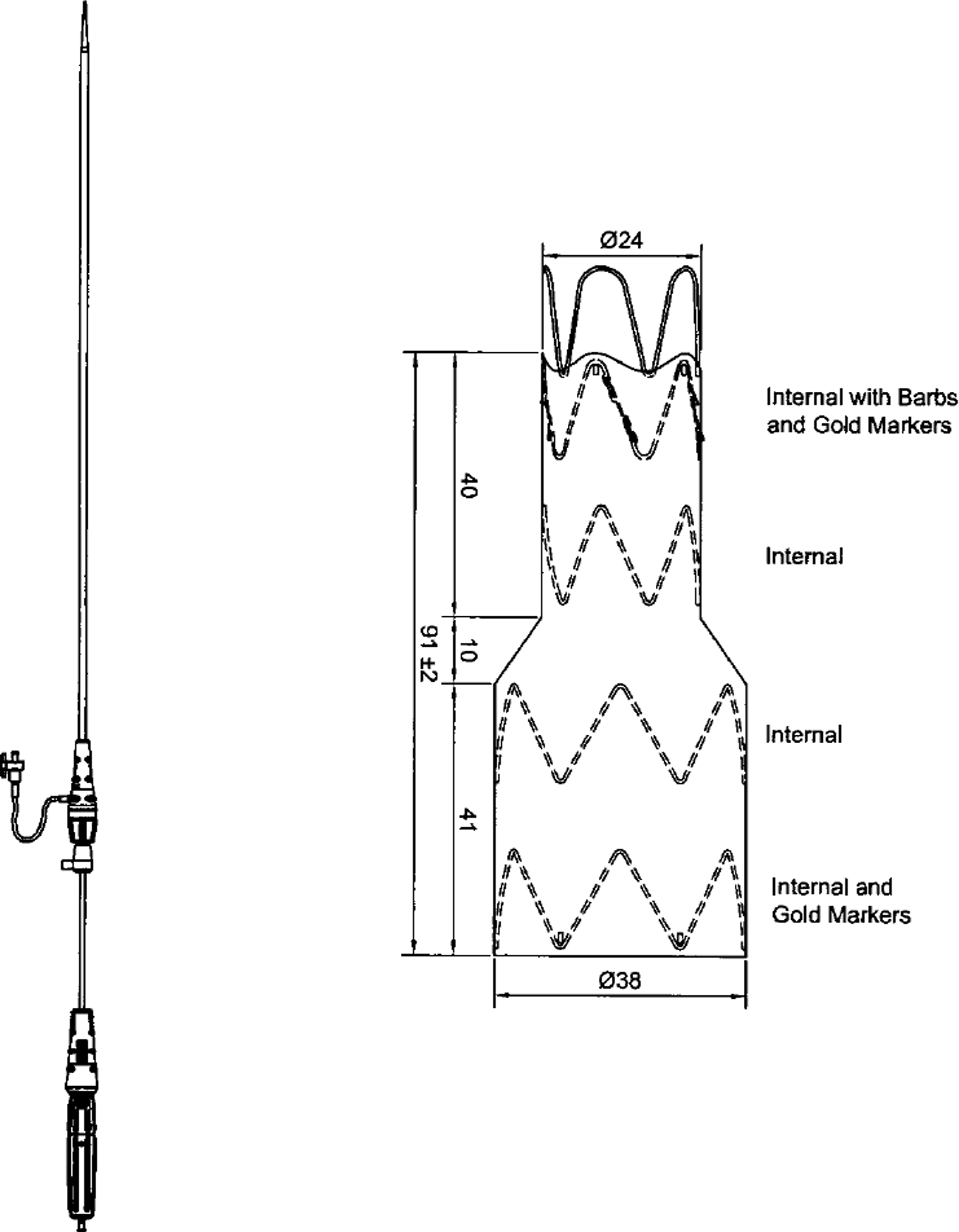
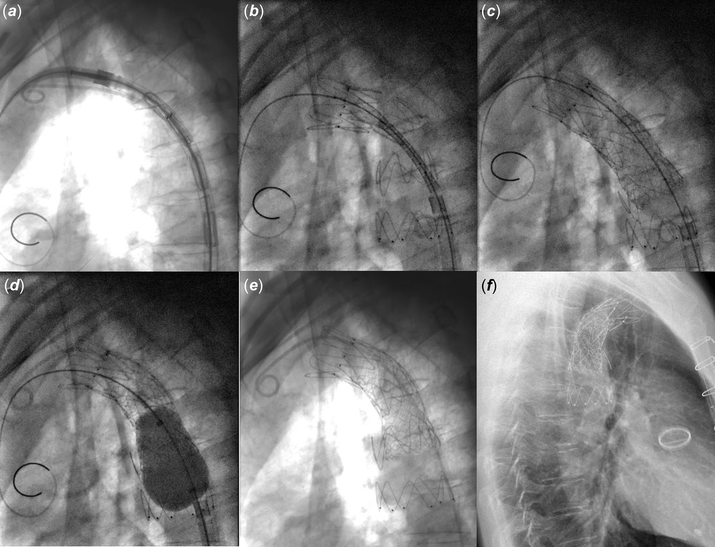
Fluindione was stopped five days prior and bridged with therapeutic enoxaparin (7,000 IU bid). On the day of intervention, INR was 1.3 and anti-Xa was 0.26 IU/mL. After consent, the procedure was performed under general anaesthesia with antibiotic prophylaxis and 3D fusion imaging. Through 18F femoral access secured by two Perclose ProGlide devices (Abbott, USA), a double-curve Lunderquist wire was positioned in the ascending aorta. A custom thoracic endograft was deployed through an 18F flexor sheath just distal to the left common carotid artery (Figures 2, and 3, Video 1). The residual gradient decreased to 10 mmHg, and a 58 mm OPTIMUS XL bare-metal stent (AndraTech, Germany), manually crimped onto a 24 × 60 mm BIB balloon, was then implanted across the isthmus (Figure 3) with the caudal end subsequently flared (Video 2). Final angiography showed a smooth aortic lumen without wall injury and complete resolution of the gradient (Figure 3, Video 3).
Custom-made aortic endograft from Cook Medical with a length of 91 ± 2 mm, a cranial diameter of 24 mm, and a caudal diameter of 38 mm.

Figure 2. Long description
The image shows a custom-made aortic endograft from Cook Medical. The endograft has a length of 91 millimeters plus or minus 2 millimeters, a cranial diameter of 24 millimeters, and a caudal diameter of 38 millimeters. The diagram includes labeled sections such as ‘Internal with Barbs and Gold Markers,’ ‘Internal,’ and ‘Internal and Gold Markers.’ The endograft features a series of zigzag patterns representing its internal structure.
The post-procedural course was uneventful, without vascular or neurological complications. The patient remained normotensive and free of residual gradient. Echocardiography showed favourable haemodynamics (peak velocity 2.2 m/s across the isthmus, no distal flow dampening), with preserved flow in the left common carotid artery. Anticoagulation was resumed the same evening with enoxaparin bridging until INR 2–3, and the patient was discharged in stable condition 48 h later. At a one-month follow-up, he demonstrated excellent outcomes with no pressure gradient.
Discussion
We report a 40-year-old man with recurrent coarctation of the aorta managed using a tailored hybrid endovascular approach. A custom thoracic endograft was deployed to bridge the marked proximal-to-distal diameter discrepancy, followed by a balloon-expandable stent to relieve residual stenosis and optimise luminal geometry. This strategy allowed precise adaptation to complex arch anatomy while minimising procedural risk.
Transcatheter therapies, including balloon angioplasty and stenting, have become established alternatives to redo surgery in adolescents and adults, offering shorter hospital stays and lower early morbidity. Reference Forbes, Kim and Du1–Reference Haddad, Hascoet and Karsenty3 Balloon-expandable stents reliably relieve gradients, though complications such as stent fracture, aortic injury, or access-site issues can occur. Reference Forbes, Kim and Du1–Reference Haddad, Hascoet and Karsenty3,Reference Hatoum, Haddad, Saliba and Abdel Massih5 However, in adults with complex coarctation of the aorta, rigid balloon-expandable stents may have poor wall apposition proximal and distal to the lesion, particularly when there is tortuous anatomy or significant diameter mismatch. Reference Fillinger, Greenberg, McKinsey and Chaikof6
Rather than relying solely on graft oversizing to achieve sealing across the coarctation and distal aorta, custom endografts offer several advantages: they accommodate tapered anatomies, conform to steep or angulated arches, and can exclude associated pseudoaneurysms or aneurysms. Reference Lala, Scali and Feezor4,Reference Schneider, Uebing and Shore7 Although endografts demonstrate high technical success in selected patients, Reference Lala, Scali and Feezor4,Reference Schneider, Uebing and Shore7 their broader adoption is limited by cost, radial force constraints, and availability. Hybrid strategies combining custom endografts with balloon-expandable stents may therefore represent a patient-tailored, technically rational approach for anatomically complex or high-risk recurrent coarctation of the aorta.
Conclusions
A hybrid endovascular approach using a custom thoracic endograft reinforced with a balloon-expandable stent may be a feasible option for managing recurrent coarctation of the aorta in complex adult congenital patients. This strategy could potentially address anatomical and haemodynamic challenges, accommodate diameter discrepancies, and reduce procedural risk, representing a possible alternative to redo surgery in high-risk or anatomically complex cases.
( a ) Custom thoracic endograft positioning in the distal aortic arch. ( b ) Implanted endograft. ( c ) Balloon-expandable stent deployment within the endograft. ( d ) Caudal end flaring. ( e ) Final result (lateral view). ( f ) Discharge lateral X-ray.

Figure 3. Long description
The image sequence displays various stages of thoracic endograft placement in the aortic arch. Image (a) shows the positioning of a custom thoracic endograft in the distal aortic arch. Image (b) depicts the implanted endograft. Image (c) illustrates the deployment of a balloon-expandable stent within the endograft. Image (d) captures the process of caudal end flaring. Image (e) presents the final result from a lateral view. Image (f) shows a discharge lateral X-ray.
Supplementary material
The supplementary material for this article can be found at https://doi.org/10.1017/S1047951126113419.
Acknowledgements
None.
Financial support
None.
Competing interests
S. HAULON has received proctoring and consulting fees from Cook Medical, GE Healthcare, and Bentley. All other authors have reported that they have no relationships relevant to the contents of this paper to disclose.