I. INTRODUCTION
Additive manufacturing (AM) of glasses and, in general, of silicate ceramics specifically aimed at obtaining highly porous bioactive scaffolds for bone tissue applications, is still an open issue. The first experiences were presented in the review paper by Gmeiner et al.,Reference Gmeiner, Deisinger, Schönherr, Lechner, Detsch, Boccaccini and Stampfl1 according to which the approaches can be roughly divided into technologies for indirect printing and technologies for direct printing. Indirect printing corresponds to the manufacturing of polymeric replicas, e.g., by means of stereolithography, for the gel-casting of glass powders,Reference Chopra, Mummery, Derby and Gough2,Reference Padilla, Sánchez-Salcedo and Vallet-Regí3 in analogy with what was done with other bioceramic powders.Reference Chu, Halloran, Hollister and Feinberg4,Reference Limpanuphap and Derby5 Sintering occurs after template removal and burn-out of organic additives. The direct fabrication of glass-based scaffolds, i.e., the development of a porous structure directly from glass powders combined with suitable binders, is definitely more challenging: if direct ink writing (robocasting) is quite well established,Reference Fu, Saiz and Tomsia6–Reference Eqtesadi, Motealleh, Pajares and Miranda10 other techniques, leading to more complex geometries, are still at a preliminary stage. As an example, lithography-based AM technology was applied for fabricating cellular 45S5 Bioglass® structures, whereas the biaxial strength of the manufactured samples was comparable to the flexural strength of traditionally processed Bioglass®,Reference Hench, Kokubo, Black and Hastings11 and the compressive strength of 3D scaffolds did not exceed 0.5 MPa, i.e., it remained below the values for porous Bioglass® structures obtained by means of foam replication.Reference Tesavibul, Felzmann, Gruber, Liska, Thompson, Boccaccini and Stampfl12
The control of the viscous flow of glass powders is essential for near-net shaping of highly porous glass-based components; in fact, there is a delicate trade-off between densification of struts and stabilization of the 3D structures,Reference Winkel, Meszaros, Reinsch, Müller, Travitzky, Fey, Greil and Wondraczek13 implying careful control of the firing temperature, crystallization rates, and powder size. As an example, in the previously mentioned lithography-processed Bioglass scaffolds, the final sintering temperature was reduced to 950 °C, specifically to avoid distortion of the printed partsReference Tesavibul, Felzmann, Gruber, Liska, Thompson, Boccaccini and Stampfl12; as an alternative, fine Bioglass powders could be combined with a secondary, rigid phase such as hydroxyapatite powders (according to a “compounding” approachReference Winkel, Meszaros, Reinsch, Müller, Travitzky, Fey, Greil and Wondraczek13). Indeed, rigid, secondary phases may be actually provided by the same starting glasses, when transformed into glass-ceramics.Reference Boccaccini, Chen, Lefebvre, Gremillard and Chevalier14–Reference Elsayed, Romero, Ferroni, Gardin, Zavan and Bernardo16
Crystallization of bioglasses is actually a fundamental issue since it may decrease or delay the kinetics of apatite formation,Reference Peitl, LaTorre and Hench17,Reference Peitl, Zanotto and Hench18 thus configuring an additional constraint in the heat treatments. As an example, Meszaros et al. sintered 3D-printed parts below 795 °C, to avoid crystallization effects.Reference Meszaros, Zhao, Travitzky, Fey, Greil and Wondraczek19 However, with properly designed compositions, a significant bioactivity and biocompatibility can be achieved even with highly crystallized materials (a fundamental example is that of Biosilicate® glass-ceramics, exhibiting osteoconductivity, osteoinductivity, biocompatibility, and antibacterial properties comparable to those of the “golden standard” amorphous 45S5Reference Montazerian and Zanotto20).
The sintering of fine glass particles actually stimulates their crystallization. A relatively high degree of crystallization may be achieved in very short times since the surface of glass is a preferred site for nucleation for several systems.Reference Müller, Zanotto and Fokin21–Reference Hernández-Crespo, Romero and Rincón24 An optimum balance between densification and crystallization is generally achievable only if the crystallization peak is located at a temperature suitably higher than that corresponding to the dilatometric softening point,Reference Bernardo, Scarinci, Edme, Michon and Planty25 i.e., the temperature at which viscous flow becomes appreciable.Reference Ray and Tiwari26 If the temperature difference is limited, adjacent glass particles can develop only some necking, and the resulting glass-ceramic particles possess a large amount of interconnected residual porosity.Reference Bernardo, Scarinci, Edme, Michon and Planty25 An abundant residual porosity may not represent an issue in glass-ceramics for biomedical applications, e.g., for bone tissue engineering; while macroscopic pores (typically >500 µm) favor cell ingrowth and vascularization, micro- or even nanosized pores in the cell walls favor cell attachment.Reference Hulbert, Morrison and Klawitter27
The investigation described in the present paper explores the coupling of viscous flow sintering and crystallization in complex cellular structures produced by means of different AM techniques, starting from glasses of the CaO–MgO–SiO2 system, designed to yield bioactive material even after crystallization.Reference Elsayed, Romero, Ferroni, Gardin, Zavan and Bernardo16 The balance between sintering and crystallization had to be adjusted depending on the different requirements, in terms of powder size, of the adopted manufacturing technology.
II. MATERIALS AND METHODS
A. Glass preparation
Two glasses in the CaO–MgO–SiO2 system, later referred to as WDP and WDE, were used in the experiments. WDP had a CaO:MgO:SiO2 molar ratio equal to 2:1:3, theoretically leading to 50 mol% wollastonite (W, CaO·SiO2) and 50 mol% diopside (D, CaO·MgO·2SiO2); small amounts of Na2O and P2O5 were introduced to reduce the liquidus temperature.Reference Elsayed, Romero, Ferroni, Gardin, Zavan and Bernardo16 WDE contained exclusively the main oxides, and the CaO:MgO:SiO2 molar ratio was adjusted to that of the wollastonite–diopside eutectic (∼52 mol% W, 48 mol% D; CaO:MgO:SiO2 ≅ 2:0.96:2.96).Reference Sainz, Pena, Serena and Caballero28
The glasses were obtained by firing, at 1375 °C for 1 h in platinum crucibles, mixtures of highly pure oxides, carbonates, and phosphates. The melts were quenched by direct pouring on cold steel plates. The glasses solidified in the form of irregular blocks, with plenty of cracks and pores, later subjected to dry ball milling. Finally, the obtained powders were manually sieved and separated into fractions with particle size below 25 μm, between 45 and 75 μm, and above 75 μm. Each fraction, for each glass composition, was subjected to differential thermal analyses (DTA/TGA, STA409, Netzsch Gerätebau GmbH, Selb, Germany, operating at 10 °C/min heating rate). Some fragments were kept apart to perform the dilatometric analysis (402E Netzsch Gerätebau GmbH, Selb, Germany). The composition of the glasses was assessed by means of X-ray fluorescence (XRF) spectroscopy (Panalytical MagixPro, Malvern Panalytical GmbH, Kassel, Germany).
B. AM and sintering of glass-ceramics
Highly porous reticulated scaffolds were fabricated by using both 3D printing (3DP) and digital light processing (DLP). Powders in the 45–100 µm size range were used for 3DP manufacturing experiments (RX-1, ExOne GmbH, Gersthofen, Augsburg, Germany). The main printing parameters were layer thickness = 100 µm; powder bed relative packing density = 50 vol%; saturation = 150% (ratio between volume of binder-printed and volume of pores in the powder bed). For DLP, glass powders in the 20 µm size range were mixed, with a solid load of 60 wt%, into a commercial photocurable acrylic polymer. The glass filled polymer resin mixture was printed using the visible light range between 400 and 500 nm of the DLP printer (3DLPrinter-HD 2.0, Robofactory, Venice, Italy). The 3D-printed structures were cleaned in an ultrasonic bath with isopropanol for 3 min, to remove the uncured resin and then heat-treated at a heating rate of 1 °C/min up to 500 °C with a holding time of 3 h to decompose the polymer, then at 800–1100 °C, with 5 °C/min, for 1 h.
C. Characterization
Microstructural characterization was conducted by means of optical stereomicroscopy (AxioCam ERc 5s Microscope Camera, Carl Zeiss Microscopy, Thornwood, NY) and scanning electron microscopy (FEI Quanta 200, ESEM, Eindhoven, Netherlands) equipped with EDS. The geometric density of the scaffolds was measured using a digital caliper and weighed with an analytical balance. The apparent densities were measured by the Archimedes’ method (according to ISO 3233-1:2013), and true densities were measured by means of a gas pycnometer (Micromeritics AccuPyc 1330, Micromeritics, Norcross, Georgia), operating with helium gas, on the samples in bulk (3D-printed scaffold) and powder forms.
For comparison purposes, tablet-shaped samples (with a diameter of 10 mm and a thickness of 2.5 mm) were prepared by 3DP as well as by uniaxial pressing at 60 MPa. The mechanical properties of pressed and 3DP tablet-shaped samples, from powders with the same starting size, were characterized by ball-on-three-balls (B3B) tests.Reference Börger, Supancic and Danzer29 The compressive strength of sintered scaffolds was evaluated at room temperature, by means of an Instron 1121 UTM (Instron, Danvers, Massachusetts) operating with a crosshead speed of 0.5 mm/min. Each data point represents the average value of at least 10 individual tests.
The crystalline phases were identified by means of X-ray diffraction on the powdered samples (XRD; Bruker AXS D8 Advance, Karlsruhe, Germany), supported by data from PDF-2 database (ICDD-International Center for Diffraction Data, Newtown Square, PA) and Match! program package (Crystal Impact GbR, Bonn, Germany).
Simulated body fluid (SBF) tests were performed according to ISO 23317. 3D-printed and the pressed samples were left immersed in SBF for 4 weeks and the ionic concentration of the solution was measured every 7 days by ICP-OES (Optima 3000, Perkin-Elmer, Waltham, Massachusetts). The solution was never refreshed.
The dissolution of the samples in blood pH conditions was simulated by dissolution measurements according to DIN EN ISO 10993-14 standard but applied to 3D-printed and pressed discs instead of granules. Briefly, the samples were immersed in 20 mL of Tris–HCl solution for each 1 g of the sample, and the sample holder was left at 37 °C under a continuous circular movement. The pH was adjusted at pH = 7.4 by adding HCl to the Tris buffer solution. The total dissolution and ionic concentration of the solution were measured by ICP-OES every week (168 h) for a total of 9 weeks. The solution was completely replenished after each measurement.
III. RESULTS AND DISCUSSION
A. Assessment of sinter-crystallization window
The two glass formulations here tested have been developed based on previous experiments on the fabrication of highly porous bioactive glass-ceramics. More specifically, WDP glass had already been proposed for the development of highly porous glass-ceramic foams, by “inorganic gel casting” (i.e., direct foaming of glass suspensions, in alkaline medium, followed by sinter-crystallization)Reference Elsayed, Romero, Ferroni, Gardin, Zavan and Bernardo16 as well as for reticulated scaffolds, by direct ink writing of silicone-based pastes.Reference Elsayed, Colombo and Bernardo30 The biocompatibility and bioactivity of WDP samples were verified by cell tests on foams.Reference Elsayed, Romero, Ferroni, Gardin, Zavan and Bernardo16
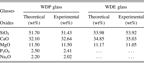
Formulation WDE represents an effort to simplify the glass formulation for obtaining wollastonite–diopside glass-ceramics (by the removal of Na2O and P2O5). The biocompatibility and bioactivity, for this composition, had already been assessed in the literature.Reference Sainz, Pena, Serena and Caballero28 The data reported in Table I demonstrate that, based on XRF analysis, the produced glasses matched nearly perfectly the theoretical, expected compositions.
Comparison between theoretical and experimental glass formulations.

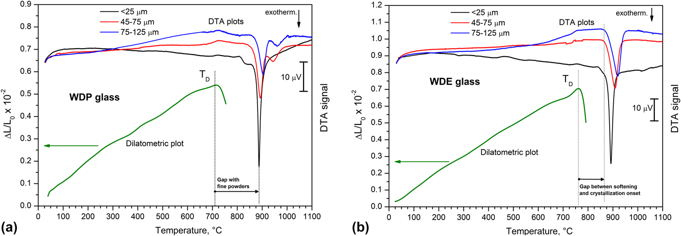
The thermal analysis of the two glasses is shown in Fig. 1. For both compositions, we can observe their sensitivity to surface nucleation since the exothermic crystallization peaks were more pronounced and appeared at a lower temperature, with decreasing powder size. The surface nucleation was not surprising, considering the expected crystal phases, i.e., wollastonite and diopside.Reference Karamanov and Pelino31 The main difference between the two glasses concerns the sinter-crystallization window.Reference Bernardo, Scarinci, Edme, Michon and Planty25 In fact, despite similarities in the temperature at which the crystallization peaks occur, WDE softened at much higher temperature than WDP, affecting the balance between viscous flow sintering (a significant viscous flow is active at a temperature of 50–100 °C above the dilatometric softening pointReference Ray and Tiwari26) and crystallization. Based on these considerations, we can posit that firing WDP at 800 °C would lead to an amorphous material, at least for coarser powders, since this temperature is well above the dilatometric point (710 °C), but still far from the crystallization temperature. On the contrary, WDE would densify well above 800 °C (the dilatometric point being located at about 760 °C), with possible overlapping with crystallization, (we can note an onset in the crystallization peak at about 860 °C, even for coarse powders).
Assessment of sinter-crystallization windows by comparison of dilatometric and DTA curves of the studied glasses: (a) WDP; (b) WDE.

B. Preliminary densification studies on pressed and printed powders
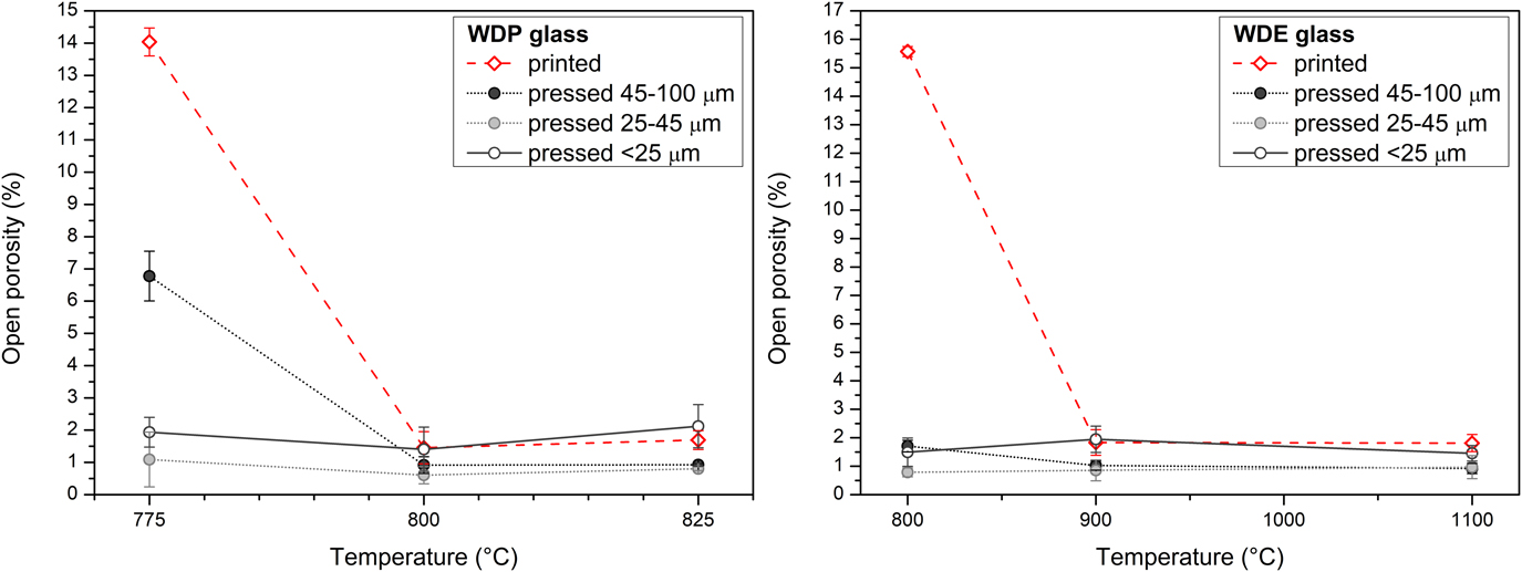
Dilatometric and DTA plots provided a first overview of the different balances between sintering and crystallization in the two starting glasses. Preliminary sintering studies were carried out to understand the impact on processing. In particular, powder-based 3DP implied the use of quite coarse particles, in the 45–100 μm range, to optimize the powder flowability during the layer deposition process, and the packing density was undoubtedly lower than that achievable, from the same powders, by uniaxial pressing (at 60 MPa). Figure 2 illustrates the resulting water absorption of tablets, printed or pressed. Operating with WDP, a slight increase of sintering temperature, from 775 to 800 °C, determined a dramatic reduction of open porosity as an effect of viscous flow, independently from powder size and packing [Fig. 2(a)]. On the contrary, the sealing of open porosity for poorly packed 3D-printed WDE powders was achieved only at 900 °C [Fig. 2(b)]. The elimination of the porosity surplus in tablets obtained from printing instead of pressing, in other words, was hindered by crystallization (crystal inclusions are known to increase significantly the apparent viscosity of glassReference Boccaccini32,Reference Müller, Eberstein, Reinsch, Schiller, Deubener and Thiel33 ).
Preliminary densification assessment of sinter-crystallized WDP and WDE glasses, by means of water absorption tests on tablets, from uniaxial pressing of powders or 3DP.

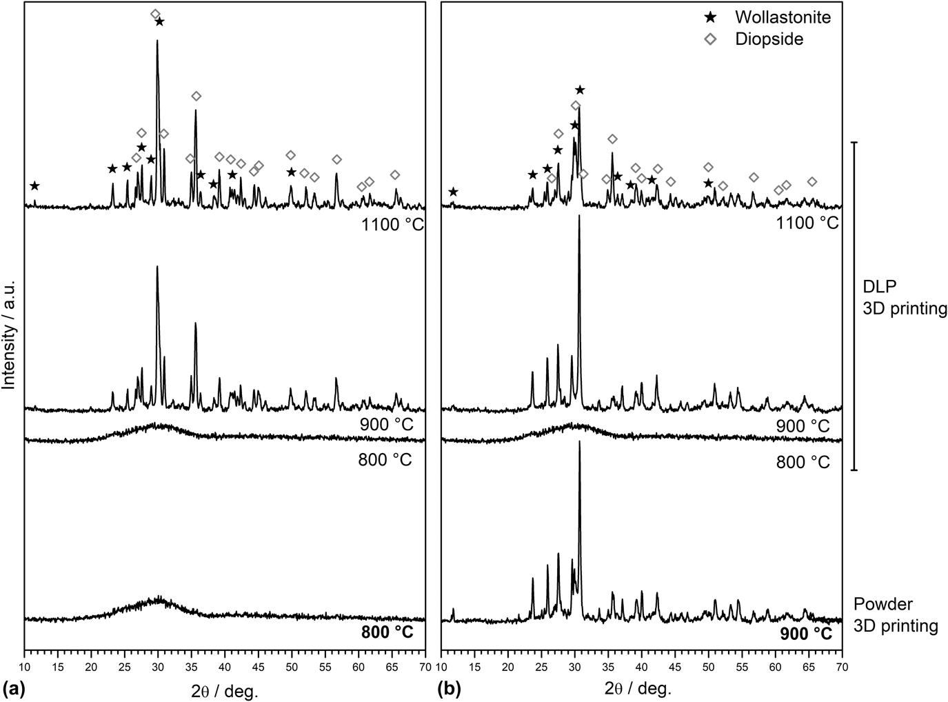
The amorphous nature of samples from WDP glass sintered at 800 °C was indeed confirmed, as shown in Fig. 3(a). The crystallization of the samples from WDE glass heated at 900 °C is also evident [see Fig. 3(b)]. The enhanced densification, with decreasing powder size, observed for WDP glass sintered below 800 °C [Fig. 2(a)] is consistent with simple viscous flow sintering, without crystallization (according to Frenkel’s model,Reference Prado, Ferreira and Zanotto34 the densification increases with decreasing powder size, at a given glass viscosity).
Mineralogical evolution, upon sintering, of printed samples from WDP (a) and WDE (b) glasses.

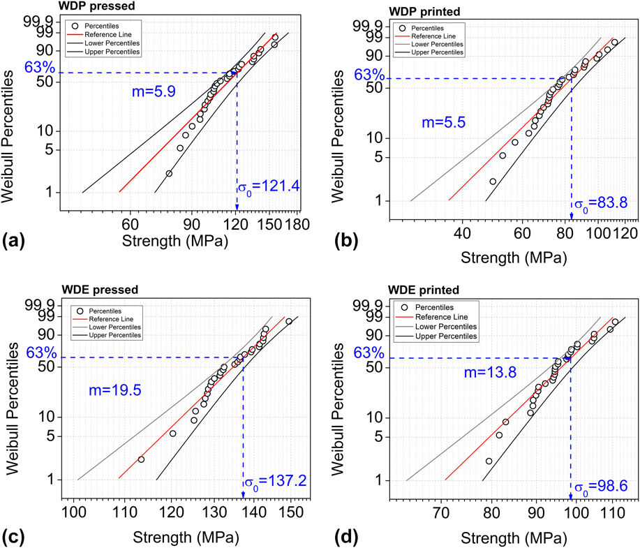
Preliminary mechanical tests were conducted for verifying the impact of 3DP on strength, besides on densification. Figure 4 shows that, with the same starting glass powder size, the enhanced overall porosity (4.6 ± 0.3 vol% for WDP printed, 4.2 ± 0.1 vol% for WDP pressed; 7.1 ± 0.5 vol% for WDE printed, 5.8 ± 0.1 vol% for WDE pressed) did not determine a substantial strength degradation, especially for WDE-based samples, which were partially crystallized (the crystallization is also testified by the enhanced reliability, expressed by Weibull’s modulus, as typically observed when passing from glasses to glass-ceramicsReference Reddy, Tulyaganov, Pascual, Kharton, Tsipis, Kolotygin and Ferreira35). In other words, 3DP, even when carried out using the technique leading to a lower packing of particles, could be considered as a quite promising method for the manufacturing of bending-dominated structures, such as reticulated scaffolds.
Assessment of the impact of printing on mechanical properties, by means of B3B tests conducted on sintered samples from: WDP, fired at 800 °C [(a) pressed; (b) 3D-printed]: WDE, fired at 900 °C [(c) pressed; (d) 3D-printed].

C. Fabrication and characterization of 3D-printed glass-ceramic scaffolds
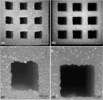
The morphology of 3D-printed scaffolds is shown in Fig. 5. We can observe that firing at 800 °C, for WDP, and at 900 °C, for WDE, did not lead to an excessive shape deformation, indicating that the processing heating schedule, selected on the basis of dilatometric and DTA studies, was appropriate. Additional tests carried out at higher temperatures with WDP glass (not shown here for brevity) caused the viscous collapse of the samples; on the contrary, WDE specimens could be heated at higher temperature without geometrically deforming, due to the growth of crystals.
Morphology of reticulated scaffolds from 3DP of WDP (a and c) and WDE (b and d) glasses.

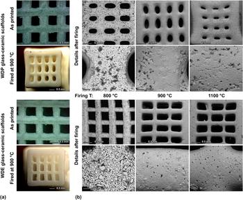
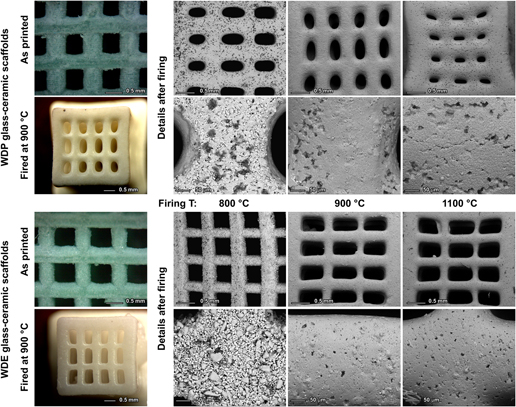
When using the DLP technology, the starting glass powders were homogeneously incorporated in the polymeric matrix subjected to UV curing and, after debinding, the scaffolds were ceramized at different temperatures, from 800 up to 1100 °C (see Fig. 6).
Microstructural evolution of samples from DLP processing: (a) optical stereomicroscopy images of glass-ceramic scaffolds before and after firing; (b) evolution of reticulated scaffolds with heating temperature (low and high magnification details).

WDP glass, fired at 800 °C, remained amorphous, as in previous experiments [see Fig. 3(a)]. Unlike the samples from 3DP, the crystallization impeded a significant viscous collapse, at 900 °C. The enhanced crystallization could be justified by the reduced powder size and consequently enhanced surface nucleation; a viscous collapse was evident only for treatments at high temperature (1100 °C). WDE had a different evolution: fine particles remained amorphous but had a limited sintering at 800 °C. The densification improved at 900 °C, with a substantial crystallization, whereas firing even at 1100 °C did not lead to significant deformation, despite a lower crystallization. The high magnification details reveal quite smooth surfaces, for WDP already at 800 °C and for WDE above 900 °C.
Quite surprisingly, WDP led to the simultaneous crystallization of wollastonite (CaSiO3, PDF #27-0088) and diopside (Mg-rich variant, Ca0.89Mg1.11Si2O6, PDF #870698), while WDE had a substantial crystallization of wollastonite at 900 °C, with limited formation of diopside even at 1100 °C. This implied a different nature of the surrounding glass phase: the viscous collapse of WDP at 1100 °C, despite high crystallization, could be ascribed to the low viscosity of the Na-phosphate residual glass phase; WDE, on the contrary, had a silicate residual glass phase.
In all cases (see Table II), the compressive (crushing) strength of the developed scaffolds compared favorably with the strength range for cancellous bone (2–12 MPa).Reference Thompson and Hench36,Reference Rahaman, Liu, Huang, Narayan and Colombo37 In addition, especially for WDE-based samples, the strength-to-density ratio was quite remarkable: if we consider a simplified model for the crushing strength, for open-cell structures, the bending strength of the solid phase is of the order of 200 MPa (according to Gibson and Ashby,Reference Gibson and Ashby38 σc ≈ σbend·0.2·[1 − P]3/2, where P is the total porosity).
Porosity and strength of samples produced by 3DP and DLP.

D. Preliminary biocompatibility tests (on 3D-printed samples)
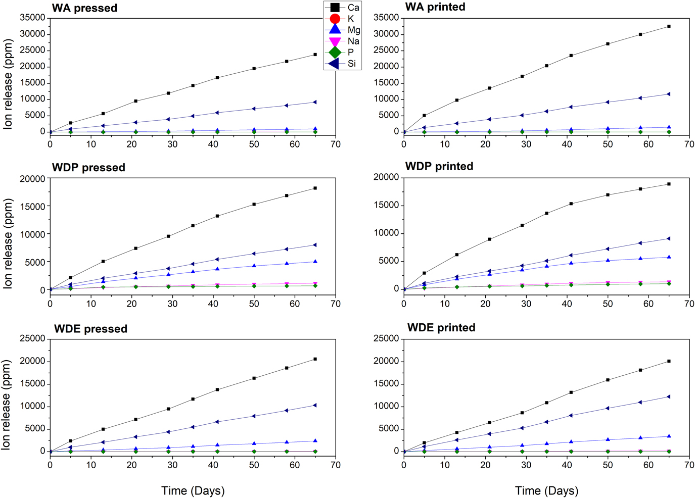
A preliminary analysis of biocompatibility was conducted on powder 3D-printed samples. The stability in conditions simulating the pH of blood was assessed by measuring the dissolution and the release of ions in a Tris–HCl buffer, and the graphs in Fig. 7 show cumulative results. Pressed and 3DP samples were compared with a wollastonite–apatite (WA) glass-ceramic standard, according to the formulation suggested by Kokubo et al.Reference Nakamura, Yamamuro, Higashi, Kokubo and Itoo39
Evolution of ion release in Tris–HCl buffer for wollastonite–diopside glass-ceramics compared to a WA glass-ceramic reference.

The motivation for the development of WA glass-ceramics was the improvement, on one hand, of the mechanical properties of the Bioglass, while maintaining its ability to bond to bone,Reference Kokubo40 and on the other hand, to enhance the resorption rate of pure apatite in vivo, which is very low.Reference Lu, Descamps, Dejou, Koubi, Hardouin, Lemaitre and Proust41 The bonding mechanism of WA to bone involves the formation of an apatite layer on the surface of the glass-ceramic, which can also be reproduced in vitro in an SBF solution. It has been shown that calcium and silicon ions play a crucial role in the precipitation of the apatite layer. Ca++ ions increase the ionic activity of apatite in solution, while Si–OH groups on the surface act as nucleation sites for the precipitation of apatite.Reference Ohtsuki, Kokubo and Yamamuro42
The mechanism is similar to the formation of apatite on the surface of NaO–CaO–SiO2–P2O5 bioglasses, and it has been shown that also many crystalline silicate ceramics have the ability to form apatite on their surface in SBF.Reference Wu and Chang43 Relevantly for the current work, this also applies to the wollastonite and diopside minerals. Indeed, in WA, calcium and silicon ions were dissolved partially from the glassy phase and partially from wollastonite. Pure wollastonite showed excellent in vitro apatite formation and, thus, good bioactivity.Reference Siriphannon, Kameshima, Yasumori, Okada and Hayashi44 However, pure wollastonite had a high dissolution rate, which caused an excessive increase of pH in the surrounding fluids.Reference Iimori, Kameshima, Okada and Hayashi45 The formulation of WA glass-ceramics, therefore, is also important to control the dissolution rate of the material.
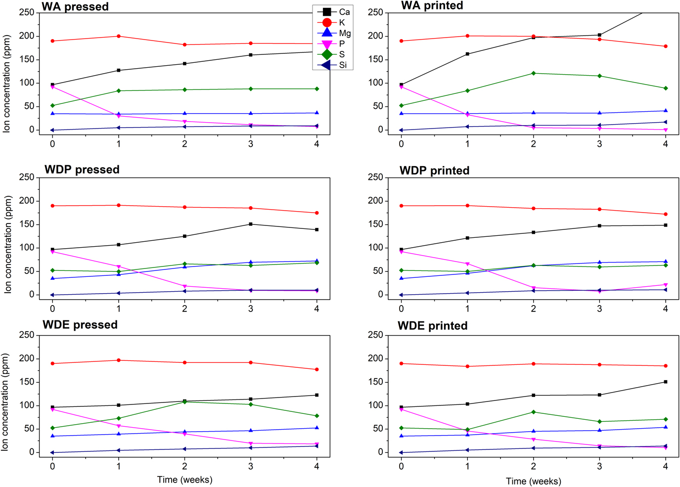
The same principle should then apply to the WD glasses and glass-ceramic object of the current study. In fact, the ionic release of both the eutectic WDE and the flux WDP materials (reported in Fig. 7) did not differ much from that of the standard WA glass-ceramic, indicating that the dissolution rate of WD materials could be suitably controlled. In particular, the Ca2+ release rate decreased compared to WA, while Si4+ release was lower for WDP (amorphous) and higher for WDE (crystallized). Interestingly, WDE glass-ceramic released more Ca2+ and Si4+ than WDP, but significantly less Mg2+. This is consistent considering the phases that were crystallized in WDE: wollastonite and diopside. Wollastonite (which is a calcium silicate), as already mentioned, has a high dissolution rate, which contributes to the increased release of Ca2+ from WDE. Diopside, however, is more stable and has a low dissolution rate.Reference Prado, Ferreira and Zanotto34 Since diopside is a calcium and magnesium silicate, the crystallization of this phase leads to a decreased Mg2+ release rate. It is also noticeable, that pressed and 3D-printed samples had a similar evolution of ion concentration for all compositions, both in Tris–HCl (Fig. 7) and in SBF (Fig. 8). This observation is related to the fact that both pressed and 3D-printed samples had similar levels of porosity (as shown in Fig. 2) and were also expected to have a similar amount of geometric surface area.
Evolution of ion concentration in SBF solution for wollastonite–diopside glass-ceramics compared to a WA glass-ceramic reference.

The concentration of the SBF solution, measured once a week for 4 weeks without refreshing, is reported in Fig. 8. We can note that the phosphorous concentration steeply decreased in the first 2 weeks. In parallel, the concentration of Ca increased due to the release of Ca from the samples, similarly to the dissolution behavior studied in Tris–HCl buffer. However, the concentration of Ca in SBF increased only slightly over time, possibly indicating the simultaneous precipitation and growth of a phase containing Ca and P.
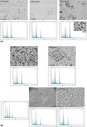
The hypothesis of precipitation of Ca and P ions and growth of a calcium phosphate phase were supported by SEM–EDX (energy-dispersive X-ray spectroscopy) investigation of the surface of the samples, shown in Fig. 9(a). Indeed, in all the samples, a phase containing Ca and P was identified on the surface. The thickness and the morphology of the developed layer varied significantly between different samples. Pressed samples from WA and WDP (that could be considered also as a model for the scaffolds, with smooth surfaces, produced by DLP) exhibited a thick layer, homogeneously covering the surface. The surface of the pressed WDE sample, instead, was covered by rounded agglomerates of nanosized needle-like crystals [resembling the structures from early stage growth of apatite,Reference Salahinejad and Vahedifard46 see top right images in Fig. 9(a)]. The printed WA samples had a very thin coating, whereas printed WDE and WDP samples formed a thick layer with some irregularities: for WDE samples, the coating was not continuous, whereas for WDP samples, there were some NaCl-rich zones [evidenced by the lighter color, bottom left image of Fig. 9(b), with related EDX spectrum], mixed with calcium phosphate agglomerates [see bottom center and right images, in Fig. 9(b), with related EDX spectra].
Comparison of the mineral deposits on glass-ceramic samples of different formulations after immersion in SBF, by means of SEM coupled with energy-dispersive X-ray spectroscopy: (a) pressed samples; (b) printed samples (3DP).

The validation of the bioactivity of all the produced samples, by cell culture studies, will be the focus of further investigations. The observed mineralization of the surfaces, with SBF immersion, and the similarity with an already proven bioactive glass-ceramic, in any case, make the developed scaffolds promising components for bone tissue engineering. The coupling of sinter-crystallization with AM can be tuned on several levels (compositions, powder size, and printing technique) so that a wide range of values for different properties can be achieved.
IV. CONCLUSIONS
We may conclude the following:
(1) The sinter-crystallization of two glasses, leading to wollastonite–diopside glass-ceramics, was successfully tuned to suit the use of two different AM techniques, such as 3DP and DLP;
(2) The different requirements of different 3DP technologies, in terms of powder size, and the sensitivity of the adopted glasses to surface nucleation, conditioned the firing temperature: samples produced by 3DP, operating with coarser powders leading to limited crystallization, were more appropriate for low temperature firing (800–900 °C); samples produced by DLP, operating with finer powders more prone to crystallization, could be fired at higher temperature;
(3) Despite the differences in manufacturing technology and microstructure, all the samples featured very good strength-to-density ratios;
(4) The observed mineralization of the surfaces of 3D-printed glass-ceramics, after immersion in SBF, and the similarities with already validated bioactive glass-ceramics, in terms of ionic releases, make the developed scaffolds promising components for bone tissue engineering.
ACKNOWLEDGMENTS
Hamada Elsayed gratefully acknowledges the financial support of the Cultural Affairs and Missions Sector, Egypt. The authors thank Mr. Carlo Castegini and Ms. Ilaria Baesso (University of Padova) for experimental assistance.