Case report 1
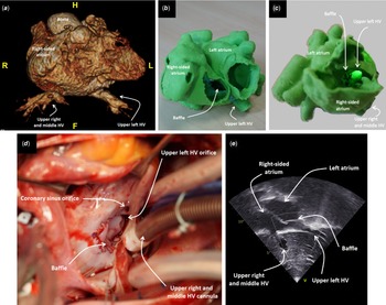
A 6-year-old boy from Guinea was referred to our institution in March 2021 via the Enfance-et-Vie charity for tetralogy of Fallot repair, and his pulsed oxygen saturation was 85%. Echocardiography and contrast-enhanced CT showed features of left isomerism (bilateral bilobed lung, midline liver, and polysplenia) and double-outlet right ventricle with severe infundibular stenosis {A,D,D}. Azygos continuation of the inferior caval vein, left atrial isomerism and a large inferior atrial septal defect were observed. In addition, the right and middle upper hepatic veins drained into the right-sided atrium while the left hepatic vein, which was rather large, drained separately into the left-sided atrium called left atrium in the setting of left atrial isomerism (Fig. 1a).
Contrast-enhanced CT scan 3D reconstruction ( a ), 3D printing of the atria with a baffle sutured in situ seen through the tricuspid valve orifice from an anterior view ( b ) and from a right lateral view with resection of the anterior wall of the right-sided atrium ( c ), procedural photography ( d ), and postoperative transthoracic echocardiography ( e ) showing left HV rerouting to the right-sided atrium. Of note, the tendon of Todaro was absent. CT scan = CT scan; HV = hepatic vein(s).

According to the 3D reconstruction of the CT scan, the left hepatic vein was inferior and connected to the left-sided atrium, although its course was from left to right: the most conceivable surgical correction was therefore a baffle repair and not the left hepatic vein reconnection. To simulate baffle repair, 3D printing was performed. A baffle was sutured into the imprinted heart, closing the atrial septal defect and rerouting the left hepatic vein into the right-sided atrium (Fig. 1b and Supplementary Video S1).
Complete repair was uneventfully performed. Cardiopulmonary bypass was instituted between the superior caval vein and the ascending aorta. Drainage of the right and middle hepatic vein was performed while the intrapericardial left hepatic vein was snared during cardiopulmonary bypass. Surgical repair consisted of ventricular septal defect closure, infundibular resection, and patch augmentation of the main pulmonary artery. A PhotoFix® bovine pericardial patch (CryoLife, Kennesaw, GA, USA) was used for the intra-atrial baffle, leaving the coronary sinus on the right side (Fig. 1c and d). The patient was discharged from hospital 11 days after surgery and flew back home 3 weeks later.
Case report 2
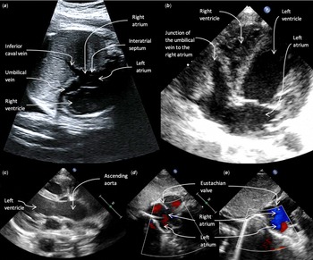
A 3.2-kg boy was diagnosed prenatally with ductus venosus agenesis, with an inferior caval vein found to connect normally to the heart (Fig. 2a). The mother was treated with digoxin at 30 weeks’ gestation for a resolving foetal episode of supra-ventricular tachycardia. The child was born at 36 weeks 5 days in the setting of premature rupture of membranes with good immediate adaptation despite moderate respiratory distress requiring continuous positive airway pressure for 3 days. Antibiotics were stopped on day 2 for negative blood cultures. The neonate had persistent episodes of desaturation. Mean pulsed oxygen saturation at 1 month was 90% on 0.5 L/min oxygen and 83% on room air.
Foetal ultrasound at 28 weeks 6 days gestation showing direct entry of a patent umbilical vein and inferior caval vein into the right atrium ( a) , ultrasound-modified 4-chamber view at 3 days of age revealing the arrival of the umbilical vein to the right atrium associated with an anterior displacement of the tricuspid valve annulus and a hypertrophied right ventricle, as well as a dilatation of the left ventricle (end-diastolic diameter 25 mm, Z-score + 3.4 SD) ( b ), confirmed on parasternal long-axis view with aortic dilatation (sinus diameter 16.4 mm, Z-score + 8.3 SD, tubular ascending aorta diameter 13.5 mm, Z-score + 6.3 SD) ( c ), At rest in the supine position ( d ), an interatrial shunt was exclusively right-to-left (red on colour Doppler), while in the upright position ( e ), the shunt was reversed (blue on colour Doppler), without evidence for pulmonary hypertension. Note the orientation of the inferior caval vein towards the atrial septum ( a ) and the prominent Eustachian valve ( d and e ). Of note, the tubular ascending aorta diameter was 17 mm at 6 months of age, Z-score decreasing to + 4.9 SD. SD = standard deviations.

Abdominal ultrasound at birth confirmed the ductus venosus agenesis with a hypoplastic left portal branch. There was left hepatic hyper-arterialisation but no portosystemic shunt. The umbilical vein was directly connected to the right atrium confirmed on CT scan (Supplementary Figure S2). Initial echocardiography showed an abnormal anterior displacement of the plan of the tricuspid valve without significant tricuspid regurgitation, left ventricle, and aortic root dilatation, and atrial septal aneurysm with left-to-right atrial shunt (Fig. 2b and c). Venous injections of microbubbles from the upper and lower extremities showed passage of the bubbles into the left heart before and after 3 cardiac cycles. A chest CT scan excluded pulmonary arteriovenous malformations and revealed 2 left and right lung nodules consistent with pulmonary hamartoma. Lung perfusion scintigraphy showed an extrapulmonary shunt. During upright position and crying, echocardiography showed an inversion of the atrial shunt with a concomitant drop of the pulsed oxygen saturation to 70%, which resolved after recumbency at rest to at least 90% (Fig. 2d and e), fulfilling the diagnostic criteria for a platypnea-orthodeoxia syndrome. Haemoglobin was always within normal range during the first 2 months of life. Mean saturation progressively improved to 88% on room air at 4 months and 91% at 6 months with normal development and growth, associated with the decrease in ascending aorta diameter Z-score (Fig. 2). Six-month follow-up abdominal ultrasound scan revealed heterogenous and atrophic left liver parenchyma with microcalcifications.
Discussion
Extracardiac or intracardiac right-to-left shunts are potential causes of refractory cyanosis, apart from haemoglobin disorders.Reference Porepa, Benson, Manson and Friedman1 The presence of an anomalous hepatic venous return is exceedingly unusual in the setting of biventricular repair of CHD.Reference Stoller, Hoffman, White and Mee2 It represents nevertheless a well-known issue in patients with heterotaxy syndrome who undergo the Fontan pathway, when the hepatic veins are separated from the inferior caval vein and are not redirected towards the pulmonary circulation inducing the development of pulmonary arteriovenous malformations.Reference Srivastava, Preminger and Lock3 These can also be favoured by a portosystemic shunt: chest CT scan and abdominal Doppler ultrasound are very useful in this context and lung perfusion scintigraphy can help to exclude an intrapulmonary shunt, besides the ultrasound microbubble test.Reference Rodríguez-Roisin and Krowka4
Although anomalous hepatic venous connections are expected with left atrial isomerism given the usual azygos continuation of the inferior caval vein, multiple hepatic drainage as in the first case is very rare: the 3 hepatic veins usually merge at the diaphragmatic level before draining into a right-sided, left-sided or common atrium. To reverse pulmonary arteriovenous malformations and cyanosis, diversion of hepatic flow from the left to the right atrium with a baffle was depicted in literature, but the coronary sinus still drained into the left atrium causing some level of desaturation.Reference Lee, Menkis and Rosenberg5 Section and reimplantation of the common hepatic vein orifice, or intra-atrial implantation of a prosthetic vascular graft conduit were also reported, but these were not deemed possible in the first patient, respectively because of the distance between the culprit hepatic vein and the right-sided atrium, in addition to the risks of thrombosis and infection.Reference Stoller, Hoffman, White and Mee2,Reference Johnson, Schamberger, Brown and Girod6 Intracardiac rerouting of a single hepatic vein without conduit, as described here, seems therefore an interesting alternative and has not yet been reported.
Pulmonary arteriovenous malformations can also be associated with cyanosis only while standing, in the setting of platypnea-orthodeoxia syndrome. The latter is characterised by dyspnoea and arterial desaturation, defined by a drop in oxygen saturation of more than 5% while in the upright position, which improves upon recumbency, often associated with an extracardiac or intracardiac shunt including atrial septal defect. This syndrome is a well-known cause of cyanosis in the elderly, when the ascending aorta is dilated, or after thoracic surgery, causing a preferential blood flow across the atrial defect with or without increased right atrial pressure.Reference Agrawal, Palkar and Talwar7 This syndrome has not been depicted in younger children, when the upright position echocardiography is rarely performed.Reference Cetiner, Uyan and Celiker8 Although normally caused by acquired disorders, a rare example of foetal disorder leading to neonatal platypnea-orthodeoxia has been described here.
In the second case, 2 mechanisms may explain the right-to-left shunt mainly during upright position and crying: intermittently high right atrial pressure functionally and abnormal cardiac anatomy. Functionally, the increased venous return and right atrial pressure during the first-phase deep inspiration of crying followed by increased intrathoracic pressure during the second-phase strain may explain the inversion of the shunt observed in the present report.Reference Dinwiddie, Pitcher-Wilmott, Schwartz, Shaffer and Fox9 Additionally, right atrial pressure has been shown to rise during the first seconds after standing up independently from intrathoracic pressures.Reference Sprangers, Wesseling, Imholz, Imholz and Wieling10 Moreover, the ductus venosus plays the role of an antenatal flow regulator allowing full development of the hepatic venous vascularisation and limiting the blood arriving to the heart: its absence may have caused the left portal agenesis and the antenatal overflow from the umbilical vein to the right cardiac chambers directly and to the left heart through the patent foramen ovale.Reference Pacheco, Brandão, Montenegro and Matias11 The resulting antenatal global heart dilatation led to significant ventricular hypertrophy with persisting neonatal right ventricular hypertrophy, despite the resolution of overflow with umbilical vein closure, and decreased compliance which could have contributed to the baseline increased right atrial pressure. However, invasive pressure and compliance assessment were not realised as the newborn was clinically well apart from cyanosis and improved progressively.
Anatomically, the ductus venosus agenesis associated with the abnormal umbilical vein to the right atrium induced aortic dilatation and tricuspid valve deformation, altogether causing the atrial septum tilting with flow redirection towards the defect favoured by the prominent Eustachian valve as reported in literature.Reference Agrawal, Palkar and Talwar7 Indeed, a counter-clockwise heart rotation with atrial septal plane horizontalisation has been suggested in literature as the classical anatomical deformation explaining platypnea-orthodeoxia in adults bringing the interatrial septum closer to the opening of the inferior caval vein, a similar orientation of the inferior caval vein perpendicular to the septum was found in the current study (Fig. 2a).Reference Zanchetta, Rigatelli and Ho12 Further, traumatic tricuspid valve and papillary muscle distortion causing tricuspid regurgitation has been associated with platypnea-orthodeoxia, although no significant tricuspid regurgitation was noticed in the present second case.Reference Hsu, Leu, Lu, Yu and Hsu13 Besides, in literature, a newborn with direct umbilical vein to the right atrium and ductus venosus agenesis had no reported cyanosis for at least 2 months after birth but there was no tricuspid deformation.Reference Fa and Brewer14 The progressive resolution of the relative aortic dilatation and tricuspid distortion and of the right ventricular hypertrophy allowed improvement of saturation. Percutaneous atrial shunt closure could be suggested in the absence of spontaneous closure in case of persistent cyanosis or significant left-to-right shunt after potential normalisation of the tricuspid and aortic abnormalities.
Eventually, heterotaxy including interruption of the inferior caval vein, as in the first case, and abnormal direct umbilical vein to the right atrium, as in the second case, can be associated leading to poor outcome.Reference Jouk and Champetier15 Indeed, outcomes after prenatal diagnosis of ductus venosus agenesis depend on the presence of other malformations: partial portal vein agenesis is usually benign when isolated.Reference Nagy, Cernea and Dijmarescu16 To our knowledge, this is the first description of a platypnea-orthodeoxia syndrome in the neonate.
Supplementary material
The supplementary material for this article can be found at https://doi.org/10.1017/S1047951123003712.
Acknowledgements
The authors thank the patients and their family for consenting to the article publication with imaging. The authors also thank Thameur Rakza, Marine Goutner, Charlotte Despatures, Clémence Etienne, Evelyne Lesénéchal, Aurélie Delay, Alexandre Delarue, Pierre-Alexandre Fontanges, Caroline Chopinet, Christos Gkizas, Benjamin Longère, Ariana Simeone, Frédéric Gottrand, Sara Costantini, Gabriella Ricciardi, and the antenatal teams for their help in investigations and surgery.
Author contribution
All authors took part in the diagnosis. Data collection was performed by Saïd Bichali, Jérôme Soquet, Ivan Bouzguenda, and Héloïse Lérisson. The manuscript was written by Saïd Bichali and Jérôme Soquet. All authors reviewed, read, and approved the final manuscript.
Financial support
This research received no specific grant from any funding agency, commercial, or not-for-profit sectors.
Competing interests
None.
Ethical standards
The authors assert that all procedures contributing to this work comply with the ethical standards of the relevant national guidelines on human experimentation and with the Helsinki Declaration of 1975, as revised in 2008, and have been approved by the institutional committees.
Informed written consent was obtained from the parents or legal guardian of all individual participants included in the study. The participants when appropriate and their parents or legal guardian have consented to the submission of the case report to the journal.