We present a newborn with an unusual variant of tetralogy of Fallot with pulmonary atresia, with a right pulmonary artery arising directly from ascending aorta, and a left pulmonary artery arising from the right coronary artery via an indirect aortopulmonary collateral artery. This case not only demonstrates an unusual combination of pulmonary blood supply in the setting of tetralogy of Fallot with pulmonary atresia, but also represents a rare finding of a fistulous origin of the collateral arteries from the coronary arteries that, to the best of our knowledge, has not previously been reported in association with the origin of the right pulmonary artery from the aorta.
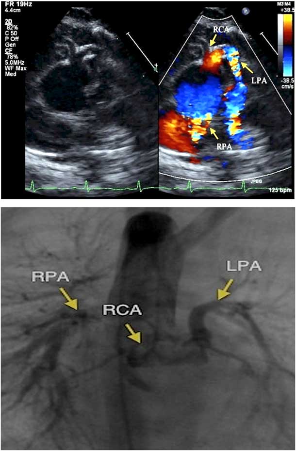
The child was born at term via spontaneous vaginal delivery with no complications. He was found to have oxygen saturations in the mid-80s. Subsequent transthoracic echocardiogram revealed usual atrial arrangement, concordant atrioventricular connections, right-hand topology, single overriding arterial trunk with an atretic pulmonary valve, and a right aortic arch with mirror image head and neck vessel branching. There was a large perimembranous ventricular septal defect with anterior–superior deviation of the outlet septum. Echocardiogram suggested an atretic right ventricular outflow tract anterior to the arterial trunk. The intrapericardial pulmonary arteries were non-confluent. The right pulmonary artery arose from the posterior aspect of the ascending aorta, above the sinuses, with moderate dilation. There was an indirect aortopulmonary collateral from a conal branch of the right coronary artery that connected to a diminutive pulmonary trunk, with a severely hypoplastic left pulmonary artery (Fig 1a and Supplementary Movie S1). Direct aortopulmonary collaterals also arose from the descending aorta, providing additional supply to the left lung. The right and left coronary arteries arose normally.
Top Image: Echocardiogram (parasternal short axis) shows a right pulmonary artery (RPA) arising directly from the posterior aspect of the ascending aorta and a pulmonary trunk being fed by a conal branch of the right coronary artery (RCA), via fistulous connection, continuing as a left pulmonary artery (LPA). Bottom image: Angiogram in the ascending aorta confirms a LPA arising from the RCA and a RPA arising from the posterior aspect of the ascending aorta.
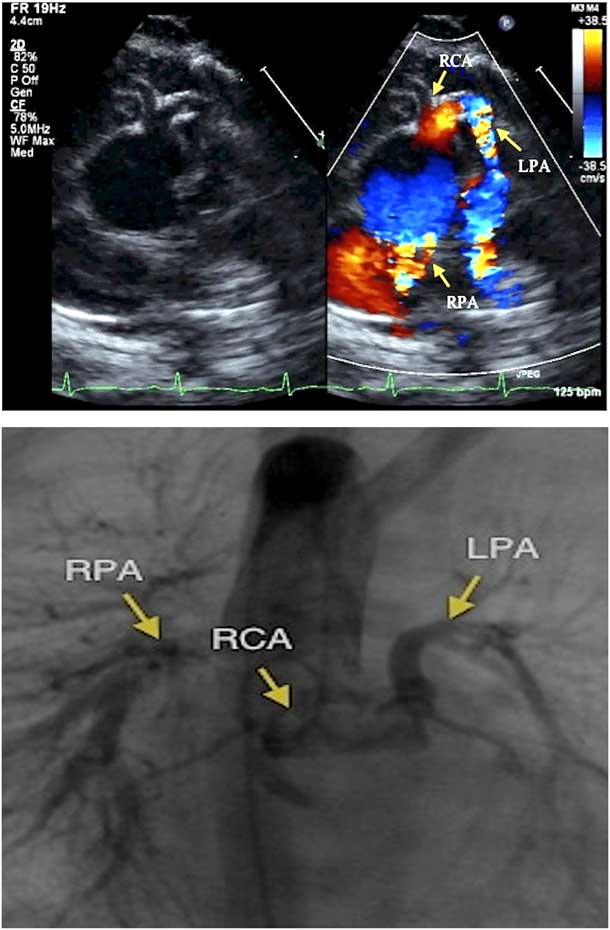
Angiography confirmed the echocardiographic findings. The right pulmonary artery arose from the posterior and rightward aspect of the ascending aorta with normal size and branching (Fig 1b and Supplementary Movie S2). The child underwent surgical repair at seven months—ventricular septal defect closure, right ventricle to pulmonary artery aortic homograft, and pulmonary artery unifocalisation. Surgical inspection revealed an atretic right ventricular outflow tract and pulmonary annulus below the hypoplastic pulmonary trunk.
Discussion
The presence of a membranous to outlet ventricular septal defect and an overriding arterial trunk, with absent antegrade flow from the right ventricle and discontinuous pulmonary arteries is consistent with tetralogy of Fallot with pulmonary atresia. The presence of an atretic right ventricular outflow tract and pulmonary annulus confirmed the completion of aortopulmonary septation, with division of the distal and intermediate parts of the developing outflow tract, but at the expense of the pulmonary component. Supply of the pulmonary arteries in our patient is especially interesting, given its implications on the embryogenesis of the discontinuous pulmonary arteries, suggesting abnormal protrusion of the dorsal wall of the aortic sac rather than the involvement of the sixth pharyngeal arch arteries.
In tetralogy of Fallot with pulmonary atresia, the branch pulmonary arteries can be discontinuous, but one of them usually remains connected to a remnant of the pulmonary trunk. Each lung is supplied by the branch pulmonary arteries via bilateral arterial ducts, or one lung can be supplied by a systemic to pulmonary artery collateral, while the other lung connects to an arterial duct. Systemic to pulmonary artery collaterals arise primarily from the descending aorta, but can originate from the ascending aorta and its branches.Reference Baker and Anderson 1 These collaterals are thought to be remnants of the splanchnic vascular system, which supplies the primordial lung beds. They regress later in foetal life unless the central pulmonary arteries fail to develop.Reference Liberthson, Sagar and Berkoben 2 With the non-confluent pulmonary arteries, pulmonary blood supply can also be supplied by coronary artery to pulmonary artery collaterals, but this is rare with tetralogy of Fallot with pulmonary atresia. Coronary aortopulmonary collaterals typically connect to confluent pulmonary arteries, instead of individual branch pulmonary arteries. In addition, coronary to pulmonary artery collaterals typically originate from the left coronary, not the right.Reference Amin, McElhinney and Reddy 3 , Reference Collison, Dagar and Kaushal 4
Recent studies of animal models have concluded the extrapericardial aortic sac is embedded within the pharyngeal mesenchyme, giving rise cranially to the third and fourth, and caudally to the sixth arch arteries. The developing extrapericardial pulmonary arteries are thought to originate from the mid-ventral portions of the sixth arch arteries. Definitive intrapericardial pulmonary and aortic outflow channels consist of three segments: intrapericardial arterial trunks distal to sinotubular junctions, arterial roots with their valves, and subvalvar ventricular outflow tracts. Fusion of the outflow tract cushion is thought to be required for formation of separate aortic and pulmonary roots, whereas protrusion of the dorsal wall of the aortic sac is responsible for intrapericardial separation of the aortic and pulmonary channels.Reference Anderson, Chaudhry and Mohun 5
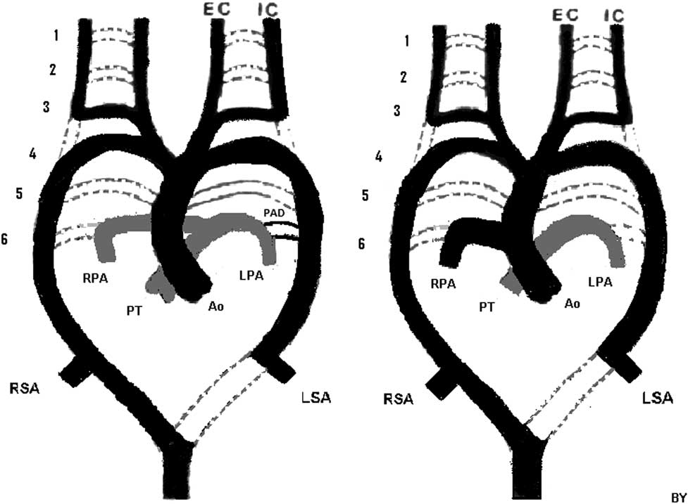
Anomalous origin of one branch pulmonary artery, arising directly from the ascending aorta, has been previously described, and is generally believed to result from incomplete leftward migration of the right pulmonary artery perhaps because of failure of migration of the right sixth pharyngeal arch.Reference Kutsche and Van Mierop 6 – Reference Odell and Smith 8 The sixth pharyngeal arches are responsible for connecting the primitive pulmonary arteries to the dorsal aorta (Fig 2a). The proximal left sixth arch is thought to connect the left central pulmonary arteries to the right pulmonary artery, while the distal part becomes the arterial duct.Reference Anderson, Chaudhry and Mohun 5 , Reference Bamforth, Chaudhry and Bennett 9
Left: Diagram demonstrating normal embryologic vascular development with a right aortic arch and a left-sided patent arterial duct. Right: Diagram demonstrating embryologic vascular development of our patient. Note that, in our patient, the distal part of the sixth arch did not appear to exist. Ao=aorta; EC=external carotid; IC=internal carotid; LPA=left pulmonary artery; LSA=left subclavian artery; PAD=patent arterial duct; PT=pulmonary trunk; RPA=right pulmonary artery, RSA=right subclavian artery.
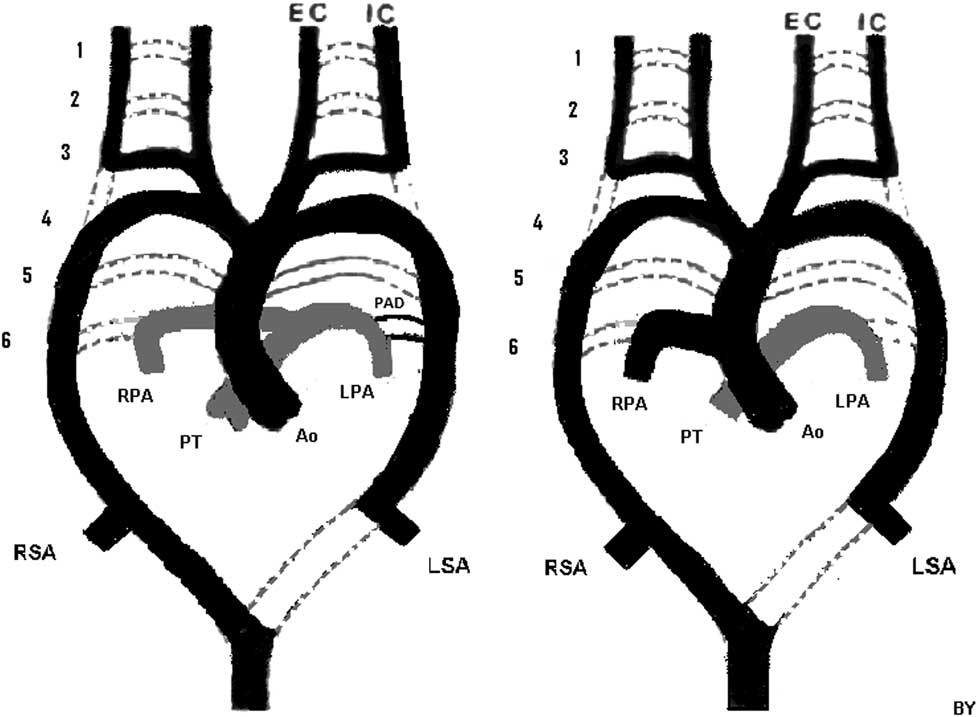
In our patient, the anomalous origin of the right pulmonary artery from the proximal ascending aorta, with no connection to the pulmonary trunk, might be because of incomplete migration of the right sixth arch towards the left sixth arch and the pulmonary trunk (Fig 2b). Given that the sixth arch arteries are themselves extrapericardial, however, migration failure of the sixth arch artery does not provide a satisfactory explanation for this anatomy. Instead, it is most consistent with abnormal migration of the aortopulmonary septum, also known as abnormal protrusion of the dorsal wall of the aortic sac. This abnormal migration leaves the right pulmonary artery in continuity with the intrapericardial aorta, instead of placing the right pulmonary artery in continuity with the aortic component of the extrapericardial aortic sac, following the separation of the aortic and pulmonary roots.
Acknowledgement
The authors thank Dr. Wyman W. Lai for his assistance with the echocardiographic images and manuscript preparation.
Conflicts of Interest
None.
Supplementary material
To view supplementary material for this article, please visit http://dx.doi.org/10.1017/S1047951114000341