INTRODUCTION
Human babesiosis is a tick-borne zoonosis caused by different species of Babesia. It is considered to be an emerging disease in the USA where B. microti is most often identified as the causative agent of the disease (Kjemtrup and Conrad, 2000). In Europe, human babesiosis occurs sporadically and most cases are attributed to the bovine parasite, B. divergens (see Kjemtrup and Conrad, 2000; Zintl et al. 2003; Gray, 2004). Recently, several new operational taxonomic units (OTUs) of Babesia (Babesia WA1, Babesia MO1, Babesia EU1, B. divergens-like parasite), causing human babesiosis in the USA or Europe, have been described (Quick et al. 1993; Herwaldt et al. 1996, 2003, 2004). In the last decade, the discovery of new zoonotic babesiae has been assisted via the use of molecular methods for the diagnosis of acute human babesiosis. Also, the use of such methods is providing important insights into enzootic Babesia not considered previously in Europe. For instance, molecular studies have shown that the tick Ixodes ricinus serves as a vector for B. microti, B. divergens and probably Babesia EU1 in Europe (Duh et al. 2001; Skotarczak and Cichocka, 2001; Foppa et al. 2002; Gray et al. 2002b; Kalman et al. 2003) and defined the reservoir hosts for B. microti and Babesia EU1 in Europe (Duh et al. 2003, 2005; Zintl et al. 2003; Karbowiak, 2004). Given the sporadic nature of human babesiosis in Europe, there are many questions regarding the transmission cycles of zoonotic Babesia (Kjemtrup and Conrad, 2000; Gray et al. 2002a; Hunfeld and Brade, 2004). Based on the few sero-epidemiological studies conducted to date, it has been suggested that, in Europe, human infections with Babesia may occur more often than previously documented (Foppa et al. 2002; Hunfeld et al. 2002). Therefore, further sero-epidemiological and molecular epidemiological studies are needed to establish the distribution, prevalence and medical relevance of zoonotic Babesia in different countries in Europe.
Indirect fluorescence immunoassay (IFA) kits for the detection of IgM and IgG serum antibodies against B. microti are commercially available (Fuller Laboratories, Focus (MRL) Diagnostics); they include reactive human serum as a positive control and instructions for the interpretation of positive results. However, no such tests have been developed or critically evaluated for the detection of specific serum antibodies against B. divergens in humans. For most sero-epidemiological studies, the IFA developed for the routine detection of anti-B. divergens antibodies (IgG) in cattle was adapted for the testing of human sera (Gray and Kaye, 1991; Hunfeld et al. 2002; Zintl et al. 2003). The aim of the present study was to determine whether the latter IFA is appropriate for the testing of human sera.
MATERIALS AND METHODS
Serum samples
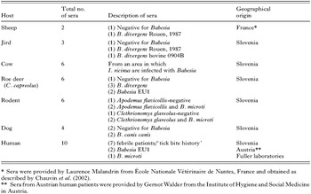
Serum samples were obtained from 7 different hosts including sheep, jird (Meriones unguiculatus), cattle, roe deer (Capreolus capreolus), wild rodents, dogs and humans (Table 1). The sheep and jird were experimentally infected with human or bovine B. divergens isolates, but other hosts acquired Babesia infection naturally.
Table 1. Description of sera used in this study (The species of Babesia were determined using a molecular approach.)

Propagation of Babesia divergens in jirds
Blood from jirds infected either with a bovine (0904B) or a human (Rouen, 1987) line of B. divergens were kindly provided by Laurence Malandrin. Both lines were adapted to in vitro cultures. To verify its virulence, each Babesia line was inoculated intraperitoneally into jirds (Malandrin et al. 2004). To produce antigen from each Babesia line, blood (400 μl; diluted 1[ratio ]600 in phosphate-buffered saline, pH 7·2; PBS) from an infected jird was inoculated into a parasite-free jird. After 5–7 days of infection, when a parasitaemia reached 30–40%, whole blood was taken from jirds and the B. divergens-infected erythrocytes were used for the preparation of the antigen.
IFAs for the detection of anti-Babesia divergens or anti-B. microti serum antibodies
The (15-well) slides for the B. divergens IFA were prepared essentially as described previously (Gray and Kaye, 1991; Hunfeld et al. 2002), but reducing the incubation times in IFA.
After extensive washing in PBS, the erythrocytes from parasitaemic jirds were suspended in this buffer (3-times the original blood volume). Each well was then filled with this suspension and the excess aspirated, leaving the thin, evenly distributed layer of erythrocytes, comprising at least 100 parasitized cells.
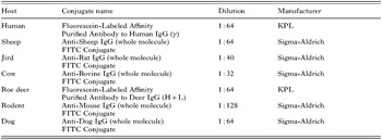
All sera were used at a dilution of 1[ratio ]16 and, in the case of seroreactivity, they were diluted serially in 2-fold steps to determine the end-point titres. Seven μl of each dilution were loaded on the slides, incubated at 37 °C in a humidity chamber for 30 min and washed 3 times in PBS. Different IgG fluorescein isothiocyanate (FITC) conjugates with different dilutions were used (see Table 2). Seven μl of FITC conjugate were applied to each well, incubated at 37 °C in a humidity chamber and washed as before. The slides were then examined for fluorescence using an Eclipse 80i microscope (Nikon Instruments) using a 400× magnification and photographed employing a digital camera CXM1200F (Nikon Instruments).

A commercially available B. microti IFA IgG Antibody Kit (Cat. No. BMG-120, Fuller Laboratories) was also used. The assay was carried out as described by the manufacturer. Slides were examined for the fluorescence and photographed in the same manner.
Blood smears and molecular methods
Thin smears were prepared from the blood of some animals and humans included in the study (see Table 3). The smears were air-dried, stained using the Diff-Quik Staining Set (Dade AG, Switzerland) and examined light microscopically at a 1000× magnification. For all samples, the presence or absence of Babesia was verified by PCR-coupled sequencing as described previously (Duh et al. 2003, 2005). In brief, a 407–435 bp portion of the small subunit of nuclear ribosomal RNA (18S rRNA) was amplified, column-purified and sequenced from both strands using a cycle sequencing kit (BigDye Terminator Cycle Sequencing Ready Kit, ABI PRISM, Applied Biosystems). The presence of Babesia EU1 in roe deer was confirmed after TOPO TA-cloning (Invitrogen) and sequencing of the complete 18S rRNA.
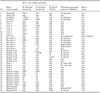
Table 3. Comparison of bovine and human Babesia divergens isolates in IFA used for the detection of B. divergens antibodies in sera from different host species (The results are compared with those achieved using molecular techniques and/or by blood smear examination. Legend: neg – negative result; pos – positive result; NA – not available; PC – positive control; NC – negative control; Sheep PC – inoculated with human B. divergens isolate; Jird PC1 – inoculated with human B. divergens isolate; Jird PC2 – inoculated with bovine B. divergens isolate; FCS – fetal calf serum; Af – Apodemus flavicollis, Cg – Clethrionomys glareolus; SLO – patient from Slovenia; AU – patient from Austria; Human PC1 – human patient infected with Babesia EU1; Human PC2 – reactive human serum as positive control from Babesia microti IFA IgG Antibody Kit (Fuller Laboratories).)

RESULTS
The 38 sera from different hosts were tested using the commercially and/or ‘in-house’ IFA assays for the detection of antibodies against B. divergens or B. microti (Table 3). B. microti-positive results (low titres) were obtained exclusively in the rodents with B. microti infection, as confirmed by molecular methods. The comparison of the 2 B. divergens antigens (human or bovine origin) in the ‘in-house’ IFA revealed the same number of ‘positive’ and ‘negative’ samples, but the end-point titres differed by up to 3-fold in naturally infected animals or humans and by up to 4-fold in experimentally infected animals. Higher end-point titres were obtained using the antigen prepared from the B. divergens of human origin.
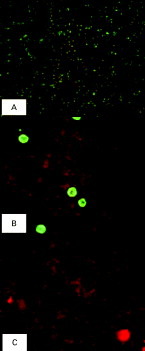
All (positive) control sera were determined as being positive using the specific fluorescence pattern shown for B. divergens (see Fig. 1). A positive reaction was represented by apple-green dots (~3 μm) of fluorescence within infected erythrocytes. The same fluorescence pattern was obtained using different test sera. This pattern differed from the only one described previously for B. divergens (see Hunfeld and Brade, 2004) in the size and number of fluorescence dots. The authors (Hunfeld and Brade, 2004) provided a photograph in which multiple, larger fluorescence dots are visible, resembling the pattern observed for non-specific fluorescence in this study (see Fig. 1). The pattern also differed from that characteristic for B. microti (cf. protocols from Fuller Laboratories).

Fig. 1. Fluorescence pattern obtained with IFA using a Babesia divergens isolate of human origin. Serum sample (no. 29, Table 3) was tested. Photomicrographs were taken using a Nikon Digital Camera (CXM1200F) at 400× magnification. (A) Small apple-green fluorescence dots indicating positive reaction; (B) non-specific fluorescence; (C) negative control.
Mostly, no serological cross-reactivity between B. divergens and B. microti (Table 3) was observed. The exception was a single serum sample from a rodent (no. 20, see Table 3). However, the serological cross-reactivity between B. divergens and Babesia EU1 was recorded for all 3 sera from the hosts infected with Babesia EU1 (as confirmed using molecular techniques). A serum from a human patient (no. 37, see Table 3) and sera from 2 roe deer (nos. 16 and 17, see Table 3) reacted to both B. divergens antigens used in the ‘in-house’ IFA.
DISCUSSION
Since B. divergens is a bovine parasite, the aim of most sero-epidemiological studies in Europe has been to establish its prevalence in cattle. IFA and ELISA have proved to be relatively sensitive, specific and reproducible employing (as an antigen) B. divergens derived from cattle or propagated in jirds (Gray and Kaye, 1991; Precigout et al. 1991; L'Hostis and Chauvin, 1999; Zintl et al. 2003). Although these tests have been extensively and successfully used for almost a decade, they have only recently been applied in a sero-epidemiological survey of B. divergens in a human population in Europe (Hunfeld et al. 2002). The results of the present study demonstrate that the evaluation and standardization of B. divergens IFA for human sera testing is critical. Utilizing antigens of B. divergens (of human or bovine origin), it was shown that, although the positive results of IFA using either of the antigens were concordant, the antibody titres differed. Without exception, the antigen obtained from human B. divergens isolate (Rouen, 1987) produced 2- to 4-fold higher end-point titres using the sera included. In a study of the seroprevalence of Babesia in humans in Germany, Hunfeld et al. (2002) selected a cut-off of 1[ratio ]128 using the bovine B. divergens antigen in the IFA. Based on the present study, the end-point titres in their study could have been even higher than described. Importantly, for the comparison of the results with future sero-epidemiological studies in Europe, it is recommended that B. divergens antigens of human and bovine origins should be employed (separately). Seroconversion or a 4-fold rise in IgG antibody titre is usually considered to indicate an acute infection when using serological detection methods. Given the difference in antibody titre between the bovine and human B. divergens antigens shown in the present study, it is proposed that only one of the antigens should be employed for testing human sera consecutively to avoid false negative or positive results.
Although the IFA is a useful serological method, the interpretation of the results is subjective and depends on the experience of the microscopist. Without the availability of a positive control serum or the corresponding fluorescence pattern, even an experienced microscopist will encounter difficulties in defining a positive result. Since the description of the first case of human babesiosis in Europe in 1957, only 23 cases have been attributed to B. divergens to date (Skrabalo and Deanovic, 1957; Kjemtrup and Conrad, 2000; Hunfeld and Brade, 2004). Given the small number of such cases, it is difficult to obtain positive human control serum with specific antibodies against B. divergens. Precise descriptions and photographs of fluorescence patterns will therefore assist researchers in interpreting B. divergens IFA results in human babesiosis cases. Herein, the photomicrographs as well as the description of fluorescence of B. divergens-positive sera from different hosts are recorded. Interestingly, the fluorescence pattern of B. divergens in our study differs from that of B. microti and from that published recently for B. divergens (see Hunfeld and Brade, 2004).
In this study, the sera from humans and various animals were tested. The sera from 6 cows and 5 human patients from Slovenia included in the study only had a serological response to B. divergens. No parasites or their DNA were detected in these samples using other approaches, which indicates that these cows and human patients most likely had a past infection with B. divergens. These findings suggest that IFA is a useful method for retrospective epidemiological surveys. Nonetheless, when using IFA for assessing the seroprevalence of B. divergens in humans, the serological cross-reactivity (as described herein) between different OTUs of Babesia needs to be considered. It has been reported previously that serological cross-reactivity between the two most important zoonotic Babesia (i.e. B. microti and B. divergens) is limited (see Zintl et al. 2003). However, Herwald et al. (2003) demonstrated cross-reactivity between B. divergens and Babesia EU1 in sera from 2 human patients diagnosed with babesiosis using a B. divergens antigen of bovine origin. Their results are supported herein, in that the serum from a Babesia EU1-infected patient (confirmed using molecular tools) reacted with the B. divergens antigen (of human origin). Also, it was shown here that the sera from roe deer infected with Babesia EU1 cross-reacted with both B. divergens antigens used herein. The Babesia EU1 infection of roe deer was confirmed by molecular methods, cloning and sequencing the complete 18S rRNA gene (Duh et al. 2005). Based on these findings, cross-reactivity between B. divergens and Babesia EU1 needs to be considered in future studies. Since attempts to isolate Babesia EU1 have been unsuccessful, no Babesia EU1 antigen has been available for serological studies in humans.
In conclusion, this study showed that the evaluation and standardization of a B. divergens IFA for testing human sera is critical and that different B. divergens antigens provide different end-point titres of antibodies. These aspects together with the serological cross-reactivity between B. divergens and Babesia EU1 need to be taken into account when interpreting IFA results.
We are grateful to Laurence Malandrin and Gernot Walder for providing us with both B. divergens isolates and Babesia EU1 positive human sera, respectively. We acknowledge with thanks the help of Tereza Rojko and Stanka Lotrič-Furlan from the Department of Infectious Diseases and Febrile Ilnesses, University Medical Centre Ljubljana, who supplied the human sera from Slovenian patients. We would also like to thank Janez Posedi from Veterinary Faculty in Ljubljana for providing us with the bovine serum samples.