Personal protective equipment (PPE) is essential to protect healthcare personnel and to prevent transmission of pathogens. However, in practice, personnel often use incorrect technique for donning and doffing PPE,Reference Tomas, Kundrapu and Thota 1 , Reference Zellmer, van Hoof and Safdar 2 leading to an increased risk for contamination of skin and clothing.Reference Tomas, Kundrapu and Thota 1 We and others have recently reported that simulations with fluorescent lotions or powders can be useful in training personnel in correct use of PPE.Reference Tomas, Kundrapu and Thota 1 , Reference Clay, O’Shea and Fletcher 3 – Reference Cassir, Boudjema, Roux, Reynier and Brouqui 4 The major value of using fluorescent lotions and powders for training is that they provide personnel with immediate visual feedback on skin and clothing contamination that can be used to correct deficiencies in technique.
Although fluorescent lotions or powders are useful tools, they have some limitations. Clothing, skin particulates, and dust can fluoresce under black light.Reference Clay, O’Shea and Fletcher 3 To avoid false-positive results, training of the staff evaluating contamination and assessment of personnel for fluorescence prior to each simulation is recommended. Using a colored rather than a white fluorescent tracer may also reduce the potential for false-positive readings.Reference Clay, O’Shea and Fletcher 3 Routine testing using fluorescent lotions or powders also does not provide images that document sites of contamination. Such images would be of value to provide documentation of training results and to provide a means to compare and validate findings obtained from multiple centers or from different personnel. Due to limitations of fluorescent tracers, we tested the hypothesis that a novel marker using retroreflective microspheres visualized using flash photography could provide an effective means to assess skin contamination of personnel during simulations of contaminated PPE removal.
METHODS
The Cleveland VA Medical Center’s Institutional Review Board approved the study protocol. During simulations of contaminated glove removal by healthcare personnel, we compared the frequency of contamination of the hands and wrists with bacteriophage MS2 and a reflective marker visualized using flash photography. Bacteriophage MS2 15597-B1 (American Type Culture Collection, Manassas, VA) is a nonpathogenic, nonenveloped RNA virus commonly used to study the spread of pathogens.Reference Tamimi, Carlino, Edmonds and Gerba 5 Bacteriophage MS2 was propagated in Escherichia coli 15597 as previously described.Reference Tomas, Kundrapu and Thota 1 In a previous study involving simulations of contaminated glove removal, we demonstrated that the frequency of skin contamination was similar for bacteriophage MS2 and a fluorescent lotion (52% and 58%, respectively).Reference Tomas, Kundrapu and Thota 1
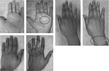
The reflective marker (3M, St. Paul, MN) contains ~60 µM diameter retroreflective microspheres with a refractive index of ~1.7–2 suspended in water that are designed to reflect incident light back to the source. The reflective marker can be visualized using flash photography; it is not visible in a picture taken with the flash off but is visible when the flash is on. Typical images of reflective marker contamination visualized on skin with flash photography but not visible in pictures taken with no flash are shown in Figure 1. Retroreflective microspheres may be composed of a variety of materials including glass, synthetic resins, or ceramic material; the retroreflective microspheres used in the current study were composed of soda lime glass. Retroreflective microspheres have previously been used to increase the visibility of traffic signs and clothing when light is shone upon them at night, but their use has not previously been reported for healthcare applications.
Contamination of skin with reflective marker visualized by flash photography. Left panels: without flash; right panels: with flash; circles indicate contamination.

In total, 50 simulations were performed in a research laboratory. Healthcare personnel donned and doffed contact isolation gowns (Safety Plus polyethylene gown, TIDI Products, Neenah, WI) and nitrile gloves (Denville Scientific, Metuchen, NJ) in their usual manner with no instruction on technique. Gloved hands were inoculated with 0.5 mL of phosphate-buffered saline (PBS) containing 107 plaque-forming units of MS2 and 0.5 mL of reflective marker. Study participants rubbed both solutions over their gloved hands until dry and then removed their gloves and gowns in their usual manner. Hand and wrist and clothing contamination with the reflective marker was then assessed using flash photography (ie, pictures were taken with and without flash). Volunteers then wiped both hands and wrists with a sterile, premoistened 10×10-cm (ie, 4×4 inch) gauze pad that was placed into a sterile container containing 10 mL of PBS and shaken in a vortex mixer for 1 minute to elute the bacteriophage. Aliquots of each elutant were serially diluted and cultured to quantify virus particles.Reference Tomas, Kundrapu and Thota 1 Agreement between hand and wrist contamination detected by fluorescent lotion and bacteriophage MS2 was assessed using Cohen’s κ.Reference Cohen 6 , Reference Landis and Koch 7 Data were analyzed using R statistics version 3.1.1.
Interrater reliability of contamination assessments was tested by having 2 research assistants assess 25 paired photographs taken with and without flash; agreement was assessed using Cohen’s κ. During the assessment, images of positive and negative controls were available for reference.
RESULTS
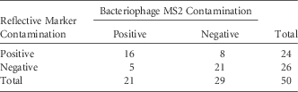
Table 1 shows the frequencies of contamination of hands and/or wrists with the reflective marker and with bacteriophage MS2. Of 50 simulations, 16 (32%) resulted in contamination of the hands and/or wrists with both bacteriophage MS2 and the reflective marker, 5 (10%) resulted in contamination with MS2 alone, and 8 (16%) showed contamination with reflective marker alone. The mean bacteriophage recovery for contaminated simulations was 0.74±0.9 log10 PFU (95% confidence interval [CI], 0.49–1.0). There was no significant difference in the overall percentage of contamination with the reflective marker and bacteriophage MS2 (48% vs 42%; P=.69). The Cohen’s κ of contamination assessed using the reflective marker and bacteriophage was 0.47, indicating moderate agreement based on Landis and Koch ranges.Reference Cohen 6 , Reference Landis and Koch 7
Comparison of Bacteriophage MS2 and Reflective Marker Contamination

Overall, 5 pictures without contamination and 19 pictures with contamination were correctly identified by the raters. Only 1 picture with contamination was read as positive by one rater and negative by the other. The inter-rater reliability in assessing contamination with reflective marker had a Cohen’s κ of 0.88 (95% CI, 0.6–1.0), indicating almost perfect agreement based on Landis and Koch ranges.Reference Cohen 6 , Reference Landis and Koch 7 No marker was visible on photographs taken with no flash. In addition, no false-positive readings were obtained on flash-photograph images of hands and wrists that had no exposure to the marker.
DISCUSSION
To effectively train personnel in the correct use of PPE, there is a need for simple tools that provide immediate visual feedback on skin contamination that can be used to correct deficiencies in technique. Our results suggest that the reflective marker tested here could provide one such a tool. In simulations of contaminated glove removal, we found that the frequency of contamination of hands and/or wrists was similar for the reflective marker and the nonpathogenic virus bacteriophage MS2. The marker was easily visualized with very good interrater reliability of contamination assessments. The likelihood of false-positive results appeared to be low, as no positive results were obtained when unexposed skin was assessed by flash photography. Finally, the photographic images provided using the reflective marker method could be valuable to document training and to validate results.
Our study has some limitations. The number of assessments was small, and we did not test the use of the reflective marker in simulations of full-body coverage PPE used in the care of patients with suspected or confirmed infection due to potentially fatal pathogens. We also did not assess whether training with the reflective marker would be effective in reducing contamination of personnel. In addition, we did not compare the reflective marker to a fluorescent lotion. However, the frequency of contamination with the reflective marker in the current study is similar to previous results with a fluorescent lotion.Reference Tomas, Kundrapu and Thota 1 Finally, we did not observe PPE donning and doffing to identify deficiencies in technique that might result in contamination.
ACKNOWLEDGMENTS
We thank 3M for providing the reflective surface markers and Marco Bommarito, PhD, from 3M for helpful discussions regarding the use of the marker. 3M did not have any role in the design of the study or interpretation of the results and did not provide funding.
Financial support: This work was supported by the Department of Veterans Affairs.
Potential conflicts of interest: C.J.D. has received research grants from STERIS, Clorox, Merck, AvidBiotics, and GOJO and has served on scientific advisory boards for 3M and Seres. All other authors report no conflicts of interest relevant to this article.