Clinical presentation and investigations
A 7-year-old girl with type 1 neurofibromatosis was referred to our tertiary children’s hospital with high blood pressure, intermittent leg pains, and diminished femoral pulses. On physical examination, she was not breathless and had no audible heart murmurs, but her femoral pulses were very weak. Her liver was not enlarged, and there were no other signs of right heart failure.
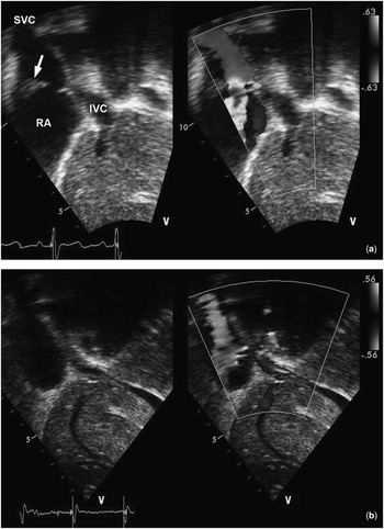
Echocardiography demonstrated a hypoplastic abdominal aorta, with a marked anterograde diastolic tail, suggestive of significant coarctation of the thoracic aorta. It also showed a membrane running across the posterior aspect of the right atrium, with a 5.7 mm opening into a larger more anterior chamber (Fig 1a). The inferior vena cava was almost completely occluded and there appeared to be collateral veins draining into the right atrium. A pressure gradient of 7.8 mmHg was estimated between the two right atrial compartments.
Echocardiogram showing the right atrial membrane ( a ), arrow and the right atrium following surgical resection of the membrane ( b ).

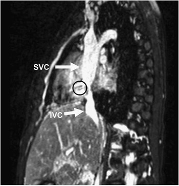
To further elucidate the findings, magnetic resonance imaging was performed. This confirmed the presence of a severe long-segment coarctation of the proximal descending aorta, with enlarged collateral vessels and post-stenotic dilatation of the aorta. It also confirmed the presence of cor triatriatum dexter, with severe narrowing of the inferior vena cava and venous return via the azygos and hemiazygos veins to the superior vena cava and the brachiocephalic veins, respectively (Fig 2).
Magnetic resonance imaging depicting the narrow inferior vena cava (IVC), large superior vena cava (SVC), and the right atrial membrane (encircled), with a small opening leading into the lower right atrial compartment.

In view of its location and extent, a decision was made to stent the coarctation segment via a right femoral artery catheter. This was successfully achieved using an 8 Zig 34 mm covered stent (NuMED Inc., Hopkinton, New York, United States of America). The pull-back gradient following stent placement was 3 mmHg, as opposed to a pre-procedural gradient of 48 mmHg.
Further catheter intervention was undertaken to dilate the stent, 15 months after the initial stent placement. On this occasion, echocardiography demonstrated increasing obstruction to the systemic venous return. Hence, the child was booked for surgical resection of the right atrial membrane.
Surgical intervention
The surgical approach was via a mid-line sternotomy. One striking finding on opening the pericardium was a severely dilated superior vena cava, more than twice the size of the inferior vena cava. Cardiopulmonary bypass was established using standard arterial and bicaval cannulas. The right atrium was incised following aortic cross-clamp and cardioplegia.
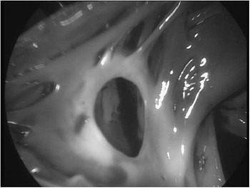
The entire posterior wall and part of the lateral wall of the right atrium were covered by thick fibro-muscular tissue, extending unto the Eustachian valve of the inferior vena cava and the coronary sinus. Superiorly, the fibrous tissue attached to the pectinate muscles of the right atrium, dividing the atrium in the process, with the superior vena cava emptying into the superior and more lateral chamber. There was a small, ∼6 mm, opening in the fibrous tissue providing the only communication between the two right atrial compartments (Fig 3). The inferior vena cava orifice was almost entirely occluded by the lesion.
The right atrial membrane as seen intra-operatively. Note the small opening between the two right atrial compartments.

The fibro-muscular tissue was extensively resected, the inferior venous cannula was temporarily removed, and intermittent circulatory arrest was employed to enable access into the inferior vena cava, where further resection of the thick fibrous tissue was carried out. Specimens of the tissue were sent to histology.
The child was uneventfully weaned from bypass and was transferred to the intensive care unit following chest closure.
Post-operative course
The immediate post-operative period was unremarkable. The patient was extubated a few hours after surgery, without any complications, and discharged home 2 days later.
Echocardiography at discharge showed a very good outcome, with no residual membrane within the right atrium, or the caval veins, and normal heart function (Fig 1b).
Histology sections of the tissue samples showed myocardium with overlying endocardium. The myocardium was fibrotic, whereas the endocardium showed marked thickening by fibro-elastic tissue. There were foci of myxoid tissue, adipose tissue, and a few lymphocyte collections. The appearances were consistent with an atrial membrane derived from endocardium.
More than 30 days after discharge, the child was re-admitted with large pericardial and right pleural effusions, requiring drainage via a mini-sternotomy. However, because of profound low cardiac output syndrome, extracorporeal membrane oxygenation was instituted, with complete recovery after 1 week of support.
After 2 years of the procedure, the patient remains well with no complaints and normal activity level, and the latest echocardiography shows no significant obstruction to the systemic venous return and no residual intra-atrial membrane.
Discussion
Atrial partitioning by fibro-elastic tissue or membrane is mainly associated with the left atrium and accounts for about 0.1% of all congenital anomalies.Reference Bin Mohd Zainudin, Koh and Mokhtar 1 Similar to the left atrium, the presence of a membrane in the right atrium has its roots in the early development of the heart. The sino-atrial orifice, which is formed when the right horn of the sinus venosus connects with the embryonic right atrium, is flanked on both sides by valvular folds – the right and left sinus venosus valves. The right sinus venosus valve, at a stage, divides the right atrium into two, forming a sheet that directs flow of oxygenated blood from the inferior vena cava to the left atrium, via the foramen ovale. This valve usually regresses by the 12th week of gestation, leaving behind the crista terminalis superiorly, the Eustachian valve of the inferior vena cava, and the thebesian valve of the coronary sinus. Failure of the right sinus venosus valve to regress, either totally or in part, leads to formation of cor triatriatum dexter.
Cor triatriatum dexter can occur in isolated forms, but has also been reported in association with other heart anomalies, mainly of the right heart, such as Ebstein’s anomaly,Reference Trakhtenbroit, Majid and Rokey 2 , Reference Gerlis and Anderson 3 pulmonary stenosis or atresia,Reference Aldawoodi, Arora and Kumar 4 and tetralogy of Fallot.Reference Udovičić, Biočić, Vincelj, Crnogorac, Sakić and Starčević 5 To the best of our knowledge, coexistence of cor triatriatum dexter with a typical left heart lesion like coarctation of the aorta, as seen in our case, has not been reported previously.
The clinical manifestation of cor triatriatum dexter depends, to a large extent, on the degree of obstruction to the systemic venous return. Hence, in the past, most less-symptomatic cases were only identified at necropsy. However, over the last couple of decades, the number of incidental findings has increased thanks to the availability of more sophisticated diagnostic methods.Reference Dobbertin, Warnes and Seward 11 , Reference Yarrabolu, Simpson, Virani, Arora, Navarijo and Stainback 12 In fact, there have been reports of prenatal diagnosis of cor triatriatum dexter.Reference Vigna, De Paola, Cignini and Padula 6
Symptomatic cases of cor triatriatum dexter are mainly treated by surgical resection of the fibro-muscular lesion on cardiopulmonary bypass.Reference Mazzucco, Bortolotti, Gallucci, Del Torso and Pellegrino 7 , Reference Ott, Cooley, Angelini and Leachman 8 There have been occasional reports of successful balloon dilatation of the membrane,Reference Savas, Samyn, Schreiber, Hauser and O'Neill 9 , Reference Bisinov, Dieter, Ballantyne, Wolff and Stein 10 but this, in our opinion, can only be employed in a certain group of patients, mostly those with less obstructive lesions and straightforward anatomy. In the more complex cases, like ours, where there is severe occlusion of the systemic veins, with large collateral vessels, surgery remains the most viable treatment option.
The extensive fibrous tissue found in our patient’s right atrium, and especially in the inferior vena cava, at the time of surgery initially fuelled speculations about a possible link with her type 1 neurofibromatosis. However, histology results obtained from all tissue samples were consistent with endocardium-derived membrane.
There has not been any established link between cor triatriatum and type 1 neurofibromatosis, and hence the coexistence of these two diagnoses, as seen in our patient, is most likely coincidental rather than causal.
Our patient did present with a late post-operative complication in the form of large pericardial and pleural effusions, more than 30 days after discharge. Following fluid drainage via a mini-sternotomy, she was in low cardiac output and required cardiac support for a few days. We do not have a clear explanation for this, although similar cases of “post-pericardiotomy syndrome” with massive pericardial effusion have been described, with patients presenting up to 6 weeks after initial surgery.Reference Imazio, Brucato and Rovere 13 It is also known that patients with large pericardial effusions are at a higher risk of developing low cardiac output or, in some cases, cardiac arrest on induction of general anaesthesia. We thought this was a case of severe post-pericardiotomy syndrome, from which the patient fully recovered after a few days of support.
Acknowledgement
The authors thank Mr Nicholas Geddes for processing the images used in this article.
Financial Support
This research received no specific grant from any funding agency, commercial or not-for-profit sectors.
Conflicts of Interest
None.