Brief report
A 28-year-old, Caucasian woman with a history of tricuspid atresia, ventricular septal defect, and normally related great arteries was palliated with a bidirectional Glenn and an atrial-pulmonary-type Fontan operation in early childhood. She was found to be newly hypoxaemic (room air oxygen saturation 85%); because of a desire to conceive, it was felt necessary to investigate into a possible right-to-left shunt.
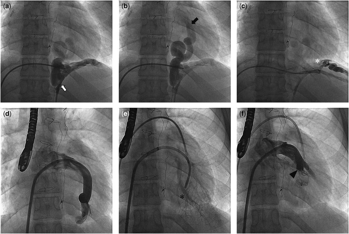
Angiography revealed a large, tortuous collateral vessel with drainage from the left hepatic, phrenic, and splenic veins (Figs 1a, b and c). This venous channel continued cephalad through the diaphragm, ultimately draining into the left lower pulmonary vein. As there were multiple tributaries to this collateral, closing the channel at its origin would be ineffective in eliminating all the tributaries, especially those from the splenic vein. Advancing a wire and a catheter from the inferior vena cava through the tributaries into the common channel was unsuccessful.
( a ) Origin of collateral vessel from the left hepatic vein with pronounced toruosity (white arrow). ( b ) Cephalad course of the collateral through the diaphragm draining into the left lower pulmonary vein (black arrow). ( c ) Another tributary to the collateral from the splenic vein (white asterisk). ( d ) Engagement of the collateral vessel after trans-septal puncture with a multipurpose guide showing entry into the left lower pulmonary vein. ( e ) Delineation of pulmonary venous return (black asterisk). ( f ) Occlusion of the collateral with an Amplatzer Vascular Plug II device (black arrowhead).

We opted to close this collateral at the site of drainage in the pulmonary vein. Trans-septal puncture with a Brockenbrough needle (Medtronic, Minneapolis, Minnesota, United States of America) through an 8F SL2 (Medtronic) sheath was performed using a standard technique under fluoroscopic and transoesophageal echocardiogram guidance. The left lower pulmonary vein was engaged with a Goodale Lubin (Medtronic) catheter, and then exchanged for an 8F multipurpose guide catheter (Cordis, Miami, Florida, United States of America) over an 0.035’ Magic Torque (Boston Scientific, Boston, Massachusetts, United States of America) guide wire (Fig 1d). Additional left femoral venous access was obtained to measure pulmonary capillary wedge pressures and Fontan pressures during occlusion. Angiography through this demonstrated that closure of this channel would not occlude any normal pulmonary venous return (Fig 1e). A 14-mm Amplatzer Vascular Plug II (St. Jude Medical, Plymouth, Minnesota, United States of America) was positioned to occlude the channel just proximal to the site of entry into the pulmonary vein (Fig 1f). The plug was placed as close to the point of entry into the pulmonary vein as possible to minimise chances of any thrombosis on the systemic side of the device. Fontan haemodynamics were repeated before device release, demonstrating unchanged filling pressures. Her saturations normalised without any increase in baseline Fontan pressures or evidence of cardiac output decrease. Anticoagulation with warfarin was carried out for 3 months with strict instructions regarding contraception until it was stopped. The patient has now been followed-up for 16 months after the procedure, and has had oxygen saturations recorded in the range of 95–100%.
Veno-venous collaterals in Fontan patients are a commonly encountered problem, and transcatheter closure has been widely described.Reference Sugiyama, Yoo, Williams and Benson 1 Usually the closure device/coils are deployed at the site of origin of the collateral channels; however, in some cases, especially with collaterals arising from infra-diaphragmatic sources such as the left hepatic vein, the anatomy may not be conducive to deploy devices. Such a case has been described by Kuo involving a venous collateral arising from the left hepatic vein with a right-angle turn, which did not allow catheter advancement to the collateral channel.Reference Kuo 2 This venous channel was accessed via a transhepatic puncture, and the channel was successfully closed with an Amplatzer Vascular Plug II device.
In our patient, the collateral channel appeared to arise from the left hepatic vein, but also had contributions from the phrenic vein and splenic veins. There were multiple “hairpin” turns that made catheter advancement through the collateral and into the common draining source impossible. In addition, the risk of incomplete closure or recurrence was significant; furthermore, we wanted to minimise the risk of thrombosis of the hepato-portal system as a complication of attempted coil/device embolisation of this channel. For these reasons, we decided to attempt closure at the site of drainage in the left lower pulmonary vein. This necessitated a trans-septal puncture. This patient’s atrial pulmonary type Fontan palliation allowed for straightforward trans-septal puncture; however, it may be possible to extrapolate this approach to lateral tunnel and extracardiac Fontan circuits as well. Uhm et alReference Uhm, Kim, Joung, Pak and Lee 3 have also published a series of eight patients with either lateral tunnel or extracardiac-type Fontan circuits who underwent trans-septal puncture for electrophysiology procedures. The techniques utilised include using the Brokenbrough needle or a radiofrequency trans-septal needle with a snare to limit translation of the needle along the conduit wall. In the Gore-tex extracardiac conduits, balloon dilation of the transconduit puncture was also needed. Although in most case series these techniques have been reported as safe, cyanosis from right-to-left shunting from the puncture is possible. Such a case was described by McCready et al,Reference McCready, Moon and Chow 4 which necessitated closure of the shunt with an Amplatzer Cribriform Occluder (St. Jude Medical).
Thus, we propose trans-septal approach as an alternative when closure of infra-diaphragmatic collaterals near the source presents difficulties as in our case.
Acknowledgement
The authors would like to thank to William S. McMahon, MD; Department of Pediatric Cardiology, University of Alabama at Birmingham.
Supplementary material
To view supplementary material for this article, please visit https://doi.org/10.1017/S1047951117000476