INTRODUCTION
Intensity-modulated radiotherapy (IMRT) in patients with cancer of the cervix or endometrium potentially reduces severity of acute/late side-effects compared with conformal radiotherapyReference Ahmad, Hoogeman, Quint, Mens, de Pree and Heijmen 1 but involves steeper dose-gradients less forgiving of organ motion. This increases risk of both overdose to organs at risk and geographical miss of planned target volume (PTV).Reference Taylor and Powell 2 Bladder filling for pelvic radiotherapy treatment is common practice to displace small bowel and rectum from high-dose target volumesReference Lim, Kelly and Stewart 3 and reduce PTV motion. Achieving consistent inter-fractional bladder volume is challenging, even using pre-defined fluid-loading guidelines, or ‘drinking protocols’.Reference Bondar, Hoogeman and Mens 4 Significant inter-fraction volume variation is common, along with a trend for bladder volume to decrease as treatment progresses.Reference Ahmad, Hoogeman, Quint, Mens, de Pree and Heijmen 1 Daily cone-beam computed tomography (CBCT) to monitor bladder fill and the resultant impact on PTV has been advocated for this patient group,Reference Haripotepornkul, Nath, Scanderbeg, Saenz and Yashar 5 while informative, this increases concomitant radiation dose, increasing risk of inducing secondary malignancies. We investigated the use of ultrasound bladder volume scanning as an alternative, non-invasive method of verifying bladder volume in this cohort before treatment without use of ionising radiation.Reference Ung, White, Mathlum and Lynch 6
MATERIALS AND METHODS
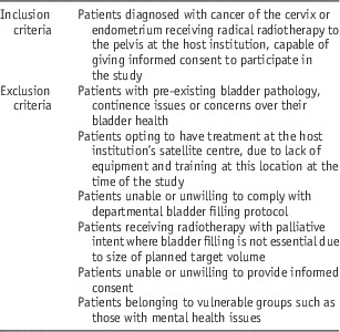
Ethical approval was granted following review by South West Frenchay Research Ethics Committee (reference 15/SW/0262). Participants were prospectively recruited over 9 months from a convenience sample of eligible patients (Table 1) receiving radical IMRT to the cervix or endometrium. All patients were supplied with a detailed study information leaflet well in advance of their first treatment to allow consideration of participation without duress.
Study eligibility criteria

20/33 (61%) of eligible patients were recruited; nine with cancer of the cervix (three post-hysterectomy) and 11 with cancer of the endometrium (eight post-hysterectomy). One patient (P10) withdrew consent to participate after their first week of treatment, declining to undergo daily ultrasound. We acknowledge as a study limitation that disease staging and grading varies greatly within the cohort, as does age range (25–79 years). Recruitment of a more homogenous population was not possible within the study time constraint of 12 months.
Treatment delivery
All patients were planned and treated supine, with standard headrest and indexed kneefix and footstocks. An iX series Varian (Varian Medical Systems Inc., Palo Alto, California, USA) linear accelerator was used to deliver 6 MV step-and-shoot IMRT with a seven beam arrangement. No routine bowel preparation was used, but glycerin rectal suppositories were given to remove gas when visualised on pre-treatment cone-beam computed tomography (CT). The host institution is currently investigating use of microenemas for routine bowel preparation in this cohort.
Acquisition of bladder scans
The ultrasound bladder volume scanner used during this study was the Cubescan BioCon-700 (Mcube Technology Co., Ltd, Seoul, South Korea). This model was selected due to already being in routine clinical use on the wards in the host institution, meaning the equipment could be redeployed if needed. Healthy staff volunteers were used during staff training sessions lasting ~30 minutes, following which, their competency was formally documented. Volunteers voided their bladder, drank 600 mL of water and then waited 40 minutes before ultrasound bladder volume scanning.
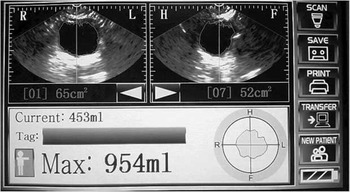
To perform a bladder volume ultrasound scan using the BioCon-700, gel is applied to the lower abdomen, and gentle pressure is applied with the probe. The probe is initially positioned midway between umbilicus and symphysis pubis, angled at ~45° inferiorly. The pre-scan view function on this model of bladder scanner enables bladder location before volume reading, indicating quality of capture and allowing optimisation of technique (Figure 1). The scanner uses an algorithm to derive bladder volume. ‘Male’ setting was used for post-hysterectomy patients, as the ‘female’ mode detects and compensates for the presence of a uterus.
Example of bladder capture on the BioCon-700. Image used with kind permission of De Smit Medical Systems Ltd. Bristol, UK/ Mcube Technology Co., Ltd. Seoul, Korea.

Achieving the planned volume range
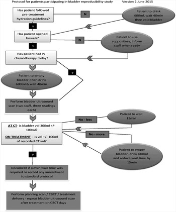
Patients were asked to achieve a bladder fill of 300±100 mL at CT, and then reproduce their planned CT volume ±100 mL during treatment, with all patients initially following a standard fluid-loading protocol (Figure 2). If bladder volume in the planned range was not achieved despite following the protocol, patients then modified their bladder volume (based on the ultrasound readings) until a volume in the acceptable range was achieved. Any required modifications to the standard protocol were recorded on the patient’s individual datasheet, for example ‘needed to increase fluid-loading intake by 100–700 mL to achieve planned fill’. Patients could decline to adjust their bladder volume at any stage. The only patient who declined to adjust volumes subsequently withdrew from the study in the first week (P10). Volume modification took place a maximum of three times in an attempt to minimise inconvenience to patients. If the required volume was still not achieved at this stage, CBCT was then performed to assess the bladder volume impact on PTV. Partial bladder voids were permitted if patients were physically capable. No fluid loading was performed following intravenous chemotherapy. Patients were initially bladder scanned and then corrective action taken based on the result.
Study protocol.

Method comparison
Two experienced radiographers contoured the outer bladder wall on treatment CBCTs, using planning software (Pinnacle; Phillips UK Ltd, Guildford, UK) to calculate bladder volume. A random sample of these contours (n=30) were retrospectively reviewed by both radiographers to address risk of intra-observer variation. No adjustments to original contours were performed. Inter-observer variation was evaluated using the Bland–Altman method.Reference Bland and Altman 7 The mean of the two contours for each individual scan (CBCTMean) was used as the standard measure to compare with the proposed new method (ultrasound bladder volume scanning). Three ultrasound bladder volume scans were performed in rapid succession by two different staff members (to reduce both intra- and inter-observer variation) to assess bladder volume before each treatment. The mean of these six readings (USMean) was compared using the Bland–Altman methodReference Bland and Altman 7 with the standard measure (CBCTMean) for 255 datasets. Statistical analysis was performed using MedCalc for Windows (Version 14.8.1) for Bland–Altman analysis. Additional graphs and tables were generated in Microsoft Excel (Microsoft, 2010).
Focus group methodology
Three months following introduction of ultrasound bladder volume scanning into practice, two staff focus groups (n=5) were selected by a facilitator from a convenience sample based on staff availability and skill mix. This included staff from both pre-treatment and treatment floor, although one staff member from pre-treatment failed to attend the second group, citing staffing issues. Informed consent for anonymous participation in a focus group was formally obtained. The questions for discussion at both sessions were identical, and were provided to participants in advance in an attempt to facilitate deeper reflection; however, we acknowledge that this degree of structuring during sessions may have restricted depth of discussion. Tape recordings were transcribed verbatim and coded into themes by the researcher. Transcripts were verified by the facilitator pre- and post-coding, and independently peer-reviewed by a colleague not involved in the groups. Participants were identified during sessions by number only.
RESULTS
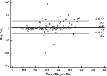
The two radiographers who performed bladder contouring for the standard measure were referred to as Rad X and Rad Y . Inter-observer variation for Rad X and Rad Y bladder contouring was calculated using the Bland–Altman methodReference Bland and Altman 7 for 255 datasets (Figure 3). The 95% limits of agreement and confidence levels are indicated, demonstrating strong agreement, with the mean of the differences 0·8±13·2 mL (1 SD).
Bland–Altman comparison of inter-observer variation for bladder contouring.

The radiographers performing the ultrasound readings before each treatment were referred to as Rad A and Rad B , however, due to staffing constraints, it was unfeasible for Rad A and Rad B to be the same two staff members daily. This means statistical analysis of inter-observer reliability is not possible, which we acknowledge as a study limitation. Intra-observer variation was calculated from 541 ultrasound readings, giving a co-efficient of variation 6·6%.
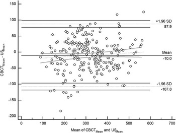
To compare the standard measure (measuring bladder volume from CT/CBCT in planning software) against the new method (measuring bladder volume using a bladder volume ultrasound scanner), the Bland–Altman method was used to compare CBCTMean – USMean against mean of USMean and CBCTMean (Figure 4). This gave a mean of the differences −10±49·92 mL (1 SD).
Bland–Altman comparison of cone-beam computed tomography (CBCT) with ultrasound showing regression line of differences and confidence intervals.

A total of 277 comparisons between the standard measure (the mean of two bladder volumes contoured on CT or CBCT) and the mean of the corresponding six readings from the ultrasound volume scanner were performed. Of these 277 comparisons, 255 were performed during treatment, comparing USMean with CBCTMean. A further 17 comparisons were made using the data collected before CT planning scans in pre-treatment, comparing the bladder volume on the planning CT scan with the mean of the six ultrasound readings taken immediately before planning CT (CTMean with CTUSMean). Mean USMean was 323 mL, mean CBCTMean 313 mL. Mean CTUSMean was 301 mL, mean CTMean was 332 mL. The 17 pre-treatment datasets provided too few comparisons to generate a meaningful Bland–Altman plot, but demonstrated a mean of differences −30·4±77·7 mL (1 SD); greater bias with more variance than USMean compared with CBCTMean.
Failure to perform or record 16 ultrasound readings during fractions where CBCT was performed meant omission of 9 USMean to CBCTMean comparisons. The reasons for failing to either perform scans or record results were not annotated by treatment staff. Two patients (P16 and P20) had superior PTVs, meaning ultrasound and CBCT comparisons were not possible during most fractions, as limitations on cone-beam scan length omitted the inferior bladder.
We set ‘a priori’ limitsReference Giavarina 8 of pre-treatment planned bladder volume ±100 mL of CT planned volume during this study. This volume range was considered both realistic for patients to achieve daily with minimal distress, and clinically acceptable regarding the potential impact on PTV based on previous clinical experience in the host institution. The majority of values when comparing the mean contoured bladder volumes from cone-beam CT with the mean bladder scanner readings were within the 95% confidence interval, meaning the methods are clinically interchangeable for the stated confidence levels. With the Bland–Altman method intended for comparison of two sets of independent data obtained on one occasion, validity may be compromised where repeated measures from individuals are performed.Reference Myles 9 Using the same data for multiple measurements per subject gives a mean of the differences −10±50·6 mL (1 SD).
Despite our initial study protocol including a post-treatment bladder ultrasound volume, this measurement was abandoned early in the study. P1–3 repeatedly declined this measurement due to a strong desire to urinate. We concluded any benefit of data collection for this measurement was outweighed by potential for patient distress.
Bladder volume versus time trends
Planned CT bladder volumes differed greatly throughout the cohort. The mean bladder volume was 320 mL, with a range of 161–508 mL (1 SD 111 mL). Three patients achieved sub-optimal planned fills of 190, 170 and 161 mL (P7, P10, P17). Repeated attempts to increase bladder volumes in these patients were unsuccessful (either by increasing fluid intake or time waited following fluid intake) and clinical decisions were made to accept these volumes, providing planning dose constraints could be met.
The ultrasound measurements recorded immediately before 479 fractions of treatments were analysed to determine changes in bladder volume over the course of treatment. These did not include the measurements recorded during corrective action to achieve planned bladder fill. Time and resource constraints during this study meant analysis of this volume of data was unfeasible, although this data could form the basis for future research. A total of 109 readings were missing from datasheets, with no explanation for omissions annotated by treatment staff.
Patients modified their bladder volumes up to a maximum of three times, with four modifications necessary for P15 on one occasion. This was a clinical decision due to known significant PTV displacements from underfilling. Of 479 fractions, 67 (14%) were delivered despite the bladder volume being outside the patient’s planned range. In these cases, clinical decisions were made to accept these volumes and CBCT performed to assess the impact of altered bladder volume on PTV. P10 withdrew consent to participate in week 1, achieving planned volume for 66% of treatment before withdrawal. P15 only achieved planned bladder fill for 44% of treatment due to non-compliance with drinking protocol. P18 only achieved bladder volumes within range for 36% of treatments, attributed to an excessive planned bladder fill of 508 mL. Volumes within patients’ planned range were achieved for 85% of all treatments.
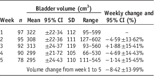
The population mean treatment bladder volume was 309 mL, with a range 66–602 mL (1 SD, 111 mL). Table 2 shows the mean cohort bladder volume variation over treatment. All patients followed pre-hydration guidelines to optimise hydration before attendance, then the drinking protocol on arrival (Figure 2). Fluid intake or time waited post-fluid loading was altered as per protocol to aid consistency of fill; however, the data regarding specific individual modifications was not analysed due to time constraints. Formal analysis of biofeedback was also not performed during this study. Patients were asked discreetly before entering the treatment room if they had followed protocol and felt as if they had a ‘comfortably full’ bladder. This was only used as a crude indicator, as it was noted by treatment staff on datasheets that most patients reported some degree of compromise in their ability to detect urinary urgency.
Variation in mean cohort bladder volumes over treatment

Focus group findings
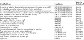
Analysis of data from the two staff focus groups identified a number of recurrent issues which emerged during implementation of ultrasound bladder scanning. These were grouped into six closely related themes, namely machine workflow, scanner availability, staff training, patient-specific factors, staff opinion, and impact on treatment verification and accuracy (Table 3). The issues identified by staff could be classed as either benefits or limitations when introducing ultrasound volume scanning into routine practice. Classing the broader themes in this simplistic manner was not possible, with several themes having both positive and negative aspects.
Issues and themes identified during focus groups

Abbreviations: CBCT, cone-beam computed tomography; CT, computed tomography.
DISCUSSION
Use of the ultrasound bladder volume scanner improved inter-fraction bladder variation, with only an 8% mean cohort volume reduction from week 1 to 5. Consistently poor filling in two patients was attributed to non-compliance with drinking protocol (P15) and excessive fill at CT making treatment volumes unachievable (P18). Staff experience with the ultrasound volume scanner was generally positive, with similar benefits and limitations being raised during both focus groups:

Wallice et al.Reference Wallice, Hickman, Zarkar and Webster 10 reduced requirements for CBCT from 18 to 2% by introducing bladder volume scanning. A reduction in requirement for CBCT was not quantified during our study due to absence of a control group. Retrospective data before implementation could be compared with this cohort in further research, however with 47% of treatments requiring at least one intervention after ultrasound to achieve acceptable bladder volume, we conclude that daily use of the BioCon-700 removed the requirement to repeat CBCT purely to verify bladder volume.
Mortenson et al.Reference Mortensen, Jakobsen, Edmund, Juhler-Nøttrup and Nordentoft 11 stated contouring outer bladder wall introduces systematic error, as ultrasound volume scanners only measure the fluid inside the bladder. Ahmad et al.Reference Ahmad, Hoogeman, Quint, Mens, de Pree and Heijmen 1 demonstrated statistically insignificant volume variation when comparing inner/outer bladder wall contours. During our bladder contouring, Rad Y verified the outer bladder wall contours created by Rad X with adjustments being made as necessary. We acknowledge that bladder wall contouring ideally should be performed independently, but time and resource restrictions made this unfeasible. Measuring voided urine to compare volume with ultrasound readings was also not performed during our study. This method is advocated as ‘gold standard’ by some,Reference Ahmad, Hoogeman, Quint, Mens, de Pree and Heijmen 1 however Small et al.Reference Small, Watson and McConnachie 12 stated even when measuring catheterised volumes, this method may have variable degree of accuracy. There is also potential for residual bladder volume to remain post-void in some individuals, adversely affecting accuracy of measurements.Reference Hynds, McGarry and Mitchell 13 In addition to concerns over accuracy, we felt that requesting our patients to void into receptacles had potential for causing distress, due to both female physical limitations and the requirement for repeat voids while optimising bladder volume using our protocol.
When comparing our work with literature in the area, as experienced by Haworth et al.,Reference Haworth, Paneghel and Bressel 14 heavy departmental workload frequently impacted adversely on our quality of data collection. Direct comparisons with other papers are mostly confounded due to study variations; most detail experience with prostate patients, with differing methodologies and statistical analyses. We took a pro-active corrective approach, using ultrasound volume scanning to facilitate modification of bladder volume before treatment. Comparisons with volume versus time trends from other studies are futile; we actively corrected volumes, whereas other studies acknowledged volume variations without necessarily rectifying them.Reference Ahmad, Hoogeman, Quint, Mens, de Pree and Heijmen 1 Our mean intra-patient standard deviation of 67 mL is smaller compared with other studies: 168 mL,Reference Ahmad, Hoogeman, Quint, Mens, de Pree and Heijmen 1 134 mL,Reference Hynds, McGarry and Mitchell 13 171 mLReference O’Doherty, McNair and Norman 15 ; although comparisons are again confounded by our volume corrections. Ahmad et al.,Reference Ahmad, Hoogeman, Quint, Mens, de Pree and Heijmen 1 reported mean of the differences −16±67 mL (1 SD) in 24 patients with cancer of the cervix using the BVI 6400 Bladderscan (Verathon Inc., Bothell, Washington, USA). We encountered smaller differences with less variance, −10±49·92 mL (1 SD) with a similar sample size (n=20). The reasons for this are unverified, although we suggest that the pre-scan feature on the BioCon-700 improved our ability to accurately identify and capture the full bladder volume, supported by the focus groups:

Bladder volume scanners were originally designed to detect residual volume in urinary retention rather than to accurately measure larger volumes; Stam et al.Reference Stam, van Lin, van der Vight, Kaanders and Visser 16 evaluated this concern, comparing the BVI 3000 Bladderscan (Verathon Inc.) with CT scans in 26 prostate patients, demonstrating strong correlation (R=0·95). Modern volume scanners are capable of detecting volumes up to 999 mL, meaning functionality has undoubtedly evolved. Wallice et al.Reference Wallice, Hickman, Zarkar and Webster 10 suggested scanner accuracy as ‘appropriate for radiotherapy purpose’, namely verification of approximate bladder volume confirming adherence to drinking protocol. Ung et al.Reference Ung, White, Mathlum and Lynch 6 agreed accuracy is acceptable in detection of large inter-fraction variation. The focus groups agreed the bladder volume scanner was appropriate for purpose:

Focus group discussion suggested measurements in certain patients were less straightforward than others:

Ahmad et al.Reference Ahmad, Hoogeman, Quint, Mens, de Pree and Heijmen 1 reported scanning difficulties in one patient due to obesity, leading to study exclusion. Despite known limitations of diagnostic ultrasound in obesity due to impedance of sound waves,Reference Alagiakrishnan and Valpreda 17 locating and measuring bladders in larger patients did not prove overly problematic during our study. Reduced accuracy of bladder volume scanning in post-operative patients was not definitively demonstrated, despite post-operative patients being excluded in other studies.Reference Stam, van Lin, van der Vight, Kaanders and Visser 16 The BioCon-700 user manual states, ‘Conditions to degrade the accuracy of the measurement: – when a patient has had supra-pubic or pelvic surgery’. Our results indicate the scanner largely remained fit for purpose even in our post-surgical patients; further work could seek to quantify this.
Hynds et al.Reference Hynds, McGarry and Mitchell 13 suggested a major flaw with drinking protocols is assumption of constant bladder fill after a set fluid intake. Ahmad et al.Reference Ahmad, Hoogeman, Quint, Mens, de Pree and Heijmen 1 predicted inflow rates of 9 mL/minute, while O’Doherty et al.Reference O’Doherty, McNair and Norman 15 demonstrated significant inflow variation over 35 minutes ranging from 0·9 to 9 mL/minute, supporting the hypothesis that rate of filling is individual, a finding reflected during the focus groups:

We found compliance with drinking protocol was not a guarantee of reproducible volume. Ung et al.Reference Ung, White, Mathlum and Lynch 6 suggested some causative factors for bladder volume variation during radiotherapy as individual patient hydration status, diurnal fluid balance variation, adequate pre-drinking void, varied renal re-absorption and impact of radiotherapy on inflow. Our study took a similar approach to O’Doherty et al.,Reference O’Doherty, McNair and Norman 15 and also followed the recommendations in Taylor and PowellReference Taylor and Powell 2 that drinking protocols be tailored to individuals. Compliance with drinking protocol was not formally assessed, although staff occasionally perceived lack of compliance when ultrasound demonstrated an empty bladder despite urinary urgency:

Post-operative patients informally mentioned reduced perception of desire to urinate, with several admitting reduction of water intake over continence concerns. Chang et al.Reference Chang, Yoon and Cha 18 noted individual biofeedback greatly varied even before onset of side-effects. We suggest conducting patient focus groups to further investigate compliance and give deeper insight into patient experience with drinking protocols, especially given that our healthy staff volunteers found the protocol physically challenging to follow during training sessions.
Nakamura et al.Reference Nakamura, Shikama and Takahashi 19 reported significantly greater inter-fractional bladder volume variations with larger planned volumes. The failure of P18 to reproduce planned volume on treatment supports the importance of achieving an appropriate bladder volume for the planning CT scan. O’Doherty et al.Reference O’Doherty, McNair and Norman 15 stated while smaller bladder volumes are more reproducible, volumes <250 mL made meeting planning constraints challenging, albeit in prostate patients. The lowest volume acceptable in our study protocol was 200 mL, however three volumes <200 mL (190/170/161 mL, P7, P10, P17) were accepted following clinical decisions based on physical inability to hold greater volumes. The permitted treatment volume range of CT±100 mL resulted in the lowest treatment bladder volume of 66 mL; this is undeniably low for a ‘comfortably full’ bladder protocol. We advocate caution when ultrasound volume scanning smaller bladders, and feel it is desirable to achieve daily volumes equivalent to or higher than planned volume wherever possible. Since the conclusion of this study, we have increased our lower bladder volume limit to a minimum CT fill of 250 mL, and a minimum fill of 50 mL below planned CT volume during treatment.
The focus groups indicated staff generally found the scanner intuitive:

The training requirements in order to achieve competency in use of bladder volume scanners appear controversial in the literature. Ung et al.Reference Ung, White, Mathlum and Lynch 6 suggested minimal training is needed, but then found lack of operator consistency compromised results. O’Shea et al.Reference O’Shea, Armstrong, O’Hara, O’Neill and Thirion 20 emphasised importance of training for reproducibility, but only used one individual to collect readings, risking bias. Ahmad et al.Reference Ahmad, Hoogeman, Quint, Mens, de Pree and Heijmen 1 used one ‘experienced’ individual to perform scans, but omitted the details of the training given to operate the scannner. Stam et al.Reference Stam, van Lin, van der Vight, Kaanders and Visser 16 suggested competency with bladder volume scanning was achievable within 15 minutes, in keeping with our findings of 30 minutes. We found that poor communication over the required level of competency, along with an initial lack of trained staff caused issues during implementation:

Despite some participants stating they initially lacked formal training, attributed to staffing issues, most staff felt confident in use of the scanner once it had been demonstrated by other trained staff members:

A key future training requirement was the need to develop confidence identifying empty bladders, acknowledged in Ahmad et al.Reference Ahmad, Hoogeman, Quint, Mens, de Pree and Heijmen 1 as being challenging. Staff frequently perceived a technique failure when scanning patients with empty bladders, leading to distress:

A period of supervised scans, as advocated in Hynds et al.Reference Hynds, McGarry and Mitchell 13 may have enhanced staff experience and improved confidence in use of the equipment:

Despite training issues, overall experience with the scanner was positive:

Timing issues related to implementing bladder volume scanning were not quantified during this study. Staff in both groups perceived that bladder scanning required additional time, but also recognised that our standard practice (performing CBCT, and then discovering unacceptable bladder volume) was ultimately more time-consuming:

Bladder scanning in a separate room to improve throughput on the treatment machine was revealed as common practice during the focus groups, but was poorly documented, possibly explaining some volume discrepancies between ultrasound/CBCT measurements:

O’Shea et al.Reference O’Shea, Armstrong, O’Hara, O’Neill and Thirion 20 reported that the average time to acquire bladder ultrasound volume measurements was <1 minute per patient. We found that staff initially lacked the confidence to acquire results this rapidly, but agree this time is realistic once familiar with ultrasound volume scanning technique.
Conclusions
Use of the Cubescan BioCon-700 allowed patients to modify their bladder volumes before radiotherapy treatment, resulting in greater inter-fraction consistency. Performing bladder volume scans allowed verification of planned bladder volume before any exposure to ionising radiation. Our cohort demonstrated minimal reduction in mean bladder volume over treatment of only 8·4% from week 1 to 5. We advocate commencing radiotherapy with a standard drinking protocol, and then making individual modifications to this protocol where required to compensate for patient-specific factors. Fluid loading is not required on fractions where delivery of intravenous chemotherapy has taken place.
The BioCon-700 was demonstrated to be clinically interchangeable with the standard measure for the stated limits of agreement, with a mean of the differences −10±49·92 mL (1 SD). Bladder volume scanning presented a non-invasive, well-tolerated method to verify inter-fraction bladder volume. Staff found use of the bladder scanner to be straightforward, with the technique being smoothly implemented into practice. Daily ultrasound bladder volume scanning is now performed as standard for all patients undergoing radiotherapy to the cervix or endometrium, with the intention of expanding its use for other pelvic radiotherapy patients in future.
Acknowledgements
Thanks to the consultants, radiographers and physicists of the Gynaecological Radiotherapy Group at Gloucestershire Oncology Centre, especially Dr Audrey Cook, Dr Radhika Counsell, Emma Charman, Clare Salmon, Naomi Bulmer, Ruth Bees and Sarah Burns. The Radiotherapy and Oncology team at Sheffield Hallam University for their help and guidance at all stages, in particular Dr Robert Appleyard. The Research and Development Team at Gloucestershire Hospitals NHS Foundation Trust, in particular Mark Walker for assistance during ethical approval, and Chris Foy for his statistical input. All those in the Radiotherapy team at Cheltenham General Hospital who helped with recruitment, data collection and participation in focus groups.
Financial support
This research received no specific grant from any funding agency, commercial or not-for-profit sectors. This study was sponsored by Sheffield Hallam University (design and management) and by Gloucestershire Hospitals NHS Foundation Trust (conduct). The sponsors have had no direct involvement in collection, analysis and interpretation of data, manuscript writing or the decision to submit the manuscript for publication, bar the support provided by the University during completion of a Master’s degree. The authors would like to thank Gloucestershire Fund for Oncology Centre Users and Supporters (FOCUS) who originally funded the purchase of the BioCon-700 scanner used during this study.
Conflicts of Interest
No conflicts of interest are declared.
Ethical Standards
The author asserts that all procedures contributing to this work comply with the ethical standards of the relevant national guidelines on human experimentation (NHS Health Research Authority) and with the Helsinki Declaration of 1975, as revised in 2008, and has been approved by the institutional committee (South West – Frenchay Research Ethics Committee, 15/SW/0262).