Introduction
The Caucasus is known as one of the few biodiversity hotspots in Europe (Williams et al. Reference Williams, Zazanashvili, Sanadiradze and Kandaurov2006), characterised by rich flora and fauna. The Caucasus Biodiversity Hotspot includes the territories of Armenia, Azerbaijan, Georgia (also known as Transcaucasia or the South Caucasus), and parts of Russia (represented by Ciscaucasia or the North Caucasus), Turkey, and Iran.
The land mollusc’s fauna of the Caucasus (318 species of Gastropoda, according to Walther et al. Reference Walther, Kijashko, Harutyunova, Mumladze, Neiber and Hausdorf2014) has been intensively studied over the last decades (Hausdorf Reference Hausdorf2000, Reference Hausdorf2001; Neiber et al. Reference Neiber and Hausdorf2015; Reference Neiber, Walther and Hausdorf2017; Reference Neiber, Walther, Kijashko, Mumladze and Hausdorf2022 etc.). At the same time, the nematode fauna associated with gastropods of this region has remained largely understudied.
Since 2017, several regions of the North Caucasus (a.k.a. Ciscaucasia) in the Russian Federation have been surveyed for the presence of gastropod-associated nematodes. Ciscaucasia is a vast territory with diverse landscapes (from plains to mountains) and climates (from semi-arid to warm and humid), and the gastropod and nematode faunas vary predictably from place to place. The areas chosen for the survey were those with the richest gastropod populations, including the Stavropol Upland and western and central parts of Krasnodar Krai, covering areas of different altitude, climate, and vegetation. Several similar surveys have been carried out in Europe over the last decade, and a number of new species have been described that originated from European land gastropods (Ivanova et al. Reference Ivanova, Panayotova-Pencheva and Spiridonov2013; Nermut’ et al. Reference Nermut’, Půža and Mráček2016a, Reference Nermut’, Půža, Mekete and Mráčekb, Reference Neiber, Walther and Hausdorf2017; Singh et al. Reference Singh, Couvreur, Decraemer and Bert2019; Ivanova et al. Reference Ivanova, Clausi, Sparacio and Spiridonov2019; Andrus et al. Reference Andrus, Rae and Wade2022). The pattern of gastropod–nematode associations was recorded in each study, and the set of taxa generally replicated that described by H. Mengert in Germany almost 70 years ago, and later described by S. Morand in France (Mengert Reference Mengert1953; Morand Reference Morand1988). To simplify, each survey recorded one to three species of opportunistic parasites of the genus Pellioditis Dougherty, 1953 (until the recent nomenclatural act by Tandigan de Ley et al. Reference Tandingan De Ley, Kiontke, Bert, Sudhaus and Fitch2023 considered as Phasmarhabditis Andrássy, Reference Andrássy1976); a common ectoparasite, Alloionema appendiculatum Schneider, Reference Schneider1859, and a number of much rarer and less abundant internal parasites such as Angiostoma Dujardin, 1845/Agfa Chitwood, 1935/Nemhelix Morand & Petter, 1986.
As the Caucasus region is considered to be an area of significant endemism (Williams et al. Reference Williams, Zazanashvili, Sanadiradze and Kandaurov2006), we expected the local nematode fauna to be distinctive. For comparative analysis, gastropod-associated nematodes from the Republic of Georgia (belonging to the Transcaucasian geographical region) were included in the analysis.
Materials and methods
Areas surveyed and gastropod material examined
Adygea
The Republic of Adygea is an enclave within the boundaries of Krasnodar Krai, located in its central region, south of the city of Krasnodar. The gastropods were collected in the deciduous foothill forests surrounding the Granite Gorge formed by the Belaya River (Nickel settlement, Merkulaevka village, by the Sibirka stream, 44°10’41'' N, 40°09’21'' E). In total, 533 individual gastropods identified as Deroceras caucasicum (Simroth, Reference Simroth1901), Deroceras sp., Helix albescens Rossmässler, 1839, Hygromiidae gen. sp., Fruticicola fruticum (O.F. Müller, Reference Müller1774), Krynickillus melanocephalus Kaleniczenko, Reference Kaleniczenko1851, Limacus flavus (Linnaeus, Reference Linnaeus1758), Metalimax varius (O. Boettger, 1824), Micropontica sp., Monacha cartusiana (O.F. Müller, Reference Müller1774), Oxychilus sp., Oxychilus translucidus (Mortillet, Reference Mortillet1854), Parmacella ibera Eichwald, Reference Eichwald1841, Pomatias rivulare (Eichwald, Reference Eichwald1829), Serrulina serrulata L. Pfeiffer, Reference Pfeiffer1847, and Scrobifera sp. were examined for the presence of parasites.
Krasnodar krai
In the Guam Gorge or Guamka (44°14’12''N 39°54’26'' E), in the upper reaches of the Kurdzhips River, collections were made in the deciduous foothill forests. In total, 245 individuals (Acrotoma sp., Caucasicola raddei (Kobelt, Reference Kobelt1880), Caucasigena sp., Clausiliidae spp., Deroceras sp., Helicopsis dejecta (Rossmässler, Reference Rossmässler1838), Helicopsis sp., Helix lucorum (Linnaeus, Reference Linnaeus1758), Fruticicola fruticum, Pomatias rivulare, Krynickillus melanocephalus, Serrulina serrulata, Quadriduplicata sp.) were collected.
In Goryachiy Klyuch (Hot Spring) (44ᵒ42’06” N 39ᵒ13’02” E), a settlement with a balneological resort with thermal springs in the rocky valley of the Psekups River, 147 gastropod specimens (Acrotoma sp., Boetgerilla pallens Simroth, 1912, Clausiliidae spp., Metalimax varius, Oxychilus sp., O. translucidus) were collected from forested areas outside the populated area.
In the Bolshoi Utrish nature reserve, near Anapa and the Black Sea coast at its eastern point 44ᵒ43’52” N 37ᵒ25’30”E, gastropods were collected in the foothill forest of oak, beech, pine, and juniper at the first level of the Navagir ridge (150 asl); in total, 17 specimens were collected (Deroceras sp., Clausiliidae spp., Monacha cartusiana, Fruticicola fruticum).
Stavropol krai
Collections were made in Georgievsk, Terskiy, and Pyatigorsk. In Georgievsk district (44°11’295'' N, 43°30’500'' E) collections were made on the banks of the Podkumok River at 239 m asl, in the ash/hornbeam forest, at Safonova dacha (44°11’02'' N 43°30’13''E), and in city parks. In total, 192 gastropod specimens were collected and dissected (Deroceras caucasicum, D. reticulatum (O.F. Müller, Reference Müller1774), Chondrula tridens (O.F. Müller, Reference Müller1774), Circassina frutis (L. Pfeiffer,Reference Pfeiffer1859), Clausiliidae spp., Helix sp., Hygromiidae gen. sp., Monacha sp., P. rivulare, Quadriplicata sp., Xerolenta obvia (Menke, Reference Menke1828), Zonitidae gen. sp.).
At Terskiy (44°11’40'' N 43°18’49'' E) collections were made on roadsides and in wild vegetation near agricultural plots. In total, 36 specimens (D. reticulatum, Caucasigena rengarteni (Lindholm, Reference Lindholm1913), Circassina frutis, Cochlodina laminata (Montagu, 1803), H. albescens, Hygromiidae gen. sp., L. flavus, Serrulina serrulata, Vitrinidae gen. sp., Zonitidae gen. sp.) were dissected. In Pyatigorsk (44°02’56'' N 43°04’14'' E) collections were made on the wooded slopes of Mashuk Mountain and in city parks. In all, 56 specimens (Cochlodina laminata, Cochlicopidae gen. sp., D. caucasicum, Helix lucorum, H. christophi Boettger, 1881, Limacus flavus, Succinella oblonga (Draparnaud, 1801) and Xerolenta obvia) were dissected.
Nematode material
Live gastropods were brought to the lab and identified by dissection and morphological examination. Extracted parasites were preserved for morphological and molecular studies by preservation in hot 4–5% formalin and frozen for molecular work. Adult stages of Pellioditis were obtained by culturing juveniles on nutrient medium (pre-frozen gastropod flesh or pig kidney).
Nematodes preserved in formalin were processed with anhydrous glycerin for light microscopy as described by Seinhorst (Reference Seinhorst1959). Light microscopic studies and drawings were done using a Nikon Eclipse 200 microscope (Nikon Corporation, Tokyo, Japan) equipped with a drawing attachment. Scale bars are given in micrometres (μm). Abbreviations: V% – distance from anterior extremity to vulva to body length in %; a, b, c – de Man indices. Illustrations were prepared using WACOM Intuos A4 USB drawing tablet (Wacom, Kazo, Japan) and Adobe Illustrator CS5. For the SEM studies, formalin-preserved material was dehydrated in a graded ethanol series, critical point dried using an HCP-2 HITACHI dryer (Hitachi Ltd, Tokyo, Japan), mounted on aluminum stubs, and coated with gold in a BIO-RAD SC502 sputter coater (Bio-Rad Laboratories Inc., Hercules, USA). Images were taken on a Mira 3 Tescan (Tescan Orsay Holding, a.s., Brno, Czech Republic).
Crossbreeding test
One male and 2–4 females from the Gorjachiy Klyuch (thermalis) and Utrish (sindicae) populations were placed in a Petri dish with a thin layer of water and a pinhead-sized piece of pig kidney. Nematode reproduction was observed for one week.
Molecular characterisation and phylogenetic analysis
DNA extraction from the discovered nematodes was done through digestion with proteinase K in the worm-lysis solution containing mercaptoethanol (Holterman et al. Reference Holterman, Van der Wurff, Van den Elsen, Van Megen, Bongers, Holovachov, Bakker and Helder2006). Single specimens of a female, male, and infective juvenile were used. The nematodes in the solution were incubated at 65 °C for 90 min, followed by deactivation of proteinase K at 99 °C for 5 min.
Two pairs of primers were used to amplify the small subunit ribosomal DNA (SSU rDNA) (Blaxter et al. Reference Blaxter, De Ley, Garey, Liu, Scheldemann, Vierstraete, Vanfleteren, Mackey, Dorris, Frisse, Vida and Thomas1998; Medlin et al. Reference Medlin, Elwood, Stickel and Sogin1988): G18S4 (5’-GCT TGT CTC AAA GAT TAA GCC-3’) and 26R (5’-CAT TCT TGG CAA ATG CTT TCG-3’) for the 3’ portion of this sequence, and 24F (5’-AGR GGT TGA AAT YCG TGG ACC-3’) and Q39deg (5’-TAA TGA TCC WTC YGC AGG TTC ACC TAC-3’) for the 5’ flank of the SSU rDNA. The primer Q39deg was modified by V.V. Aleshin (personal communication). Polymerase chain reaction (PCR) cycling parameters for the first pair were: primary denaturation at 95 °C for 5 min followed by 35 cycles of denaturation at 94 °C for 30 s, annealing at 52 °C for 30 s, and elongation at 72 °C for 60 s followed by final elongation at 72 °C for 5 min. PCR parameters for second pair were 95 °C for 5 min followed by 35 cycles of denaturation at 94 °C for 60 s, annealing at 53 °C for 90 s, and elongation at 72 °C for 90 s, followed by final elongation at 72 °C for 5 min.
The pair of primers D2A (5’-ACA AGT ACC GTG AGG GAA AGT TG-3’) and D3B (5’-TCG GAA GGA ACC AGC TAC TA-3’) was used to obtain the D2-D3 expansion segment of the large subunit ribosomal DNA (LSU rDNA) (Nunn Reference Nunn1992) with PCR cycling parameters: primary denaturation at 95 °C for 5 min followed by 35 cycles of denaturation at 94 °C for 45 s, annealing at 50 °C for 1 min and elongation at 72 °C for 60 s. Primers CO1F1 (5’- CCT ACT ATG ATT GGT GGT TTT GGT AAT TG-3’) and CO1R2 (5’- CGT GCT TAT TTT ACT GCT GCT AC-3’) were used to obtain the sequence of the cytochrome oxidase subunit I (Cox1 mtDNA) (Kanzaki & Futai Reference Kanzaki and Futai2002). The protocol for the reaction included primary denaturation at 95 °C for 5 min followed by 35 cycles at 95 °C for 45 s, 59 °C for 50 s, 72 °C for 60 s, and final elongation at 72 °C for 5 min. To amplify the ITS rDNA region of these two species, a pair of primers TW81 (5’-GTT TCC GTA GGT GAA CCT GC-3’) and AB28 (5’-ATA TGC TTA AGT TCA GCG GGT-3’) was used. PCR cycling parameters were: primary denaturation at 95 °C for 5 min followed by 35 cycles of denaturation at 94 °C for 45 s, annealing at 56 °C for 1 min, and elongation at 72 °C for 60 s, with final elongation at 72°C for 5 min. As direct sequencing of this ITS product was impossible because of multiple peaks in the middle of the ITS1 region, the product was cloned in pGEM-T vector and transformed into JM109 High Efficiency Competent Cells (Promega®, Leiden, The Netherlands) according to the manufacturer’s instructions. PCR products from clones were sequenced with the same primers as used for primary amplification (TW81 and AB28). Five clones from each of two species were sequenced to confirm polymorphisms. Obtained sequences were deposited in NCBI Genbank. For Pellioditis thermalis n. sp. strain sindicae from Utrish, the obtained sequences were deposited as OR373882 for the partial SSU rDNA sequence; OQ102437 for the partial LSU rDNA sequence; OQ103047 for the partial sequence of Cox1 mtDNA; OQ103124 and OQ103319 for the two haplotypes of ITS rDNA. For Pellioditis thermalis n. sp. strain thermalis from Gorjachiy Klyuch, the obtained sequences were deposited as OR373881 for the partial SSU rDNA sequence; OQ102435 for the partial LSU rDNA sequence; OQ103053 for the partial sequence of Cox1 mtDNA; OQ103318 and OQ103320 for the two haplotypes of ITS rDNA. For comparative purposes and the phylogeny construction, relevant sequences from the NCBI GenBank database were used. Sequence alignments were generated using Clustal_X (Thompson et al. Reference Thompson, Gibson, Plewniak, Jeanmougin and Higgins1997) under default values for gap opening and gap extension penalties. All alignments were analysed using Mega X (Kumar et al. Reference Kumar, Stecher, Li, Knyaz and Tamura2018) for maximum parsimony (MP), neighbour joining (NJ), and maximum likelihood (ML) methods. Bayesian Analysis (BA) was performed on an IQ-TREE web server (Trifinopoulos et al. Reference Trifinopoulos, Nguyen, von Haeseler and Minh2016).
Results
We found two strains of the new species of the genus Pellioditis at two sites in Krasnodar krai (Gorjachiy Klyuch and Utrish, thermalis and sindicae, respectively), which revealed a number of morphological differences between them. The morphometric data for these strains are presented separately.
Description of Pellioditis thermalis n. sp.
Measurements
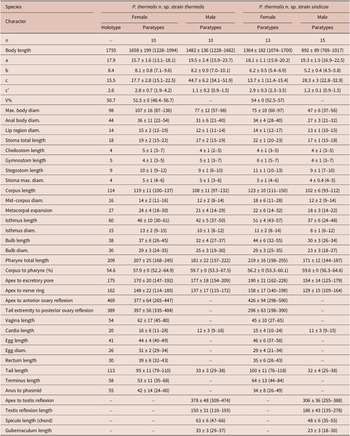
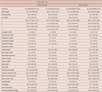
Morphometric data on adults and dauer juveniles of P. thermalis n. sp. are provided in Tables 1 & 2, respectively.
Morphometric data of adults of Pellioditis thermalis n. sp. strains thermalis and sindicae from the North Caucasus. All measurements are in μm and in the form: mean ± s.d. (range)

Morphometric data of dauer juveniles of Pellioditis thermalis n. sp. strains thermalis and sindicae from the North Caucasus. All measurements are in μm and in the form: mean ± s.d. (range)

Description
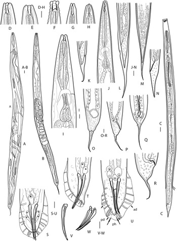
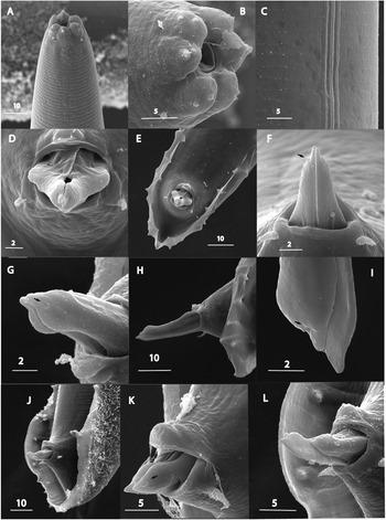
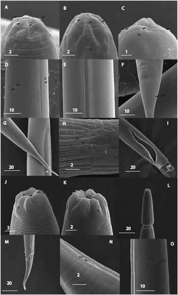
Adults. Body robust, 0.9–1.7 mm long, straight when relaxed, slightly tapering to anterior end. Cuticle about 1 μm thick, bearing rows of transversal and longitudinal striations. Lateral field c. 10–12 μm wide at mid-body, bearing three ridges and four incisions (Figure 3C). Deirid located at level of excretory pore (Figure 4E); structure identified as postdeirid located at lateral field in one specimen c. 15 μm posterior to deirid (Figures 4A and 4D). Head rounded to truncate, lip region short, sometimes slightly offset (Figures 4B and 4C). Six distinct, slightly pointed lips, each lip bearing distinct labial papilla in apical position (Figures 3A, 3B, and 4B). Four less prominent cephalic papillae in sublateral position on dorsal and subventral lips, sometimes indistinct (Figures 3B and 4B). Amphids prominent with narrow convex rim situated in sublateral position on lateral lips (Figure 4C). Mouth aperture triangular (Figure 4B). Stoma tubular, wide and short (about as long as lip region diam.) (Figures 2D–F). Cheilostom not cuticularised. Gymnostom walls parallel, thickened. Stegostom with glottoid apparatus, isomorphic, isotropic, metarhabdions thickened, with three minute warts each (Figures 2D–F). Pharyngeal collar narrow, covering two thirds of stoma length. Cheilostom: gymnostom: stegostom ratio c. 1:1:2.2. Pharyngeal collar narrow, covering two thirds of stoma length (Figures 2D–F). Pharynx muscular, differentiated into corpus expanded posteriorly (median bulb), stout, well defined isthmus and pear-shaped terminal bulb as wide as metacorpal expansion (Figure 2I). Terminal bulb with valve and haustrulum. Nerve ring surrounding middle to posterior region of isthmus. Excretory pore position at mid-isthmus. Excretory duct short. Cardia prominent, projecting into intestine. Intestine well developed, forming proventriculus at anterior. Rectal glands present.
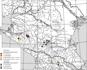
Distribution map of Pellioditis species in the Caucasus region.

Pellioditis thermalis n. sp. (A) Entire female (endotokia matricida), lateral view; (B) entire female with eggs, lateral view; (C) Infective juvenile shedding extra cuticle, lateral view; (D–H) Head (D, female, sublateral view; E, Male, subdorsal view; F, Female, matricidal hatching specimen, subdorsal view; G, Exsheathed infective juvenile, lateral view; H, ensheathed juvenile, lateral view); (I and J) Pharyngeal region, lateral view (I, female; J, exsheathed juvenile); (K–M) Ensheathed infective juvenile, tail, lateral (K–L) and dorsal (M) view; (N) exsheathed infective juvenile, tail, lateral view; (O–R) Female, tail (O, K, ventral view, P, R, lateral view); (S–U): Male, tail (S, U, ventral view and T, lateral view); (V) spicule, lateral view; (W) spicules and gubernaculum, lateral view. A, C, E–G, I–L, O, P, S–V thermalis strain; B, D, H, Q, R, W sindicae strain. Abbreviations: ad, anterior dorsal genital papilla; pd, posterior dorsal genital papilla; ph, phasmid. (Scale bars 10 μm.)

Pellioditis thermalis n. sp. SEM images. Male. (A) Sublateral view of anterior body region; (B) Head, subapical view; (C) Lateral field; (D) Cloacal region, ventral view; (E, J) Tail, ventral and sublateral view; (F, G, I) Spicule tips, dorsal and lateral view; (H, K, L) Cloacal region, lateral view. Arrow indicating hole at spicule tip. A–C and J–L sindicae strain; D–I thermalis strain. (Scale bars in μm.)

Pellioditis thermalis n. sp. SEM images. Female. (A) Sublateral view of anterior body region, arrows indicating deirid and posdeirid; (B) Head, subapical view; (C) Head, sublateral view; (D) Anterior body region showing deirid (d) and postdeirid (pd), sublateral view; (E) Anterior body region showing deirid (d) and excretory pore (eo), ventral view; (F) Vulva, subventral view; (G, H, K, L) Mid-tail region showing phasmids; (I, J) Tail. Abbreviations: a, amphid; cp, cephalic papilla; lp, labial papilla; ph, phasmid; A, D, F and J–L sindicae strain; B, C and G–I thermalis strain. (Scale bars in μm.)

Female. Body 1.0–2.0 mm long, straight after fixation. Oviparous, viviparous (Figures 2A–B). Reproductive system typical for the genus: didelphic, amphidelphic, ovaries reflected on dorsal side. Anterior and posterior branches almost equal in length. Oocytes rounded, large, arranged in two or three rows. Oviducts short. Spermathecae and uteri with numerous large sperm cells c. 10 μm in diameter. Uteri spacious. Eggs with thin smooth shell. Vagina straight, muscular, more than half corresponding diameter long. Vulva median, a wide transverse slit with thickened lips (Figure 4F). Copulatory plug present in fertilised specimens. Rectum c. corresponding diam. long (Figures 2P and 2R). Anus an arcuate slit (Figures 4I and 4J). Tail elongated, initially widely conical with whip-like terminal part set by constriction to dome-shaped with spike slightly exceeding the rounded part in length (Figures 4I, 4J, and 4L). Phasmids rod-like, protruding, located at junction of tail parts or at the base of rounded part or on terminal spike in 2 μm from to its base (Figures 4G–L). Protruding part of phasmid c. 3 μm long.
Male. Slightly shorter and slimmer than females. Corpus to pharynx ratio slightly exceeds that of females (average 59.6 vs 57.6). Testis reflexion in c. three body diam. posterior to pharynx base. Spermatocytes in testis reflexion rounded, arranged in two rows; reflexion short (c. 150 μm). Immature sperm in vas deferens c. 8–10 μm x 10–12 μm and sperm cells in ejaculatory duct c. 5 μm in diam. Anal glands large. Tail short. Bursa open, peloderan (Figures 2S–U, 3E, and 3J). Arrangement of circumcloacal papillae typical for the genus: unpaired ventral median papilla in front of anus. Two prominent circumcloacal papillae located at lateral margins of anus (Figures 3D, 3F, and 3K). Nine pairs of bursal papillae: Genital papilla (GP) formula 1 + 1 + 1 + 2/1 + 3 + phasmids (Figures 2S–U; 3E, and 3J). GP1 slightly shorter than the rest of papillae, situated latero-ventrally, distanced from following GPs. GP 2 and 3 opened on lateral edge of bursa, GP 4 and 5 grouped, and GP5 opens dorsally, GP 5 and 6 not reaching edge of bursa; GP 7–9 grouped with GP 7 and 9 not reaching edge of bursa, GP9 opening dorsally (Figures 2S–U, 3J, and 3L). Phasmids papilliform, situated closely to tail tip (Figures 2S and 2U). Tail tip not reaching the edge of bursa (Figures 2S, 2U, and 3E). Spicules nearly straight (Figures 2V, 2W, and 3H). Velum nearly indiscernible. Distal tips with holes (Figures 3G, 3I, 3K, and 3L). Manubrium not prominent, not distinctly separated from blade. Gubernaculum without processes, about half as long as spicules (Figure 2W).
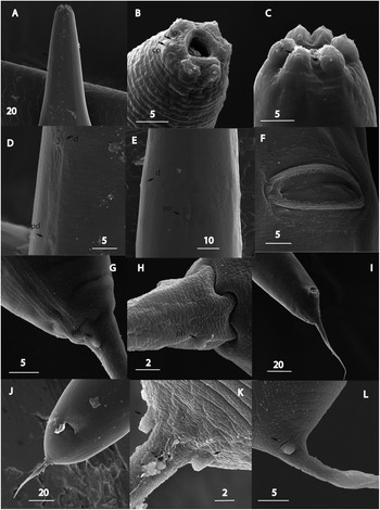
Infective juvenile (ensheathed). Body 717 μm long on average in thermalis strain and 771 μm in sindicae strain, slender, tapering gently towards head end, straight when heat killed (Figure 2C). Lateral field c. 10 μm wide at mid-body; composed of narrow central band flanked with three closely situated ridges by each side (Figure 5E) but a lateral field pattern of 10 ridges also present. Head rounded, lip region flat, not offset from body contour, cephalic cap absent, though slightly risen flaps around mouth aperture formed by lips present (Figures 2H and 5A–C). Six cephalic papillae slightly distanced from oral aperture; amphidial apertures situated at margin of second annulus (Figures 5A–C); stoma long and thin (or seven times longer than wide); cheilostom not cuticularised, gymnostom strongly cuticularised, stegostom about four times longer than gymnostom (Figure 2H). Pharynx structure typical for the genus. Nerve ring surrounding isthmus (Figure 2J). Cardia elongated, protruding into intestine (Figure 2J). Excretory pore located at nerve ring level or slightly posterior to it (Figure 5D). Deirids barely observable (Figure 5D). Hemizonid not observed. Intestine expanded at anterior and containing a mass of bacteria and collapsed posterior to expansion, filled with fat globules. Average length of genital primordium located at mid-body 49–82 μm (thermalis and sindicae strains, respectively) (Figure 2C). Tail conical, attenuated (Figures 2K–M and 5G). Rectum collapsed. Phasmids pore-like, situated in 20–29 μm posterior to anus (Figures 5F and H).
Pellioditis thermalis n. sp. SEM images. Infective juvenile. (A–C) Head, ensheathed infective juvenile, sublateral view; (D) Body region at mid-pharynx level, ensheathed juvenile, subventral view; (E) Lateral field, ensheathed juvenile; (F) Tail region, ensheathed juvenile, subventral view; (G) Tail region, ensheathed juvenile with broken tail (arrow); (H) Phasmid region, ensheathed juvenile, lateral view; (I, L, M) Infective juvenile shedding cuticle; (J, K) Head, exsheathed juvenile, sublateral view; (N) Phasmid region, exsheathed juvenile, subventral view; (O) Body region at mid-pharynx level, exsheathed juvenile, subventral view. Abbreviations: a, amphid; cp, cephalic papilla; eo, excretory opening; ph, phasmid; A, D–F, I and M thermalis strain; B, C, G, H, J–L and N-O sindicae strain. (Scale bars in μm.)

Infective juvenile (exsheathed). Similar to ensheathed dauer juvenile in internal morphology. Mean body length 644–682 μm (thermalis and sindicae strains, respectively). Cuticle without longitudinal striations (Figure 5O). Lateral fields with slightly sunken central band c. 9 μm wide at mid-body flanked by protruding ridges (Figures 5I–O). Lip region continuous with body contour, divided into six lip sectors, prominent amphid openings at base of lateral lip sectors, six papillae on top of lips (Figures 5J and 5K). Excretory pore situated anterior to basal bulb. Tail narrowly conical, half as long as in ensheathed juvenile, tip rounded (Figures 2N and 5M). Genital primordium well developed, nearly twice longer than in ensheathed juvenile. Deirids indistinct. Phasmids pore-like, distinct, located in short distance from anus to nearly mid-tail (Figure 5N).
Morphological variation between the strains
Adult nematodes of the strain thermalis differ from those of the strain sindicae by generally greater average body length (1658 μm vs 1364 μm, females and 1482 μm vs 892 μm, males). Females of the former strain differ from the latter ones by the less rounded anterior part of a tail and the slightly more posterior position of phasmids (displaced to the beginning of a tail spike vs the spike base (Figures 2O–R, 4H, and 4K), and remarkably more frequent occurrence of endotokia matricida. Males of the thermalis strain are common and only slightly slimmer and shorter than females of the same strain, whereas males of sindicae are rare and remarkably shorter and slimmer than their female counterparts. There is also a significant difference in the spicule length (average 63 μm vs 48 μm, thermalis vs sindicae, respectively). The average lengths of both ensheathed and exsheathed juveniles differ less significantly (717 μm vs 771 μm and 644 μm vs 682 μm; thermalis vs sindicae).
Taxonomic summary
Type material. Strain thermalis. Holotype female on slide accession number 1380, the paratype female, male and infective juveniles on slide accession number 1381, the paratype male and female on slide accession number 1382, two paratype males on slide accession number 1383, and two paratype males and infective juveniles on slide accession number 1384. Strain sindicae. Paratype female on slide accession number 1385, the paratype male on slide accession number 1386, the paratype male, two paratype females, and juveniles on slide accession number 1387, four paratype females and juveniles on slide accession number 1388, and the paratype male, female, and juveniles on slide accession number 1389. Slides were deposited in the Museum of the Helminthological Collections of the Centre of Parasitology at the Severtsov Institute of Ecology and Evolution, Moscow, Russian Federation.
Type host. Type host Tandonia sp.
Other hosts. Acrotoma sp., Oxychillus (translucidus?), and Deroceras sp.
Prevalence. Fourteen of 63 gastropod specimens infected (22.2%).
Type locality. Psekups River valley between Kotkh and Pshaf ridges 44ᵒ42’06” N 39ᵒ13’02”E.
Other locality. Bolshoi Utrish nature reserve by Anapa near the Black Sea coast at its east point 44ᵒ43’52” N 37ᵒ 25’30”E. Foothill forest of oak, beech, pine, and juniper at the first level of Navagir ridge (150 asl).
Etymology. The specific epithet reflects the type locality, an area rich in hot mineral springs. The strain thermalis comes from the type locality and is named after the species name. The strain sindicae, from the other locality, was named after the ancient district of Sindica on the Taman peninsula inhabited by the Circassian people.
Diagnosis and relationships
Pellioditis thermalis n. sp. is characterised by females possessing a tail about 95–100 μm long with a broadly conical to a rounded anterior part and a subulate terminal part as long as the former, prominent phasmids located at the mid-tail, equatorial vulva position, a lateral field of three ridges (four incisions), males with spicules featuring a hole at the pointed distal tip (Figures 3G, 3I, 3K, and 3L); ensheathed infective juveniles of average length 717–771 μm with the pointed tail terminus and exsheathed ones 644–682 μm with the rounded tail tip and the distinct molecular characteristics of the new species. The unusual feature of recognisable and specifically structured deirids and postdeirids was also discovered.
Adult nematodes of both sexes of the new species differ from P. quinamensis Ivanova & Spiridonov, Reference Ivanova and Spiridonov2022 in having six separate lips vs three pairs of fused lips and ensheathed infective juveniles in having a rounded vs forked tail tip.
In having a gonochoristic way of reproduction, P. thermalis n. sp. differs from hermaphroditic P. hermaphrodita (Schneider, Reference Schneider1859) Andrássy, Reference Andrássy1983 and P. californica Tandingan De Ley, Holovachov, McDonnell, Bert, Paine & De Ley, Reference Tandingan De Ley, Holovachov, Mc Donnell, Bert, Paine and De Ley2016.
By the shape of a female tail, the new species differs from all species of Pellioditis with a cupola-shaped tail joining a slender terminus, i.e., P. bonaquensis Footnote 1Nermuť, Půža, Mekete & Mráček, Reference Nermut’, Půža, Mekete and Mráček2016b , P. huizhouensis Huang et al. Reference Huang, Ye, Ren and Zhao2015, P. meridionalis Ivanova & Spiridonov, Reference Ivanova and Spiridonov2017 , P. papillosa (Schneider, Reference Schneider1866) Andrássy, Reference Andrássy1976, P. safricana Ross, Pieterse, Malan & Ivanova, Reference Ross, Pieterse, Malan and Ivanova2018, and P. zhejiangensis Zhang & Liu, Reference Zhang and Liu2020. The female tail shape of P. thermalis n. sp. differentiates it from the group of species with a conical or conoid female tail gradually tapered to a pointed terminus, i.e., P. pellio (Schneider, Reference Schneider1866), P. pelhami Tandigan de Ley, Kiontke, Bert, Sudhaus, Fitch, Reference Tandingan De Ley, Kiontke, Bert, Sudhaus and Fitch2023, P. hermaphrodita, P. californica, P. neopapillosa (Mengert in Osche, Reference Osche1952) Andrássy Reference Andrássy1983, P. tawfiki Azzam, Reference Azzam2003, P. apuliae Nermuť, Půža & Mráček, Reference Nermut’, Půža and Mráček2016a, P. kenyaensis Pieterse, Rowson, Tiedt, Malan, Haukeland & Ross, Reference Pieterse, Rowson, Tiedt, Malan, Haukeland and Ross2020, P. quinamensis, and P. villasmundi Ivanova, Clausi, Leone & Spiridonov, Reference Ivanova, Clausi, Leone and Spiridonov2023. From the following members of this group, the new species can be also distinguished by shorter female tails (vs 168 μm in P. kenyaensis; 157 μm in. P. neopapillosa; vs 152 μm in P. apuliae; vs 128 μm in P. tawfiki; vs 122 μm in P. villasmundi; vs 114 μm in P. pellio; and 108 μm in P. pelhami).
Pellioditis bohemica Nermuť, Půža, Mekete & Mráček, Reference Nermut’, Půža, Mekete and Mráček2017 was considered a junior synonym of P. pellio by Tandigan de Ley et al. (Reference Tandingan De Ley, Kiontke, Bert, Sudhaus and Fitch2023). The authors also expressed the opinion that P. tawfiki (still molecularly characterised only by the SSU sequence) represents the species of another genus, possibly Pelodera.
By the shape of the female tail, the present species is most similar to four other species from the Caucasus region, i.e., P. circassica Ivanova, Geraskina & Spiridonov, Reference Ivanova, Geraskina and Spiridonov2020, P. clausiliiae Ivanova, Geraskina & Spiridonov, Reference Ivanova, Geraskina and Spiridonov2020, P. akhaldaba Ivanova, Gorgadze, Lorthipanidze & Spiridonov, Reference Ivanova, Gorgadze, Lortkhipanidze and Spiridonov2021, and P. thesamica Gorgadze, Troccoli, Fanelli, Tarasco & De Luca, Reference Gorgadze, Troccoli, Fanelli, Tarasco and De Luca2022. While the female tails of P. circassica, P. clausiliiae and P. thesamica show little variation in shape, two discovered populations (strains ‘quick’ and ‘slow’) of P. akhaldaba show variation in female tail shape similar to P. thermalis sp. n. strains. From females of two known species from Georgia (P. akhaldaba and P. thesamica), the females of the present species can be differentiated by significantly shorter tails (95–100 μm vs 125–134 μm in P. akhaldaba and 138 μm in P. thesamica). Average female tail length of the present species is close to that of P. circassica (82 μm) and P. clausiliiae (94 μm). Females of P. thermalis sp. n. differ from the latter two species in having generally wider bodies (a = 15.7–18.1 in thermalis and sindicae vs 21.7–22 in P. circassica and P. clausiliiae). In addition, the unusually structured deirids and postdeirids that were observed in the sindicae strain have never been recorded elsewhere in the species of the genus.
Females of the present species differ from P. mairei (Maupas, Reference Maupas1919) in having a much smaller body length (vs 3400 μm). The latter species was also isolated from earthworms vs land gastropods.
Males of P. thermalis n. sp. are characterised by spicules with a mean length of 63 μm in the strain thermalis and 42 μm in sindicae and holes at their tips. In having holes in spicules, males of the new species can be compared with P. circassica, P. clausiliiae, P. akhaldaba, and P. villasmundi. From P. villasmundi, the present species differs in having significantly shorter spicules (vs 82 μm) and a different shape of the female tail. The males of the thermalis strain are comparable to the rest of species from Caucasus in the length of the spicules (vs 58 μm in P. circassica, vs 54 μm in P. clausiliiae, vs 60 μm in P. thesamica, and vs 65–67 μm in P. akhaldaba). The males of the sindicae strain differ from all mentioned species in having the shortest spicules (48 μm vs 58, 54, 65–67, 63, and 82 μm in P. circassica, P. clausiliiae, P. thesamica, P. thermalis n. sp., and P. villasmundi, respectively).
In having the same spicule length, males of P. thermalis n. sp. of the strain thermalis can be compared with P. tawfiki, but both species can be distinguished in having different female tail shape (see above) and much shorter infective juveniles (717–771 μm vs 966 μm). Spicule lengths of a similar range were recorded for P. safricana (61 μm), P. pellhami (60.4 μm), P. pellio (58.5 μm), P. papillosa (62 μm), P. neopapillosa (68 μm), and P. zhejiangensis (58 μm).
P. thermalis n. sp. differs from P. pellio and P. pelhami by having a non-furcated vs a furcated tip of the spicules and by a different shape of the female tail. Also, copulation in the present species follows a spiral rather than parallel type as was reported by Tandigan de Ley et al. (Reference Tandingan De Ley, Kiontke, Bert, Sudhaus and Fitch2023) for the former species. From P. safricana and P. zhejiangensis the present species can be distinguished by longer IJs (717 μm vs 543 μm and 610 μm) and from P. papillosa and P. neopapillosa by shorter IJs (vs 900 μm and 1010 μm). Additionally, the new species differs from P. zhejiangensis in the more anterior location of an excretory pore (shortly behind a nerve ring vs at the pharynx base). From P. huizhouensis, the present species differs in having the non-cuticularised vs cuticularised cheilostom, remarkably more anterior location of an excretory pore in females, and not-forked vs forked spicule tips.
From the recently described (under the name Phasmarhabditis) P. eagyptiaca Azzam, Reference Azzam2023 the present species can be distinguished by much shorter male tail (32–33 μm vs 63 μm) not extending vs extending beyond bursa margin and infective juveniles (717–771 μm vs 1112 μm).
Pellioditis incilaria (Yokoo & Shinohara in Shinohara & Yokoo, Reference Shinohara and Yokoo1958) has been accepted as a valid species by Tandigan de Ley et al. (Reference Tandingan De Ley, Kiontke, Bert, Sudhaus and Fitch2023) in their recent paper. However, the presence of 10 pairs of genital papillae distinguishes the males of this species from the rest of the genus.
Based on the length of infective juveniles, the new species can be compared with P. clausiliiae (742 μm), P. akhaldaba strain ‘slow’ (702 μm), P. pellio (= P. bohemica) strain 185 (691 μm), P. thesamica (670 μm), and P. villasmundi (773 μm), but it can be distinguished by the combination of features and molecular characteristics.
In addition, the new species can be distinguished by the different average length of the IJs (717–771 μm vs 839 μm in P. meridionalis; vs 900 μm in P. papillosa; vs 543 μm in P. safricana; vs 610 μm in P. zhejiangensis; vs 1232 μm in P. kenyaensis; vs 812 μm P. apuliae; vs 553 μm in P. pellio (as P. bohemica CH1); vs 902 μm in P. bonaquensis; vs 1010 μm in P. neopapillosa; vs 839 μm in P. meridionalis; vs 966 μm in P. tawfiki; vs 1098 μm in P. quinamensis.
No data on morphology and morphometrics for infective juveniles of P. pellio and P. pelhami were provided by Tandigan de Ley et al. (Reference Tandingan De Ley, Kiontke, Bert, Sudhaus and Fitch2023).
Crossbreeding test
The test has shown that the offspring have been obtained in all variants of crossbreeding.
Molecular characterisation and phylogeny of P. thermalis n. sp.
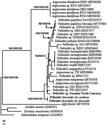
We have obtained the sequences for three loci of the P. thermalis n. sp. nuclear genome, i.e., the partial SSU rDNA, the LSU rDNA, and the ITS-region of the rDNA, and a mitochondrial taxonomic marker, the partial Cox1 mtDNA. An analysis of partial SSU rDNA revealed that P. thermalis n. sp. clustered with a weakly supported clade consisting of all known Pellioditis sequences (including undescribed species from the North Caucasus) and several Angiostoma sequences (A. margaretae, A. norvegicum, and Angiostoma sp. MF838864). Only the ‘megaclade’ containing all known Pellioditis and Angiostoma spp. SSU rDNA sequences was strongly supported (Figure 6). Phylogenetic analysis of LSU rDNA and Cox1 mtDNA was not informative for assessing the position of the new species in the genus Pellioditis due to the low number of informative positions for the former marker and scarcity of NCBI deposited data for the latter. In the LSU rDNA sequence, P. thermalis n. sp. was close to P. circassica (7 bp difference), but it showed nucleotide differences of 21–40 bp (in a 707 bp long alignment) from other species in the genus with known LSU rDNA sequences. The analysis of the Cox1 mtDNA showed that the difference between the strains of the new species was 41 bp. The partial sequence of P. thermalis n. sp. Cox1 mtDNA differed from Pellioditis spp., for which this locus is known, by 57–71 bp. Direct sequencing of the ITS rDNA amplicon was not possible as double peaks were observed after approximately 210 nucleotides in several repeated attempts. The primary PCR product was then cloned and analysed. For the strain thermalis, the analysis of five clones of primary PCR-product revealed the presence of two haplotypes, with or without the insert of 3 bp in the ITS1 rDNA. Similar to thermalis strain, two haplotypes were detected between clones (in total, four clones were examined) of sindicae strain due to the insert of 3 bp in the ITS1 rDNA.
Phylogenetic relationships of Pellioditis species from the Caucasus region and other related Pellioditis species using the SSU 18S rDNA gene as inferred from Bayesian Inference. Litoditis marina AF083021, Oscheius insectivorus AF083019 and Tokorhabditis atripennis LC639823 were used as outgroup taxa. Bootstrap values and posterior probability (as a percentage) are assigned next to the relevant nodes as MP/NJ/ML.

Clone 1 of the P. thermalis n. sp. strain sindicae ITS rDNA was almost identical to clone 1 of thermalis. These two clones differed in five positions of the alignment (data not shown): one deletion and four insertions. All these indels were located at one site in the ITS rDNA region. Clones were identical throughout the rest of the ITS region. Two other clones (clone 2 of P. thermalis n. sp. sindicae and clone 2 of the P. thermalis n. sp. thermalis) showed more pronounced nucleotide differences, e.g., with the exclusion of positions in this variable site of the ITS1 rDNA, clone 2 of P. thermalis n. sp. thermalis differed by 10 bp from clone 1 of the same strain and by 12 bp from clone 1 of strain sindicae. A comparison of the sequences obtained from both haplotypes revealed the phyletic relationship of this species with two other Caucasian species: P. akhaldaba and P. circassica. Depending on the haplotype, P. thermalis n. sp. differed from P. circassica by 42 or 51 bp and from P. akhalbada by 38 or 48 bp.
As the ITS rDNA bp differences between the thermalis and sindicae strains were much lower, we concluded that they represent only two geographically distinct populations with some morphological variability, rather than independent species. The differences between thermalis and sindicae in the ITS rDNA, currently considered the most reliable marker, were slightly above the reported intraspecific level (8 bp). At the same time, the difference in Cox1 mtDNA between these strains was very pronounced, accounting for 41 bp or 7.5% of the sequences compared. The LSU rDNA sequences obtained from both strains were found to be identical. However, the LSU rDNA gene proved to be of little value for the taxonomy of this genus, being unable to resolve relationships between Pellioditis and certain Angiostoma species (Nermut’ et al. 2016; Pieterse et al. Reference Pieterse, Rowson, Tiedt, Malan, Haukeland and Ross2020; Ivanova et al. Reference Ivanova, Geraskina and Spiridonov2020). Nematodes from both strains were shown to be able to reproduce in the interfertility test.
Other species of Pellioditis discovered in Caucasus region, and their interrelationships
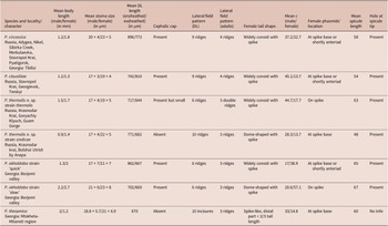
We have isolated several strains of Pellioditis in land gastropods in Caucasus region (see Table 3) and described three new species from the territory of Ciscaucasia and one from Transcaucasia. No known Pellioditis species occurring in Europe were discovered. The species distribution is shown in Figure 1.
Comparison of morphological characters of five Caucasian species of Pellioditis Dougherty, 1953

Morphological and molecular-phylogenetic analyses of Pellioditis were carried out and included yet another recently described species of the genus, P. thesamica (Figures 6–9). The ITS-based analyses carried out have placed the new species close to P. circassica and P. akhaldaba. The former species was originally described from Adygea and later discovered in the Mineralnye Vody-Pyatigorsk zone (Russia) and Tbilisi (Georgia) and the latter in the Borjomi Valley (Georgia). One of the strains of P. akhaldaba was isolated from the surface of Galleria melonella cadavers, which were used as bait for entomopathogenic nematodes. All other strains of P. circassica and P. akhaldaba were isolated from gastropods collected at lower elevations of the mountains, similar to the newly described species. Two strains of P. thermalis n. sp. were discovered from gastropods collected on different sides of the Caucasian range (westernmost spurs for the sindicae strain and northwestern ones for thermalis), although separated by no great geographical distance. Phylogenetic analysis of P. thermalis n. sp. revealed the presence of two haplotypes in each strain, with crossing affinity between them. Notable biological differences were observed between the strains: in thermalis there were almost as many males as females, with habitual matricidal hatching observed, whereas both traits were rare in sindicae.
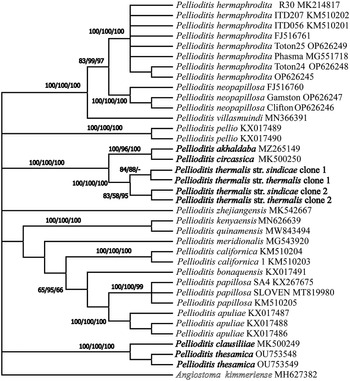
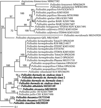
In the present study, ITS-based analyses placed P. thermalis n. sp. sp. in a subclade that forms the sister group with P. circassica and P. akhaldaba (Figures 6 and 7). The analysis also grouped two other Caucasian species, P. clausiliiae and P. thesamica, which show close relationships with the exotic P. zhejiangensis, although with weak support. Our analysis is consistent with the data of Gorgadze et al. Reference Gorgadze, Troccoli, Fanelli, Tarasco and De Luca2022 who recently described P. thesamica from eastern Georgia. Thus, the present five Pellioditis species from the Caucasus form two subclades that are closest to the European P. pellio (previously described as P. bohemica), P. neopapillosa, P. hermaphrodita, and P. villasmundi and at the same time, to Chinese P. zhejiangensis. The latter group of species represents one of the clades of Pellioditis in the evolutionary reconstruction carried out by Tandigan de Ley et al. (Reference Tandingan De Ley, Kiontke, Bert, Sudhaus and Fitch2023).
To date, the species of Pellioditis originating from the Caucasus, i.e., P. circassica, P. clausiliiae, P. akhaldaba, and P. thesamica, form a morphologically comparable group characterised by an average body length of ensheathed infective juveniles of around 700 μm, females with a broadly conical to a rounded anterior part and a subulate terminal part of the tail, and males of all but the latter species with the holed spicule tips.
Phylogenetic relationships of Pellioditis species from the Caucasus region and other related Pellioditis species using the ITS rDNA gene as inferred from the Maximum parsimony (MP, GTR+G model), Neighbour joining and Maximum likelihood (ML, T92 +G model) analyses. Angiostoma kimmeriense MH627382 was used as outgroup taxa. Bootstrap values and posterior probability (as a percentage) are assigned next to the relevant nodes as MP/NJ/ML.

Phylogenetic relationships of Pellioditis species from the Caucasus region and other related Pellioditis species using the ITS rDNA gene as inferred from Bayesian Inference. Angiostoma kimmeriense MH627382 was used as outgroup taxa. Bootstrap values and posterior probability (as a percentage) are assigned next to the relevant nodes as MP/NJ/ML.

All species of Caucasian Pellioditis were associated with more than one gastropod species, both slugs and snails, both native and alien. Snail hosts were represented by smaller species, mainly from Clausiliidae and Oxychilidae families. Infection of larger helicids was not detected.
Angiostoma
The only species of Angiostoma discovered in the Caucasus (Adygea) was A. kimmeriense Korol & Spiridonov, Reference Korol and Spiridonov1991, for which contradictory phylogenetic relationships were described earlier (Ivanova & Spiridonov Reference Ivanova and Spiridonov2018). In short, the D2–D3 and ITS-based phylogenetic analyses placed the species among the members of the Pellioditis clade. Angiostoma kimmeriense is one of the few members of the genus that retains a rhabditoid stoma as an adult. Such a rhabditoid stoma structure is apparently an ontogenetic trait for this genus, and Morand (Reference Morand1988) had shown for four species of Angiostoma that at least two first juvenile stages of these species had similar stoma structures. The position of the male phasmids of A. kimmeriense (posterior to the posteriormost genital papillae) also places it with Pellioditis sp.
The new host and locality for A. kimmeriense have been recorded. However, angiostomatids as internal parasites and large-sized nematodes unable to live in the dead host represent a further step towards parasitism compared with Pellioditis. The new host in the Caucasus was the snail species of the same genus Oxychillus as found on the Crimean peninsula and also the local endemic.
Alloionema
The genus Alloionema Schneider, 1859 belongs to the family Alloionematidae of the Panagrolaimorpha order, which, like the “Rhabditidae”, contains mainly free-living forms but also has a number of species switched to parasitism, mostly in invertebrates. The undescribed Alloionema sp. was found by the authors in Adygea in the foot of a glass snail Oxychilus sp. in 2019. Sequences for the LSU and ITS rDNA and COX1 mtDNA regions of this nematode isolate (AlloS) were obtained, and comparison with the molecular characteristics of other representatives of the Alloionema (A. appendiculatum Schneider, Reference Schneider1859, A. similis Holovachov et al. Reference Holovachov, Bostrom, Tandingan De Ley, Mc Donnell, Alvarado, Paine and De Ley2016, and A. luofuensis Huang et al. Reference Huang, Li and Zhao2016) revealed sufficient differences between them. In the phylogenetic analyses based on ITS rDNA and COX1 mtDNA regions, the North Caucasus isolate of Alloionema was in a separate position and clustered with the basal node of the A. appendiculatum + A. similis clade (Ivanova et al. Reference Ivanova, Mazakina and Spiridonov2022). All other Alloionema isolates found in RF were shown to be represented by Alloionema appendiculatum of Central and Western European origin, co-introduced with the invasive slug Arion vulgaris Moquin-Tandon, 1855 (Ivanova et al. Reference Ivanova, Mazakina and Spiridonov2022; Nermut’ et al. Reference Nermut’, Půźa and Mraček2015). Unfortunately, we were not able to collect complete morphological material from Adygea because parasitic larvae from a snail foot did not develop into free-living adults, but died. Repeated attempts to collect infected gastropods from the original site also failed, and this new species is still awaiting description. It is the only record of Alloionema sp. in the Caucasus region.
Cruznema
Cruznema Artigas, 1927 are free-living rhabditid nematodes sometimes found in association with insects and rarely with land gastropods. The species was recovered from two specimens of small dead Hygromiidae gen. sp. in the steppe vegetation adjacent to agricultural fields on the slopes of the Stavropol Upland. Once recovered, it was easily maintained on pig kidney agar. In small-scale laboratory experiments, it has shown the ability to kill Deroceras reticulatum. This is the first report of Cruznema sp. associated with land gastropods in the RF and the Caucasus region.
Rhabditophanes
Rhabditophanes sp. (Rhabditophanes Fuchs, 1930, Alloionematidae) was found in Xerolenta obvia collected in the Podkumok River valley, with its relatively dry and hot climate. The Rhabditophanes are usual associates of beetles while Alloionema and Neoalloionema are always associated with snails and slugs. The new finding supports the affinity of the genera that make up the family Alloionematidae.
Discussion
The name Phasmarhabditis was confirmed as the junior synonym of Pellioditis Dougherty, 1953 after 40 years of work (Tandigan de Ley et al. Reference Tandingan De Ley, Kiontke, Bert, Sudhaus and Fitch2023). To prove this, the authors (Tandigan de Ley et al. Reference Tandingan De Ley, Kiontke, Bert, Sudhaus and Fitch2023) presented the redescription of the re-isolated type species of the Pellioditis, Pellioditis pellio (Schneider, Reference Schneider1866), and performed molecular reconstruction of the genus. They listed 22 species in the Pellioditis, combining all valid species of Phasmarhabditis except one (they synonymised P. bohemica Nermuť, Půža, Mekete & Mráček, Reference Nermut’, Půža, Mekete and Mráček2017 with P. pellio), the newly described P. pelhami and P. pellio, P. mairei, and P. incilaria, which were listed in the Pellioditis by Sudhaus (Reference Sudhaus2011) before to the study. Of all species in the genus, P. mairei and P. incilaria have not been molecularly characterized, and their brief descriptions provide little morphological information. For the validity of P. mairei, see the opinion of W. Sudhaus (Reference Sudhaus2023), who also suggested that P. tawfiki may belong to a different genus, possibly Pelodera. While most members of the genus are known from terrestrial molluscs, the type species and P. mairei and P. pelhami were originally isolated from earthworms. At least two species (P. pellio and P. hermaphrodita) were later found in association with both types of host (Tandigan de Ley et al. Reference Tandingan De Ley, Kiontke, Bert, Sudhaus and Fitch2023). The Caucasian P. akhaldaba was isolated from a gastropod as well as the cadaver of the baited Galleria melonella. Another species of the genus was found in rotten leaves (P. huizhouensis). This shows the ecological plasticity of the members of Pellioditis. The ability of Pellioditis spp. to grow on various organic materials has also been demonstrated experimentally (Wilson et al. Reference Wilson, Glen, Pearce and Rodgers1995; Nermuť et al. Reference Nermuť, Půža and Mráček2014). While some gastropod-associated species can be pathogenic to their hosts (McDonnel et al. Reference Mc Donnell, Colton, Howe and Denver2020; Laznik et al. Reference Laznik, Majić, Stanislav Trdan, Malan, Pieterse and Ross2020), no such effect was observed in the remainder of the genus.
For the phylogenetic reconstruction of Pellioditis, Tandigan de Ley et al. (2023) carried out the concatenated analysis based on nuclear and mitochondrial genes and constructed a supermatrix. This revealed the presence of three or four clades containing members of Pellioditis (= Phasmarhabditis) as well as Angiostoma norvegicum Ross et al. 2016 as a representative of the Angiostomatidae. All species originated from the Caucasus were shown to belong to the same clade as P. pellio, P. neopapillosa, P. hermaphrodita, P. villasmundi known from Europe, and P. zhejiangensis from China. In our ITS-based analysis, five Caucasian Pellioditis sp. formed two subclades: one related to P. circassica and another related to P. clausiliiae (Figures 6 and 7). The circassica group included P. circassica, P. akhaldaba, and P. thermalis n. sp., while the clausiliiae group included P. clausiliiae and P. thesamica, i.e., each group (subclade) included species isolated on both sides of the Caucasian range.
In terms of nucleotide number, the difference between species from different subclades of Caucasian Pellioditis ranged from 66 to 104 and between species within a subclade, from 22 to 51.
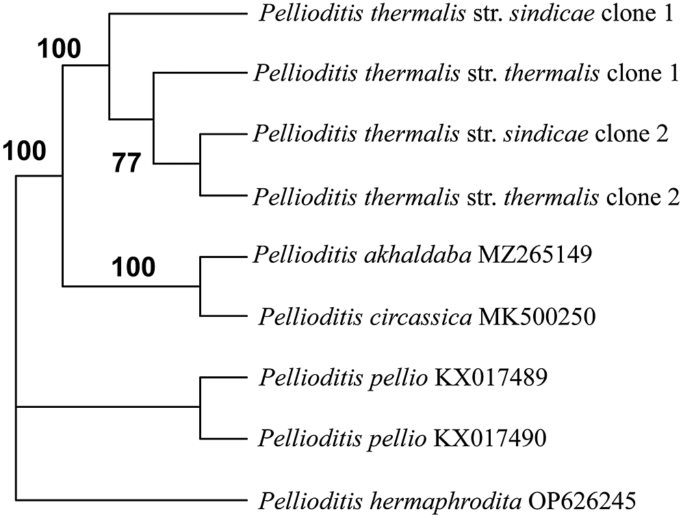
We detected the presence of two ITS rDNA haplotypes in both strains. All haplotypes grouped together in all analysis methods. At the same time, the haplotypes from different populations showed closer relationships to each other than to the haplotype from the same population (Figure 9). Hypothetically, the observed pattern could be the result of two separate evolutionary events. A population of initially non-polymorphic species was split by successive nucleotide changes in the ITS1 rDNA. After the reunion of the two separated populations and their hybridisation, a polymorphic species emerged. This polymorphic species passed through the area fragmentation, and being separated on both slopes of the Caucasian range, the populations underwent independent mutations in the ITS rDNA region. The Western Caucasus region has undergone the deep geological changes. Primarily, in the Jurassic-Eocene there was the rift basin of significant depth, but in the Middle-Late Miocene, the inversion occurred there (Nikishin et al. Reference Nikishin, Ershov and Nikishin2010), accompanied by the immediate submergence of both slopes of the Greater Caucasus. Such events must have affected the populations of hosts such as snails and slugs on both slopes of the range, increasing the number of isolation events for terrestrial molluscs and accelerating intense speciation in the genus Pellioditis.
Phylogenetic relationships between the strains of Pellioditis thermalis n. sp. and the closest Caucasian species of the genus. MP analysis of ITS rDNA.

Species radiation has been investigated for several elements of the biota, including terrestrial gastropods, within the Caucasus hotspots (Mumladze et al. Reference Mumladze, Cameron and Pokryszko2014; Neiber et al. 2015, 2017), but no data were available for the gastropod-associated nematodes prior to our study. However, the gastropod fauna of the Caucasus region has been extensively studied, and its phylogenetic relationships with gastropods of neighbouring regions and the evolutionary history of many taxa have been discussed (Hausdorf, Reference Hausdorf2000, Reference Hausdorf2001; Neiber et al. Reference Neiber, Walther, Kijashko, Mumladze and Hausdorf2022). The richness of the gastropod fauna in the Caucasus (318 species, 66% of which are endemic; Walther et al. Reference Walther, Kijashko, Harutyunova, Mumladze, Neiber and Hausdorf2014) suggests the same for the nematode fauna. Considering the number of Pellioditis species found in the Caucasus region alone, it can be assumed that the diversity of gastropods and the high percentage of the endemic species definitely contributed to the nematode diversity of the region.
Apart from the aforementioned species from Caucasus, world fauna of Pellioditis includes species from Europe (P. pellio, P. hermaphrodita, P. papillosa, P. neopapillosa, P. bonaquaensis, P. apuliae, and P. villasmundi), North America (P. californica and P. pelhami), Africa (P. tawfiki, P. safricana, P. kenyaensis, P. eagyptiaca, and likely P. mairei), and East Asia (P. incilaria, P. huizhouensis, P. meridionalis, P. zhejiangensis, and P. quinamensis). Unidentified species of Pellioditis (as Phasmarhabditis) have been recorded in Japan (Waki & Sawahata Reference Waki and Sawahata2019) and Egypt (Azzam & El-Abd Reference Azzam and El-Abd2021). According to recent research, P. californica is thought to have a cosmopolitan distribution, being isolated in the USA, Canada, UK, Ireland, Germany, and New Zealand (Tandingan De Ley et al. Reference Tandingan De Ley, Holovachov, Mc Donnell, Bert, Paine and De Ley2016; Wilson et al. Reference Wilson, Wilson, Aalders and Tourna2016; Carnaghi et al. Reference Carnaghi, Rae, Tandingan De Ley, Johnston, Kindermann, Mc Donnell, O ’Hanlon, Reich, Sheahan, Williams and Gormally2017; Andrus & Rae Reference Andrus and Rae2019, Mc Donnell et al. Reference Mc Donnell, Colton, Howe and Denver2020; Brophy et al. Reference Brophy, Howe, Denver and Luong2020; Keyte et al. Reference Keyte, Grannel, Sheehy, Shepherd and Rae2022). Pellioditis papillosa described from Europe has been reported in South Africa and the USA (Tandigan De Ley et al. 2016; Pieterse et al. Reference Pieterse, Tiedt, Malan and Ross2017), and P. hermaphrodita in the USA (Tandigan de Ley et al. Reference Tandigan de Ley, Mc Donnel, Lopez, Paine and de Ley2014), Chile (France & Gerding Reference France and Gerding2000), Egypt (Genena et al. Reference Genena, Mostafa, Fouly and Yousef2011), Iran (Karimi et al. Reference Karimi, Kharazi-Pakadel and Robert2003), and New Zealand (Wilson et al. Reference Wilson, Burch, Tourna, Aalders and Barker2012). The distribution of P. hermaphrodita is thought to be the result of the introduction of its commercialised strain, which is on sale in Europe (Howe et al. Reference Howe, Ha, Colton, Tandingan De Ley, Rae, Ross, Wilson, Nermut, Zhao, Mc Donnell and Denver2020; Schurkman et al. Reference Schurkman, Tandingan De Ley, Anesko, Paine, Mc Donnell and Dillman2022).
However, no common European species of Pellioditis has been found in the Caucasian region. Phylogenetically, the Caucasian species show more affinity in phylogenetic analysis to species from Europe than to the species of more distant origin, with the exception of P. zhejiangensis from China. Yet two Old World species from Egypt, both associated with a Mediterranean snail Eobania vermiculata, were found to group with European P. hermaphrodita (P. tawfiki) and P. neopapiilosa (P. eagyptiaca) in the ITS rDNA-based analysis (Azzam Reference Azzam2023).
The distribution of Caucasian species of Pellioditis reflects the distribution of the gastropod hosts on which the distribution of the parasites is based. The distribution/dispersal pattern of Caucasian Pellioditis shows overlapping ranges of species of the circassica and clausiliiae groups. The presence of different haplotypes could be explained in part by the lack of strict host specificity within the genus Pellioditis and the presence of two (or more) species of Pellioditis infecting a single host. The co-infection of multiple parasitic taxa in a gastropod is a common event (Morand Reference Morand1988; Singh et al. Reference Singh, Couvreur, Decraemer and Bert2019; Ivanova et al. Reference Ivanova, Geraskina and Spiridonov2020, etc.) wherein nematode parasites occupy different ecological niches within a host. However, a co-infection of two Pellioditis species has also been observed (Keyte et al. Reference Keyte, Grannel, Sheehy, Shepherd and Rae2022, own unpublished data).
According to the ITS-based analyses performed, five Pellioditis species discovered in the Caucasus region are united in two related subclades. The subclades include species from both Ciscaucasia and Transcaucasia, so the geographical barrier of the Caucasian range may or may not serve to delimit the faunas. From the other side, ancient geographical events probably formed the separate nematode faunas of Western Europe and the Caucasus region, which is reflected in the absence of species typical for Western Europe. All analyses in this study confirmed the monophyletic status of Pellioditis as a genus containing gastropod-associated species. Comparison of the morphology of species within the Caucasus group allowed us to identify certain common characters (Table 3), of which we consider the presence of holes at the tips of male spicules, the mean length of spicules, the mean length of dauer juveniles, and their lateral field pattern to be of main diagnostic value. The GP formula, the length of the bursal rays, and their position in relation to the ventral/dorsal side of the bursa are common to all these species. In general, it applies to the majority of Pellioditis species, where little variation is observed, except for P. quinamensis, which is characterised by the position of the anteriormost pair of papillae outside the bursa.
Broadly speaking, our results support the general distribution pattern of gastropod-associated nematodes described in previous studies. Compared to the diversity in the areas closest to the study area, i.e., in southern Europe, namely Bulgaria, Crimea, and Sicily, the Caucasus region shows a much higher diversity of Pellioditis species, which outweighs all other taxa found. At the same time, the numerous dissections of gastropods made in the study have shown the complete absence of Agfa Chitwood, 1935 in the Caucasian gastropods. Since Agfa species parasitise mainly in the salivary glands of larger limacid slugs, we tend to attribute this to the fact that this gastropod group is rare in the region.
Acknowledgements
All SEM studies were carried out at the Joint Usage Center ‘Instrumental Methods in Ecology’ at the A.N. Severtsov Institute of Ecology and Evolution. We thank V. Mazakina for help in the collection of gastropods.
Financial support
We acknowledge the financial support of a grant from the Russian Science Foundation 19-74-20147.
Competing interest
None.
Ethical standard
The authors assert that all procedures contributing to this work comply with the ethical standards of the relevant national and institutional guides on the care and use of laboratory animals.