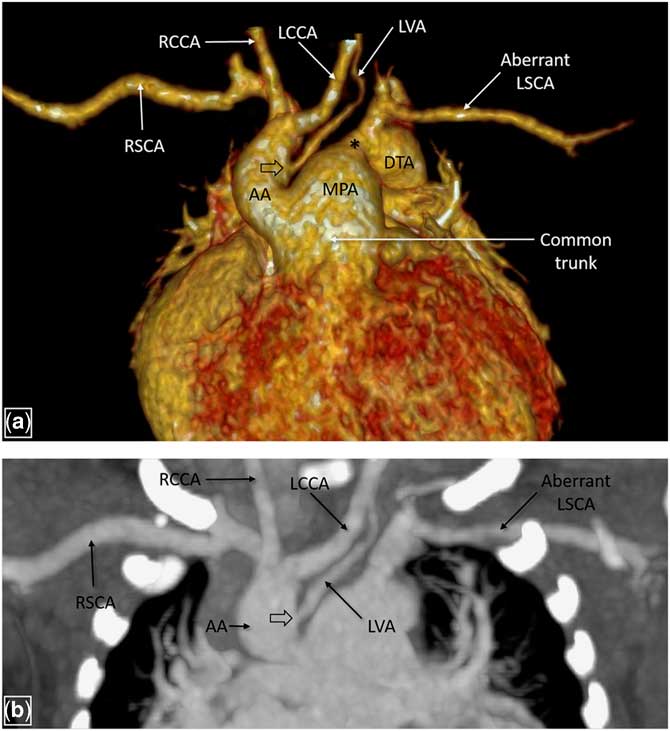
A 3-week-old boy with a diagnosis of persistent truncus arteriosus on transthoracic echocardiography underwent CT angiography to look for suspected arch anomalies. CT angiography revealed a common trunk giving rise to ascending aorta and the main pulmonary artery – type I truncus arteriosus. There was a right-sided aortic arch with interruption distal to the right subclavian artery. The descending thoracic aorta was reformed by patent ductus arteriosus and seen to give rise to the left subclavian artery (Fig 1a).
Volume rendered image of CT angiography (a) reveals presence of a common trunk giving rise to the AA and the MPA. The LVA is seen to arise from the medial aspect of the AA (indicated by block arrow), followed by the LCCA, RCCA, and RSCA. There is interruption of the arch with the DTA being reformed by patent ductus arteriosus (indicated by *). LSCA is seen to arise from the DTA. Coronal maximum intensity projection image (b) delineates the origin of the LVA along with other arch vessels. AA=ascending aorta; DTA=descending thoracic aorta; LCCA=left common carotid artery; LSCA=left subclavian artery; LVA=left vertebral artery; MPA=main pulmonary artery; RCCA=right common carotid artery; RSCA=right subclavian artery.

An incidental note was made of the left vertebral artery arising from the medial aspect of the ascending aorta as the first branch that is proximal to the origin of left common carotid artery, crossing trachea anteriorly and then ascending on the left side (Fig 1a and b). The sequence of branching of arch vessels in this case was as follows: left vertebral artery, left common carotid artery, right common carotid artery, and right subclavian artery followed by the left subclavian artery from the descending thoracic aorta.
Typically, the vertebral artery is the first branch arising from the postero-superior aspect of the first part of the ipsilateral subclavian artery. Anomalous origin of left vertebral artery, from the aortic arch, common carotid artery, external carotid artery, thyrocervical trunk, and the descending thoracic aorta have been reported previously in the literature.Reference Yuan 1 To the best of our knowledge, this is the first reported case of an anomalous left vertebral artery arising from the ascending aorta, in association with an interrupted arch and persistent truncus arteriosus.
Acknowledgements
None.
Financial support
This research received no specific grant from any funding agency, or from commercial or not-for-profit sectors.
Conflicts of interest
None.