INTRODUCTION
Xyalidae Chitwood, 1951 is a prominent family widely distributed from marine to fresh water and from the deep sea to terrestrial soil (Lorenzen, Reference Lorenzen1977). Members of Xyalidae such as the Linhystera are often dominant, occupying up to ~20% of total nematode abundance in marine sediments (Yodnarasri et al., Reference Yodnarasri, Tada and Montani2006; Kim et al., Reference Kim, Rho and Oh2013). Up to now, over 370 species belonging to more than 30 genera have been described in the family. According to faunistic studies, about 14% of deep-sea nematode species belong to Xyalidae (Miljutin et al, Reference Miljutin, Gad, Miljutina, Mokieversusky, Fonseca-Genevois and Esteves2010). In China ~20% of the roughly 200 nematode species identified from marine sediments belong to Xyalidae.
Xyalidae typically have the following characters: transversely striated cuticle; ten cephalic setae in a single circle, anterior ovary or testis to the left of the intestine, posterior testis (if present) to the right; buccal cavity conical, usually without teeth (Warwick et al., Reference Warwick, Platt and Somerfield1998). Within the family, the genus Linhystera was established by Juario (Reference Juario1974) with the type species L. problematica. Based on the redescription of L. problematica, Lorenzen (Reference Lorenzen1977) provided an emended diagnosis for Linhystera: cuticle slightly striated; (6 + 4) cephalic setae about the same length; buccal cavity minute; excretory pore and ventral gland present; two testes, with anterior testis to the left of the intestine and posterior one to the right; without gubernacular apophysis; single anterior outstretched ovary; and conico-cylindrical tail with three terminal setae. Later, Pastor de Ward (Reference Pastor de Ward1985) described L. longa, in which the posterior testis is reduced to a small refractive mass. Up to now, only these two species of Linhystera have been described, and both lack gubernacular apophysis.
During the investigation of benthic free-living nematodes in the East China Sea, we discovered two species which match with Linhystera in almost all main features except the presence of dorsal gubernacular apophysis. The two species are assigned to the genus Linhystera and the taxonomic significance of the gubernacular apophysis is discussed.
MATERIALS AND METHODS
Sediment samples were collected from the East China Sea from July to August 2012, using a 0.1 m2 Gray–Ohara box corer, and preserved with formalin (5% final concentration) on-board. In the laboratory, the fixed samples were stained with 0.1% rose Bengal for 12 h, washed on a 500 µm sieve to remove large particles and a 31 µm sieve to retain meiofauna. Ludox HS 40 was used to extract meiofauna from the remaining sediments by centrifugation. The extracted samples were sorted out under a dissecting microscope. Nematodes were transferred into 9:1 (v/v) solution of 50% alcohol–glycerol in an embryo dish to slowly evaporate to pure glycerol, and then mounted into permanent slides (Huang & Zhang, Reference Huang and Zhang2005).
The descriptions were made from glycerine mounts (Platt & Warwick, Reference Platt and Warwick1983) using a differential interference contrast (DIC) microscope (Nikon E80i). Line drawings were made with the aid of a drawing device. All measurements are in µm, and all curved structures are measured along the arc.
Abbreviations are as follows: a, body length/maximum body diameter; a.b.d., anal body diameter; b, body length/pharynx length; c, body length/tail length; c.b.d., corresponding body diameter; CSL, cephalic setae length; Spic, spicule length as arc; V: distance of vulva from the anterior end of body; V%, position of vulva from anterior end expressed as a percentage of total body length.
SYSTEMATICS
Order MONHYSTERIDA Filipjev, 1929
Family XYALIDAE Chitwood, 1951
Genus Linhystera Juario, Reference Juario1974
Linhystera breviapophysis sp. nov.
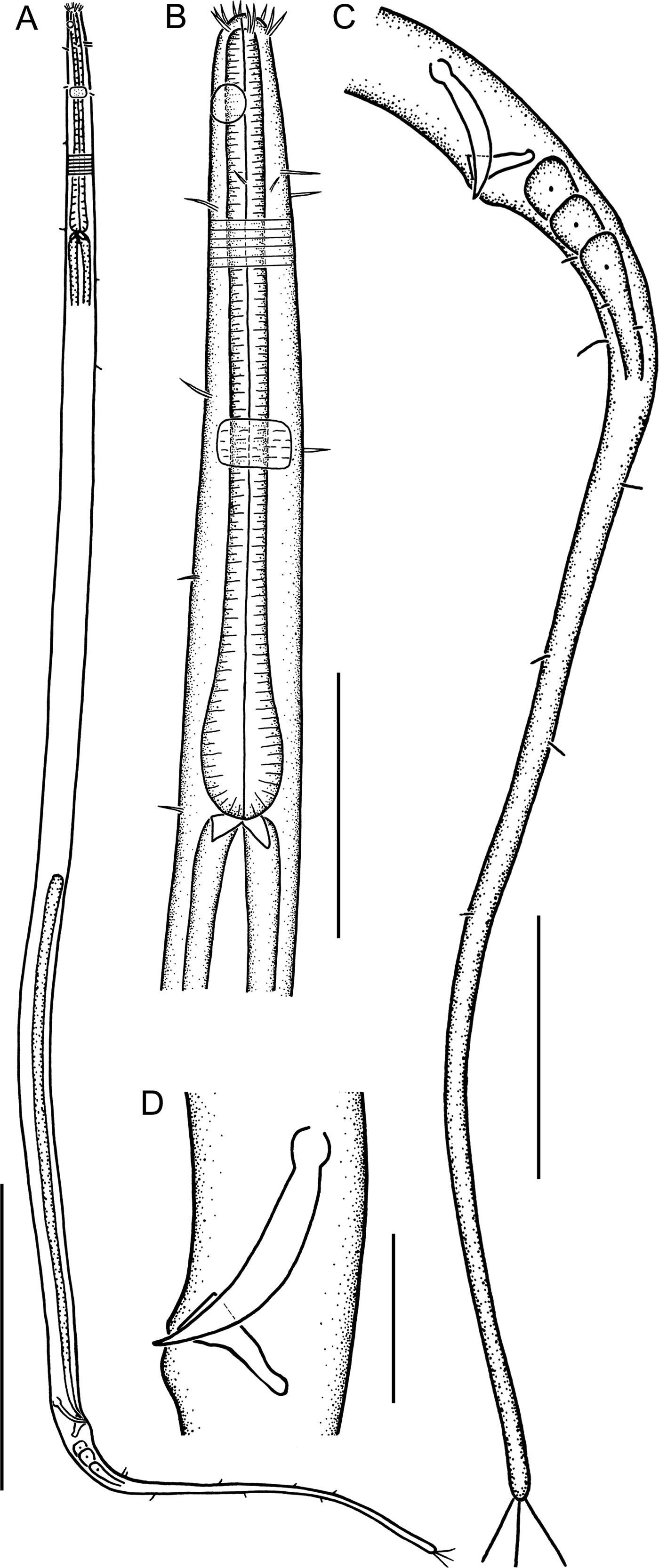
(Figures 1A–D, 3A–C; Table 1)
Linhystera breviapophysis sp. nov. (holotype): (A) overall view; (B) lateral view of anterior portion, showing the buccal cavity, cephalic and cervical setae, and amphideal fovea; (C) lateral view of posterior portion, showing the short filiform tail; (D) lateral view of copulatory apparatus, showing the spicules and gubernacular apophysis. Scale bars: A, 100 μm; B, C, 30 μm; D, 10 μm.

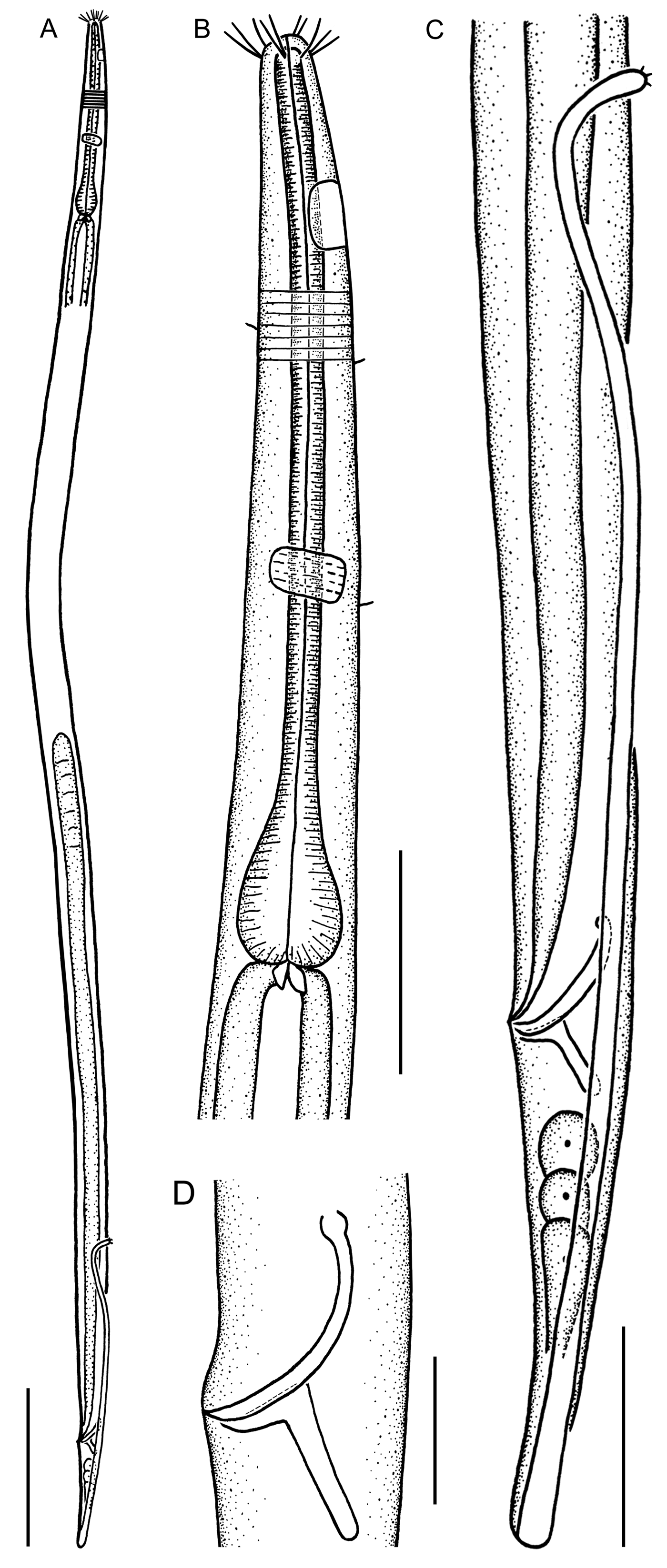
Linhystera longiapophysis sp. nov. (holotype): (A) overall view; (B) lateral view of anterior portion, showing the buccal cavity, cephalic setae, amphideal fovea, and posterior pharyngeal bulb; (C) lateral view of posterior portion, showing the long filiform tail; (D) lateral view of copulatory apparatus, showing the spicules and gubernacular apophysis. Scale bars: A, 100 μm; B, C, 30 μm; D, 10 μm.
Linhystera breviapophysis sp. nov. (A–C; holotype) and L. longiapophysis sp. nov. (D–F; holotype). (A, B, D) Lateral view of anterior region, showing the cephalic setae and amphideal fovea (arrows); (C, E, F) lateral view of cloacal region, showing the spicules and gubernacular apophysis. Scale bars: 10 μm.
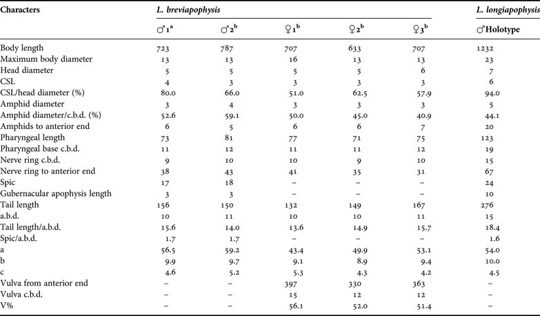
Morphometric data and individual measurements (in μm) of Linhystera breviapophysis sp. nov. and L. longiapophysis sp. nov.
a, holotype; b, paratype; –, absent.
TYPE MATERIAL
The holotype (one male on slide CJ-1-12) and four paratype specimens (one male and two females on slide CJ-1-12; one female on slide CJ-1-13) have been deposited in the Marine Biological Museum of the Chinese Academy of Sciences, which is based at the Institute of Oceanology in Qingdao.
TYPE LOCALITY AND HABITAT
Muddy sediment at the Station CJ-1 (31°41′N 122°30′E) in the East China Sea at 29.5 m deep, sediment is composed of 99.4% of silt–clay, 0.83% organic matter and salinity of 33.8. Water temperature at the sediment–water interface ~21.9°C.
ETYMOLOGY
Composition of the Latin prefix brevi (short) and the Latin noun apophysis (apophysis), referring to the relatively short gubernacular apophysis of the species.
DESCRIPTION
Males: Body cylindrical, with gradually tapering head and filiform tail; body length 723–787 μm, maximum body diameter about 13 μm (Figure 1A; Table 1). Head 5 μm wide. Cuticle with faint transverse striations throughout body, about 1 μm at intervals. Somatic setae about 4 μm long, distributed mainly in the cervical region, where a crown of seven or eight setae are distributed in the anterior quarter of cervical region and 4–6 scattered in other region. Buccal cavity minute, slit-like. Six outer labial setae and four cephalic setae located at the same circle, all about 3–4 μm long. Amphids circular, 3–4 μm in diameter, and 52.6–59.1% c.b.d., located at 5–6 μm (1.1 head diameter) to anterior body end. Pharynx cylindrical, enlarged at base, but not forming a real bulb, 73–81 μm long, occupying about 10.1% of total body length. Pharyngo-intestinal junction with small triangular cardia. Nerve ring located near the middle pharynx, 38–43 μm from anterior body end. Excretory pore and ventral gland not observed (Figure 1B). Tail filiform, 150–156 μm long and 14.0–15.6 a.b.d., with cylindrical part occupying about 71.4% of tail length. Three terminal setae, about 9 μm long. Three caudal glands in tail region. (Figure 1C).
Only one outstretched testis observed, located to the left of the intestine, about 224 μm long. Spicules paired, with a small capitulum in the proximal end, 17–18 μm long and 1.7 a.b.d. Gubernaculum with a short dorso-caudally directed apophysis, about 3 μm long. No precloacal supplements (Figure 1D).
Females: Similar to males, but slightly smaller, 633–707 μm long (Table 1). Vulva located at mid-body, 330–397 μm (51.4–56.1% body length) to anterior body end. Single anterior outstretched ovary to the left of the intestine, about 146 μm long.
DIAGNOSIS
Body length 633–787 μm. Buccal cavity slit-like. Six outer labial and four cephalic setae about 3–4 μm. Amphid circular, 3–4 μm in diameter, and located at 5–6 μm (1.1 head diameter) to anterior end. A crown of seven or eight setae distributed in the anterior quarter of cervical region. Tail short filiform, 132–167 μm long. Testis or ovary outstretched anteriorly, situated to the left of the intestine. Spicules 17–18 μm long, with a small capitulum in the proximal end. Gubernaculum with an about 3 μm long dorso-caudally directed apophysis. No precloacal supplements.
Linhystera longiapophysis sp. nov.
(Figures 2A–D, 3D–F; Table 1)
TYPE MATERIAL
The holotype (one male on slide DH2-4-01) has been deposited in the Marine Biological Museum of the Chinese Academy of Sciences, which is based at the Institute of Oceanology in Qingdao.
TYPE LOCALITY AND HABITAT
Sandy–mud sediment at Station DH2-4 (31°00′N 124°31′E) in the East China Sea at 50 m deep, sediment composed of 41.3% of silt–clay, 0.26% organic matter and a salinity of 33.2. Water temperature at the sediment–water interface ca 21.7°C.
ETYMOLOGY
Composition of the Latin adjective longus (long) and the Latin noun apophysis, referring to the very long and distinct gubernacular apophysis of the species.
DESCRIPTION
Male: Body cylindrical, with gradually tapering head and long filiform tail; body length 1232 μm, maximum body diameter 23 μm (Figure 2A; Table 1). Head 7 μm wide. Cuticle with fine transverse striations recognizable throughout body, about 2 μm at intervals. Buccal cavity minute, slit-like. Six outer labial setae and four cephalic setae located at the same circle, all about 6 μm long. Amphids elliptic, indistinct, about 9 μm long, 5 μm wide, and 44.1% c.b.d., located at 20 μm (three head diameters) from anterior body end. Pharynx cylindrical, with a posterior bulb at base, 123 μm long, occupying about 10% of total body length. Pharyngo-intestinal junction with small triangular cardia. Nerve ring located near the middle of pharynx, 67 μm distant to anterior body end. Excretory pore and ventral gland not observed (Figure 2B). Tail long and filiform, 276 μm long and 18.4 a.b.d., with cylindrical part occupying about three quarters of tail length. Three terminal setae, about 1 μm long. Three caudal glands in tail region, just behind the anus (Figure 2C).
Only one outstretched testis observed, located to the left of the intestine, about 447 μm long. Spicules paired, slender and arcuate, with a small capitulum in the proximal end, 24 μm long and 1.6 a.b.d. Gubernaculum with a long and very distinct dorso-caudally directed apophysis, about 10 μm long and perpendicular to spicules. No precloacal supplements (Figure 2D).
DIAGNOSIS
Body length about 1232 μm. Buccal cavity slit-like. Six outer labial and four cephalic setae about 6 μm. Amphids elliptic, about 9 μm long and 5 μm wide, located at 20 μm (three head diameters) from anterior end. Tail filiform, about 276 μm long. Testis outstretched anteriorly, situated to the left of the intestine. Spicules slender and arcuate, about 24 μm long, with a small capitulum in the proximal end. Gubernaculum with an about 10 μm long dorso-caudally directed apophysis perpendicular to spicules. No precloacal supplements.
DISCUSSION
Linhystera is a small genus in the family Xyalidae containing only two known species. The two new species match well with the generic diagnosis of Linhystera, as provided by Lorenzen (Reference Lorenzen1977), in all main features, except the number of testes (1 vs 2) and the presence of gubernacular apophysis. The presence of two testes is only unambiguously observed in L. problematica Juario, Reference Juario1974, while in L. longa Pastor de Ward, Reference Pastor de Ward1985 the posterior testis is reduced to a small refractive mass (Lorenzen, Reference Lorenzen1977; Pastor de Ward, Reference Pastor de Ward1985). In the two new species only the anterior testis could be recognized. On the other hand, the feature of the testes has been included as a familial character of Xyalidae: the anterior testis is located to the left of the intestine, and the posterior one (if present) to the right (Warwick et al., Reference Warwick, Platt and Somerfield1998). Thus, we suggest removing the number of testes from the generic diagnosis of Linhystera. The presence of gubernacular apophysis appears a rather impressive characteristic, differing distinctly from the two known species of Linhystera. However, the taxonomic significance of a gubernacular apophysis is not well understood. So far, it has never been a decisive feature to separate different genera. The presence or absence of a gubernacular apophysis may occur in the same genus, for instance, in the xyalid genus Theristus (Warwick et al., Reference Warwick, Platt and Somerfield1998) and in the enoplid genus Belbolla (Huang & Zhang, Reference Huang and Zhang2005). Thus, it is unconvincing to erect a new genus based mainly on the presence of a gubernacular apophysis. We suggest assigning the two new species to Linhystera at the present state of knowledge. The two new species share many characters in common with species of Amphimonhystrella (Lorenzen Reference Lorenzen1977; Warwick et al., Reference Warwick, Platt and Somerfield1998). However, Amphimonhystrella have a conical and deep buccal cavity, which is clearly different from the minute and slit-like buccal cavity in the new species. Lorenzen (Reference Lorenzen1977) also included the excretory pore and ventral gland in the generic diagnosis of Linhystera. However, these characteristics are usually rather indistinct and difficult to recognize unambiguously, in particular in small nematodes. Thus, they are seldom included in generic diagnosis (Warwick et al., Reference Warwick, Platt and Somerfield1998). We suggest excluding these characteristics from the generic diagnosis of Linhystera.
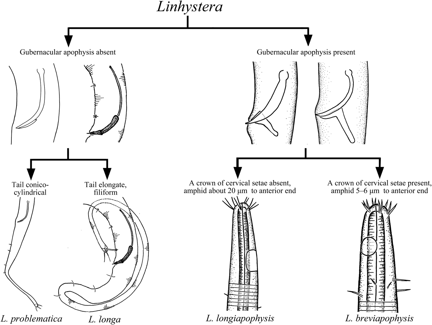
Linhystera breviapophysis and L. longiapophysis belong to a new group in the genus Linhystera due to the presence of a gubernacular apophysis. Both species are easily distinguished from L. problematica by the filiform tail (vs conico-cylindrical). Linhystera breviapophysis differs from L. longa also by the much smaller body (633–787 μm vs 1250–1320 μm) and the much shorter tail (132–167 μm vs 270–322 μm) and spicules (17–18 μm vs 51–53 μm). Linhystera longiapophysis differs from L. longa also by the lack of a crown of cervical setae (vs present) and the much shorter spicules (about 24 μm vs 51–53 μm). Linhystera longiapophysis differs from L. breviapophysis by the larger body (1232 μm vs 633–787 μm), the sparse cervical setae (vs a crown of cervical setae), the much longer and distinct gubernacular apophysis (10 μm vs 3 μm) and the longer tail (276 μm vs 132–167 μm).
Based on the generic diagnosis defined by Lorenzen (Reference Lorenzen1977) and the description of the two new species, we provide an emended diagnosis of Linhystera: Xyalidae with slightly striated cuticle; (6 + 4) cephalic setae about the same length; buccal cavity minute and slit-like; gubernacular apophysis absent or present; and conico-cylindrical tail with three terminal setae. Moreover, a pictorial dichotomous key to the four species of Linhystera is given, based on the morphological features of males (Figure 4).
Pictorial dichotomous key to Linhystera.
ACKNOWLEDGEMENT
We thank Mr Benze Shi for his help in sample processing.
FINANCIAL SUPPORT
This work was supported by the Knowledge Innovation Program of Chinese Academy of Sciences (KSCX2-YW-Z-0914), the Natural Science Foundation of China (41176107), and the Open Research Cruise offshore China by Research Vessel ‘KE XUE SAN HAO, IOCAS’.