INTRODUCTION
Within the metazoan animals lies the phylum of dicyemid mesozoans (Dicyemida Van Beneden, 1876), a poorly understood group of marine parasites that are found with high intensity in the renal appendages of benthic cephalopods. Dicyemid mesozoans are simple in morphology, comprising 8–40 cells with one central axial cell surrounded by ciliated peripheral cells (Suzuki et al. Reference Suzuki, Ogino, Tsuneki and Furuya2010). In contrast to their greatly simplified body plan, two biological features of dicyemids are quite complex and atypical of other metazoans. First, their life cycle comprises two stages of reproduction and development, the asexual vermiform and sexual infusoriform (McConnaughey, Reference McConnaughey1951; Furuya et al. Reference Furuya, Hochberg and Tsuneki2003). Second, the single dicyemid species DNA sequenced to date had a series of mitochondrial (mt) minicircles (three were characterized) that each contained a single mt gene and a non-coding region (NCR) (Watanabe et al. Reference Watanabe, Bessho, Kawasaki and Hori1999). While the presence of mt minicircles has been confirmed in a second dicyemid species (Awata et al. Reference Awata, Noto and Endoh2005) along with the suggestion of a typical metazoan high molecular weight (HMW) circular mt genome of approximately 16 kb (Boore, Reference Boore1999), neither the putative HMW molecule or other mt minicircles have been sequenced to confirm their existence or for comparisons between species to further explore this unusual genome architecture.
Chromosomal fragmentation and the occurrence of mt minicircles have been reported, either in conjunction with or in place of the typical mt genome, in few other groups. In animals, mt minicircles have been reported in several nematode species (Armstrong et al. Reference Armstrong, Blok and Phillips1999; Gibson et al. Reference Gibson, Farrugia, Barrett, Chitwood, Rowe, Subbotin and Dowton2011) as well as 15 species of sucking and chewing lice (Shao et al. Reference Shao, Kirkness and Barker2009, Reference Shao, Zhu, Barker and Herd2012; Cameron et al. Reference Cameron, Yoshizawa, Mizukoshi, Whiting and Johnson2011; Jiang et al. Reference Jiang, Barker and Shao2013; Dong et al. Reference Dong, Song, Guo, Jin, Yang, Barker and Shao2014a , Reference Dong, Song, Jin, Guo and Shao b ; Song et al. Reference Song, Barker and Shao2014). More widely among eukaryotes, minicircle molecules have been observed in the kinetoplast DNA of parasitic trypanosomatid protists (Ryan et al. Reference Ryan, Shapiro, Rauch and Englund1988; Stuart and Feagin, Reference Stuart and Feagin1992; Shapiro and Englund, Reference Shapiro and Englund1995) and their sister group diplonemid flagellates (Marande et al. Reference Marande, Lukes and Burger2005); the nuclear genomes of ciliates (Prescott, Reference Prescott1994); the chloroplast genomes of dinoflagellates (Zhang et al. Reference Zhang, Green and Cavalier-Smith1999, Reference Zhang, Cavalier-Smith and Green2002; Barbrook et al. Reference Barbrook, Symington, Nisbet, Larkum and Howe2001); and the mt genome of the fungus Spizellomyces punctatus (Burger et al. Reference Burger, Gray and Lang2003). Such a rare form of genome organization raises questions about the function of minicircle molecules, how they replicate and their evolutionary consequences, which can be explored further by examining the architecture of these minicircles between dicyemid species.
In our study, we determine the complete cytochrome c oxidase subunit I (COI) minicircle sequence for seven species of Dicyema and two species of Dicyemennea, two of the nine genera within Dicyemida, which combined, contain 90% of the nominal dicyemid species. We explore properties of the mt COI minicircle between and within dicyemid species, examine the NCR for potential secondary structures which may be involved in gene transcription or minicircle replication and discuss the evolutionary consequences of mt minicircular gene arrangement. We also estimate phylogenetic relationships of dicyemid species which we use to compare with their morphological taxonomic classification. Our study is the first to examine mt minicircle molecules from multiple dicyemid species and the first to present a hypothesis for phylogenetic relationships among dicyemids.
MATERIALS AND METHODS
Collection and identification of parasites and hosts
Ten cephalopod species were collected between July 2010 and January 2013 at six localities in eastern, southern and western Australian waters as part of a broader study on dicyemid parasite infections in Southern Hemisphere cephalopods (see Catalano et al. Reference Catalano, Whittington, Donnellan and Gillanders2014, Fig. 1). Collections were made via line fishing, SCUBA, snorkeling and from the by-catch of research prawn trawl surveys (organized by the South Australian Research and Development Institute and Western Australian Fisheries). Immediately after capture, each cephalopod was measured, weighed and tentatively identified before the left and right renal appendages were removed. Multiple glass slide smears of each renal appendage were taken and fixed in 70% ethanol before each appendage was preserved separately in 100% ethanol (Catalano and Furuya, Reference Catalano and Furuya2013). A mantle tissue sample was taken from each host individual examined and preserved in 100% ethanol for molecular genetic confirmation of host species identity (see ‘host cephalopod COI amplification and sequencing’ section). Frozen cephalopod renal appendages from the giant Australian cuttlefish, Sepia apama, collected in South Australia (SA) (February–March 1998; June, September–December 2005; February, June 2006; October 2011) and New South Wales (NSW) (July 2011; March–May 2012), were also used. Since capture, the frozen material had been stored at −80 °C at the South Australian Museum, Adelaide, SA and Cronulla Fisheries Research Centre, NSW.
(a) Nucleotide and (b) amino acid sequence of the aligned 5′ end of COI for dicyemid species showing high sequence heterogeneity. Two individuals of each species (where possible) are shown. Dashes indicate gaps for optimizing the alignment, and sequences identical or highly similar among all dicyemid species are boxed in grey. Numbers above the sequences indicate base and codon positions, numbered from the start of the COI gene for Dicyema furuyi (first 165 bp/55 codons, which are unique for this species only, are not shown). (*) signifies the start of the sequence used in our phylogenetic analyses. Methionines in the amino acid sequence are bolded to highlight the multiple possible N-terminal ends of the COI protein.

Fixed renal appendage smears from fresh cephalopod samples were stained, mounted and then examined for dicyemid parasites with a compound microscope. Comparisons were made with previously described dicyemid species as presented in the synthesis by Catalano (Reference Catalano2012) and from original descriptions. Measurements, drawings and formal descriptions were undertaken for all new dicyemid species discovered (Catalano, Reference Catalano2013a , Reference Catalano b ; Catalano and Furuya, Reference Catalano and Furuya2013). As Dicyema sp. 1 was collected from only one Octopus kaurna individual with infection exclusively by the rhombogen stage, it has not been formally described.
DNA isolation, PCR amplification and sequencing of COI minicircles
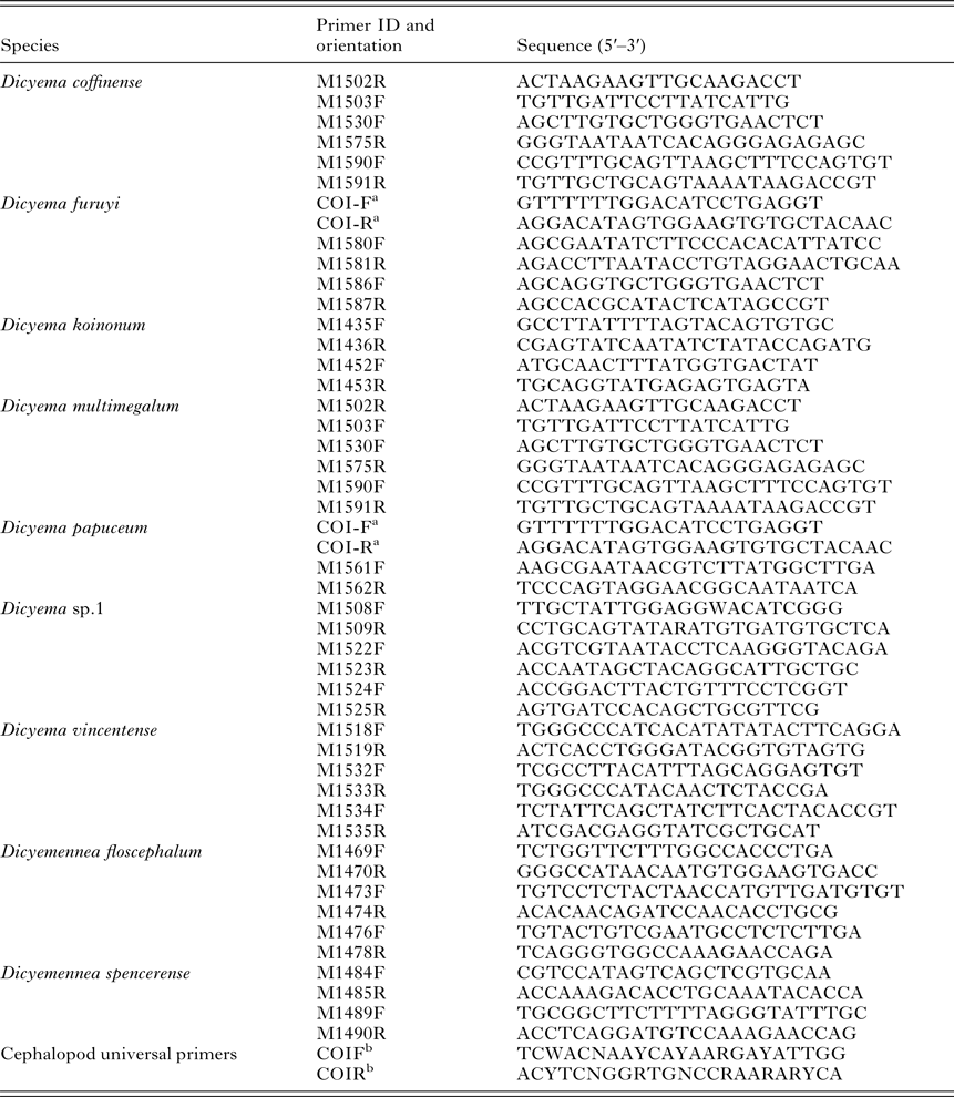
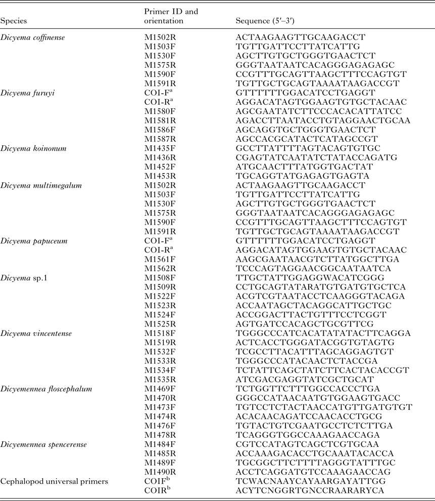
DNA was extracted from each renal appendage and mantle of each host individual separately using a Puregene DNA isolation kit (Gentra Systems) following the manufacturer's protocol for DNA purification from solid tissue. The extracted DNA was then stored at 4 °C until required. Primers from Watanabe et al. (Reference Watanabe, Bessho, Kawasaki and Hori1999) were initially used to amplify a 400 bp fragment of the COI gene (Appendix, Table A1). To test for the presence of a COI minicircle molecule, new primers were designed to the ends of the 400 bp fragment, but in an inverted orientation (Appendix, Table A1). Amplification of a product using these outward facing primers indicated a closed, circular conformation for the COI gene, as established by Watanabe et al. (Reference Watanabe, Bessho, Kawasaki and Hori1999) using genomic southern hybridization analyses. However due to high sequence heterogeneity, these outward facing primers failed to amplify most DNA extractions from dicyemid species and additional species-specific primers had to be designed iteratively based on comparisons between new and existing dicyemid COI sequence (Appendix, Table A1). A panel of the host cephalopod mantle DNA samples was screened with each primer pair to ensure they would not amplify host DNA. All generated parasite sequence data was compared to sequences deposited on GenBank to ensure dicyemid and not host or contaminating microorganisms DNA was characterized.
Amplification reactions were conducted in a volume of 25 μL with a final concentration of 1 GeneAmp PCR Buffer II, 4 mm MgCl2, 200 μ m of each dNTP, 0·2 μ m of each primer and 0·5 U of AmpliTaq® Gold DNA polymerase (Applied Biosystems). Amplifications consisted of an initial denaturation step of 95 °C for 9 min, followed by 34 cycles of PCR with the following temperature profile: denaturation at 94 °C for 45 s, annealing at 50–60 °C (depending on the primers) for 45 s and extension at 72 °C for 1 min, with an additional final extension at 72 °C for 6 min. PCR cycles were increased to 40–45 when PCR product yield was low. Amplicons were visualized on 1·5% agarose gels and purified using a Multi Screen Vacuum Manifold and 384-well Multi Screen Filter Plate (Millipore), then sequenced in both directions using the BigDye Terminator v3.1 cycle-sequencing kit (Applied Biosystems).
Host cephalopod COI amplification and sequencing
A 400 bp fragment of COI was amplified using universal primers from Darling et al. (Reference Darling, Bagley, Roman, Tepolt and Geller2008) (Appendix, Table A1). Amplification, cycling and sequencing conditions were the same as for the dicyemid minicircles, except that a 48 °C annealing temperature was used and sequencing was performed in a single direction using the forward primer only. Sequences were compared to those on GenBank to confirm host cephalopod species identification.
Secondary structure analyses of the NCR
We searched the NCR of all our dicyemid sequences for potential stem-loop structures using EMBOSS: palindrome (Faller, Reference Faller1999) with a minimum palindrome length set at 6. The maximum palindrome length and maximum gap between repeated regions varied depending on the total size of the NCR of each dicyemid species, however both conditions were set to the highest values so all potential palindromes would be identified. Three additional programs, tRNAscan-SE 1.2.1 (Lowe and Eddy, Reference Lowe and Eddy1997), ARAGORN (Laslett and Canback, Reference Laslett and Canback2004) and ARWEN (Laslett and Canback, Reference Laslett and Canback2008), were used to search for potential transfer RNA (tRNA) genes in the NCR. In particular, tRNAscan-SE uses two previously described detection programs as first-pass prefilters followed by a highly selective covariance model to identify tRNA genes (Lowe and Eddy, Reference Lowe and Eddy1997). ARAGORN and ARWEN both employ heuristic algorithms that search for hairpin structures, however whereas ARAGORN searches for tRNA and transfer-messenger (tmRNA) genes concurrently, ARWEN is specialized for finding mt tRNA genes (Laslett and Canback, Reference Laslett and Canback2004, Reference Laslett and Canback2008). These three programs were selected based on the findings of (Morrison, Reference Morrison2010) that no single computer program is necessarily capable of detecting all the tRNA genes in any given sequence.
Phylogenetic analyses of dicyemid parasite COI gene
Sequences were edited and aligned using Geneious Pro 5.3.4 (Drummond et al. Reference Drummond, Ashton, Buxton, Cheung, Cooper, Heled, Kearse, Moir, Stones-Havas, Sturrock, Thierer and Wilson2010). Due to co-infections by individuals with differing COI sequences, some base calls were ambiguous on both the forward and reverse strands. These base calls were resolved by comparison with reference haplotypes from the remainder of our data set (see Table 1 – multiple minicircles were sequenced from each dicyemid species). We designated the start of the NCR directly after the stop codon of the COI gene, with a requirement that it contained no start codon in-frame with the COI gene. However, because of the highly variable sequence composition and size of the NCR between dicyemid species, we were not able to reliably align the NCR and so it was excluded from all phylogenetic analyses. Additionally, due to uncertainty in our alignment of the 5′ end of the COI gene between dicyemid species, the first 375 bp of the sequence alignment was also excluded from phylogenetic analyses (see Fig. 1a). This excluded region is hereafter referred to as the putative 5′ section of the COI gene.
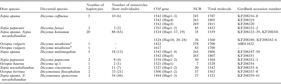
Length of the COI gene (including putative 5′ COI section), NCR and minicircle molecule (bp) for each haplotype (Hap#) recorded from all dicyemid species analysed. The number of minicircles sequenced and the number of host individuals examined for each dicyemid species are given
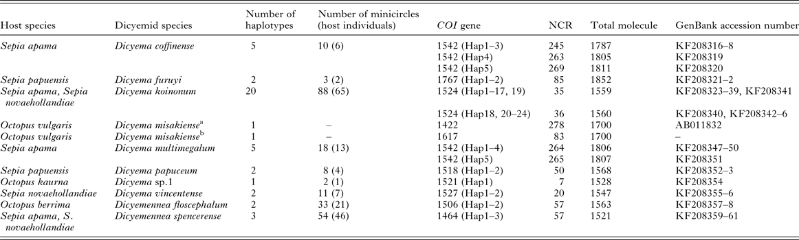
a Original D. misakiense sequence on GenBank.
b Modified D. misakiense sequence we used in our phylogenetic analyses.
Net average uncorrected sequence divergence between groups (d A ) was calculated in MEGA 6.06 (Tamura et al. Reference Tamura, Stecher, Peterson, Filipski and Kumar2013) as: d A = d XY −(d X + d Y )/2, where, d XY is the average distance between groups X and Y, and d X and d Y are the within-group mean. Phylogenetic trees were reconstructed using Maximum-likelihood (ML) (PhyML 3.0; Guindon and Gascuel, Reference Guindon and Gascuel2003; Guindon et al. Reference Guindon, Dufayard, Lefort, Anisomova, Hordijk and Gascuel2010) and Bayesian inference (BI) (MrBayes 3.2; Ronquist et al. Reference Ronquist, Teslenko, van der Mark, Ayres, Darling, Höhna, Larget, Liu, Suchard and Huelsenbeck2012).
For the ML analysis, the general time reversible (GTR) model (Rodŕiguez et al. Reference Rodŕiguez, Oliver, Maŕin and Medina1990) with a proportion of invariant sites (I) and gamma distribution for rates across sites (G) was selected as the most likely nucleotide substitution model from the Akaike Information Criteria (AIC) scores in PartitionFinder 1.0.1 (Lanfear et al. Reference Lanfear, Calcott, Ho and Guindon2012). The tree topology was optimized in order to maximize the likelihood and the best of Nearest Neighbour Interchange and Subtree Pruning and Regrafting moves used to estimate tree topologies with five random starting trees. The starting tree was estimated using BioNJ and the degree of support for internal branches was assessed by 1000 bootstrap pseudo-replications (BS).
MrBayes allows the input data to be partitioned with optimum nucleotide substitution models applied to each partition. The following nucleotide substitution models and partitioning scheme were identified as the most appropriate from the AIC scores in PartitionFinder: GTR + I + G for the first codon partition, GTR + G for the second codon partition and Hasegawa Kishino Yano (HKY) model (Hasegawa et al. Reference Hasegawa, Kishino and Yano1985) +G for the third codon partition. Two Monte Carlo Markov Chain runs of four chains each were performed simultaneously for 10 million generations, with a sample frequency every 200 generations, creating 50 000 trees. To confirm stationarity had been reached and ensure consistency between runs, the relationship between log likelihood scores and generation numbers were plotted and analysed in Tracer 1.5 (Rambaut and Drummond, Reference Rambaut and Drummond2007). We also used Tracer to determine whether our sample included sufficiently large effective sample sizes (EES) for parameter estimates and to approximate the burn-in value. Subsequently, the first 12 500 trees (25%) were discarded from the sample as the burn-in period prior to summarizing the result. A 50% majority rule consensus tree was constructed, with the robustness of nodes assessed by Bayesian posterior probability (PP) estimates.
For both analyses, published COI sequences of the lophotrochozoans (Annelida) Myzostoma seymourcollegiorum and Lumbricus terrestris, which are suggested to be the closest metazoan lineages to Dicyemida as inferred from molecular phylogenetic analyses (Kobayashi et al. Reference Kobayashi, Furuya and Holland1999, Reference Kobayashi, Furuya and Wada2009; Suzuki et al. Reference Suzuki, Ogino, Tsuneki and Furuya2010), were included as outgroups to facilitate rooting of the phylogenetic trees. The COI gene sequence of Dicyema misakiense was also included in our analyses with the following modification. Watanabe et al. (Reference Watanabe, Bessho, Kawasaki and Hori1999) reported the COI gene sequence from this dicyemid species as 1422 bp, with a 278 bp NCR (1700 bp total minicircle size). After inspection, we observed putative start codons in the NCR in-frame with the COI coding sequence. Therefore, for consistency with our edited sequences, the last 195 bp in-frame portion of the D. misakiense NCR was included as part of the COI gene, resulting in a gene size of 1617 bp and NCR size of 83 bp (see Table 1). All trees presented here were visualized and edited in FigTree 1.3.1 (Rambaut, Reference Rambaut2010), and representative sequences of all taxa and haplotypes have been deposited in GenBank (accession numbers KF208316–KF208361).
RESULTS
Dicyemid minicircles
A complete, closed minicircle was sequenced from nine dicyemid species (Table 1), with the circular nature of the molecule confirmed by successful PCR across the 3′ end of the COI gene and the 5′ end of the NCR in each case. In two cases, although parasite infection was observed from morphological examination of smears, we were not able to amplify a COI dicyemid sequence from the renal appendages of Dicyema calamaroceum infecting Sepioteuthis australis (southern calamary) and Dicyema pyjamaceum infecting Sepioloidea lineolata (striped pyjama squid) with any of our designed primers.
Each minicircle contained the COI coding sequence, putative 5′ COI section and a NCR. For all individuals of each dicyemid species except Dicyema sp. 1, more than one haplotype (as indicated by differences in nucleotide sequences) was observed.
Properties of the dicyemid COI gene: nucleotide sequence composition, amino acid translation and size
Within each dicyemid species, both the COI nucleotide and inferred amino acid sequences (excluding the putative 5′ COI section) were highly conserved (net uncorrected sequence divergence within taxa range: 0·1–0·8%). Furthermore, the length of the COI gene including the putative 5′ COI section was constant within species (Table 1). Conversely, between dicyemid species, the COI nucleotide and inferred amino acid sequences (excluding the putative 5′ COI section) were highly variable (net uncorrected sequence divergence between taxa range: 0·5–37·8%). The putative 5′ section of the COI gene, when analysed independent of the COI gene and NCR, was also highly variable between species (net uncorrected sequence divergence between taxa range: 22·9–73·6%) (Fig. 1a and b). In the putative 5′ section of the COI gene, the first ~300 bp for each dicyemid species contained multiple (3–9) in-frame methionine codons (Fig. 1b). The sequence AUA was identified as the start codon for five dicyemid species, with AUG the start codon for the remaining four dicyemid species. The length of the gene including the putative 5′ COI section varied among dicyemid species, with the shortest COI recorded from Dicyemennea spencerense (1464 bp), and the longest from Dicyema papuensis (1767 bp) (Table 1). In general, the COI gene was shorter for Dicyemennea species (1464–1506 bp), compared to Dicyema species (1518–1767 bp) (Table 1).
The non-coding region (NCR)
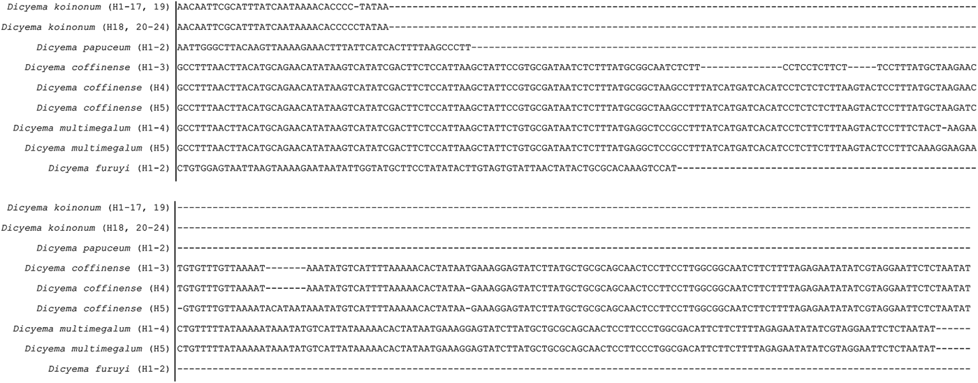
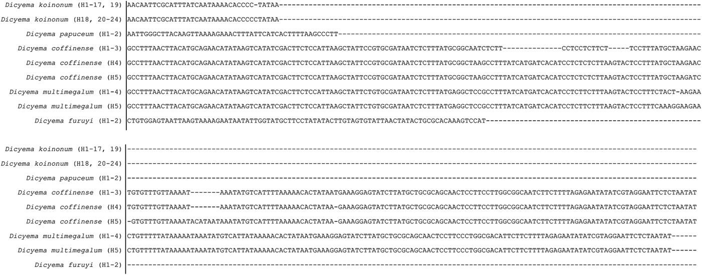
The NCR was extremely variable in nucleotide sequence and length, from 7–269 bp between dicyemid species, so much so that the region could not be aligned across species (Table 1, Fig. 2). In stark contrast with the extreme interspecies variation, nucleotide sequences of the NCR within species were invariant except for minor length variation within three species, Dicyema coffinense, Dicyema koinonum and Dicyema multimegalum (Table 1, Fig. 2).
Unaligned nucleotide sequence of the COI minicircle molecule NCR showing variability in size and sequence content between and within dicyemid species. Corresponding haplotypes for the NCR sequence of each dicyemid species are given in parenthesis.
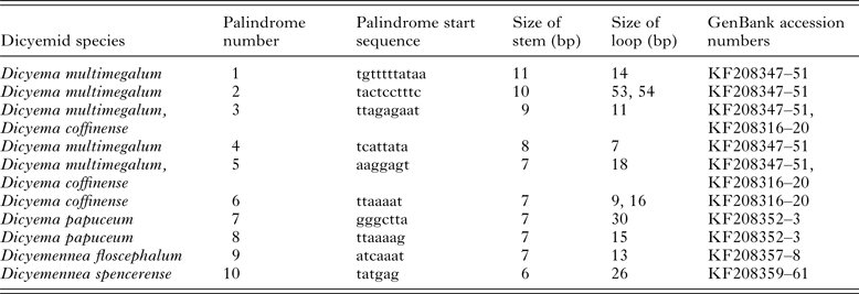
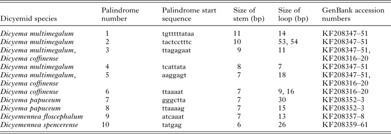
No tRNA genes were identified in any of the NCR sequences. However, palindrome repeats were identified in five species: D. coffinense, D. multimegalum, Dicyema papuceum, Dicyemennea floscephalum and D. spencerense sequences with positions of potential stems and loops annotated in GenBank submissions (Table 2). All potential stem-loop structures associated with these palindromes were unique, except for palindromes 3 and 5, which were found in both D. coffinense and D. multimegalum. The loops circumscribed by these two palindromes were of the same length and sequence composition in both D. coffinense and D. multimegalum, although they occurred at different places in the NCR for each dicyemid species (see annotated GenBank sequences KF208347–51 and KF208316–20).
Stem and loop features from palindrome sequences identified in the NCRs
Phylogenetic analysis of dicyemid COI gene
We could not confidently align the NCR or the putative 5′ section of the COI gene between species, so both were excluded from our subsequent phylogenetic analyses. Hence our phylogenetic analyses were based on a 1541 bp alignment of 47 COI haplotypes from 10 dicyemid species. After 10 million generations in the Bayesian analysis, the s.d. of split frequencies had reduced to 0·0026, with Potential Scale Reduction Factor of 1·0 for all parameters, suggesting stationarity had been reached. Additionally, from the Tracer analysis, the log likelihood scores between runs were similar with ESS > 4000 for all parameters for both runs.
The tree topology was similar between the ML and BI analyses (Fig. 3a and b, respectively). There was strong support for the majority of nodes, with BS and PP values of >90% and >0·98, respectively. Both analyses separated Dicyemennea spp. plus Dicyema sp. 1 (clade II) from all other Dicyema (clade I) with strong support (BS, 1000 replicates – Fig. 3a and PP – Fig. 3b). However, the placement of Dicyema misakiense varied, either as the sister to clade I but with low support (ML, Fig. 3a) or sister to both clades I and II with strong support (BI, Fig. 3b). Each dicyemid species formed distinct, monotypic subclades, except for D. multimegalum which rendered D. coffinense paraphyletic in the ML analysis (Fig. 3a inset) but not in the BI analysis (Fig 3b inset).
Phylogenetic analyses of the complete mt COI gene sequences (excluding the putative 5′ COI region) for representative taxa in Dicyemida and two outgroup taxa in Lophotrochozoa determined from: (a) Maximum-likelihood with bootstrap proportions (1000 pseudo-replicates); and (b) Bayesian inference with posterior probabilities. Resolution of the terminal nodes for the D. coffinense and D. multimegalum clades is shown in the inset. Dicyemid host species are listed in Table 1 along with H# (haplotype number). All sequences were determined in the present study except for sequences from D. misakiense and the two outgroup taxa.

DISCUSSION
Here we compared the complete 1·5–1·8 kb COI minicircle molecules from ten dicyemid species, comprising the full length COI gene, putative 5′ COI section and NCR. We discuss the variation in the properties of this molecule between and within dicyemid species and implications of minicircle structure for mt function in more detail below. Finally we evaluate how well the taxonomic classification of dicyemids represents an evolutionary based classification by comparisons with our phylogenetic framework estimated from our COI sequence data.
Variation in the COI gene
For invertebrate mt DNA, the typical initiation codon for translation of COI is methionine, encoded by either AUG or AUA (Elzanowski and Ostell, Reference Elzanowski and Ostell2013). Subsequently for each dicyemid species that we analysed, the first AUG or AUA in-frame codon that resulted in the longest continuous sequence terminating at a stop codon was inferred as the translation initiation site. However, it is difficult to state with certainty if this represents the true initiation site(s), as we observed multiple in-frame methionines in the putative 5′ COI section of each dicyemid minicircle (see Fig. 1b), suggesting the gene size may actually be shorter than recorded in Table 1. Other authors have also expressed uncertainty in the identification of the initiation codon in mt genes for non-parasitic invertebrates (see Cantatore et al. Reference Cantatore, Roberti, Rainaldi, Gadaleta and Saccone1989; Milbury and Gaffney, Reference Milbury and Gaffney2005). Due to the unresolved position of dicyemid parasites in the Tree of Life, it is difficult to make comparisons with closely related phyla to infer the true initiation site.
The putative 5′ section of the COI gene was also highly divergent in sequence composition between dicyemid species (net uncorrected sequence divergence between taxa range: 22·9–73·6%) (Fig. 1a). It has been suggested that the presence of asexual reproduction in the life cycle of dicyemid parasites (albeit in combination with selfing simultaneous hermaphroditism in the sexual phase) may accelerate the accumulation of mutations (Aruga et al. Reference Aruga, Odaka, Kamiya and Furuya2007), leading to a high amino acid sequence divergence rate (Paland and Lynch, Reference Paland and Lynch2006). However, it is not known if recombination or gene conversion occurs in the production of gametes in the sexual phase, which would ameliorate the impact of asexual reproduction on mutation accumulation.
Structure and function of the NCR
For dicyemid parasites, the NCR sequence and length varied greatly between species, so much so that this portion could not be aligned among dicyemid species and had to be excluded from our phylogenetic analyses. High sequence divergence and large size range differences in the NCR have been reported between lice species, with a NCR of 48 bp in Coloceras compared to 2050 bp in Pediculus (Shao et al. Reference Shao, Kirkness and Barker2009; Cameron et al. Reference Cameron, Yoshizawa, Mizukoshi, Whiting and Johnson2011).
While the gene coding region defines genome metabolic functionality by specifying proteins, the NCR can define the architecture and regulation of the genome, often harbouring the replication origin and the promoters for transcription (Le et al. Reference Le, Blair and McManus2002; Burger et al. Reference Burger, Jackson, Waller and Bullerwell2012). Although no tRNA genes were found in any of the NCRs of the ten dicyemid species we analysed, palindrome sequences with the potential to form stem-loop structures were identified in five species. Palindromic sequences have been reported in the mtDNA of various other taxa, with roles including mediating lateral gene transfer, mt recombination, chromosomal rearrangements and transcript stability (Burger et al. Reference Burger, Jackson, Waller and Bullerwell2012). Palindrome regions may be involved in similar biological processes in dicyemid parasites, or the stem-loop structure associated with them could function as the transcriptional initiation sites (Awata et al. Reference Awata, Noto and Endoh2005). Stem-loop structure may also play a role in mechanistic processes. Watanabe et al. (Reference Watanabe, Bessho, Kawasaki and Hori1999) reported one stem-loop structure in the NCR of the D. misakiense COI minicircle, although this becomes part of the 5′ region of the COI sequence with our reinterpretation of the structure of this minicircle. Uncertainty in the identification of the start codon presently confounds attempts to infer the function of minicircle components.
Evolutionary considerations
Cytoplasmic organelle genome minicircle molecules represent a rare genome structure among the eukaryotes; therefore it is interesting to consider the factors that may have given rise to their evolution (Cameron et al. Reference Cameron, Yoshizawa, Mizukoshi, Whiting and Johnson2011). The typical mt genome, whereby all the genes are linked together on a chromosome, ensures the complete genetic information is passed on to the next generation, so why do mt fragmentation and minicircle molecules occur in some organisms? In metazoan animals, mt minicircles or linear sub-genomes occur in different genomic settings, either with the co-occurrence of a typical HMW mitogenome (i.e. the dicyemid Dicyema japonicum [Awata et al. Reference Awata, Noto and Endoh2005]), or without a HMW mitogenome (i.e. human lice [Shao et al. Reference Shao, Kirkness and Barker2009]; Globodera nematodes [Gibson et al. Reference Gibson, Blok and Dowton2007a , Reference Gibson, Blok, Phillips, Hong, Kumarasinghe, Riley and Dowton b ]; cubozoan jellyfishes [Smith et al. Reference Smith, Kayal, Yanagihara, Collins, Pirro and Keeling2012]). One explanation could be to increase protein synthesis or allow differential transcription of individual mt genes. Translation on mt ribosomes (mitoribosomes) is a relatively inefficient process (Taanman, Reference Taanman1999) and in humans the two mt rRNAs can be transcribed independently of the whole genome polycistron transcript (Gelfand and Attardi, Reference Gelfand and Attardi1981). This may enable elevated production of inefficient mitoribosomes independent of other cellular metabolic requirements allowing for greater flexibility in dealing with transient stress conditions imposed by the variable physical–chemical nature of the renal sac (Schipp et al. Reference Schipp, von Boletzky and Doell1975). However, the absence of a polycistronic transcript for components the multi-subunit holoenzyme of the cytochrome c oxidase complex IV, which are encoded on separate minicircles (including COI, COII, COIII which are on separate minicircles in Dicyema misakiense [Watanabe et al. Reference Watanabe, Bessho, Kawasaki and Hori1999]), presents an issue for the control of transcription and/or translation levels to produce equivalent concentrations of the holoenzyme components, which also needs consideration for the nuclear encoded components of the translational machinery. How dicyemids have evolved efficient control of holoenzyme assembly needs experimental evaluation.
Awata et al. (Reference Awata, Noto and Endoh2005) reported a canonical mt genome in the germ line of D. japonicum, which they suggest represents the primary form of the genome that is vertically transmitted, whereas mt minicircles represent a terminally differentiated form confined to somatic cells. Minicircles appear to be generated early in dicyemid development and are then passed on through embryogenesis to daughter cells. Conceivably, because mature dicyemids comprise just a few cells (up to 40), Awata et al. (Reference Awata, Noto and Endoh2005) proposed an ‘amplification-dilution’ model in which a single initial episode of minicircle production early in embryogenesis could supply sufficient mt gene templates for transcription for subsequent cellular metabolism, consistent with their observation of greatly diminished mtDNA replication in terminally differentiated somatic cells. Thus the absence of an origin of replication for dicyemid mt minicircles may be the final step in a genome fragmentation process that could only be achieved in organisms with very small cell counts in the mature form. In human lice, nematodes and cubozoan jellyfishes where HMW mt genomes appear to be absent, structures compatible with origins of replication are present on each minicircle or linear sub-genome and not co-incidentally cell numbers in adults of these organisms are orders of magnitude larger than in dicyemids (Shao et al. Reference Shao, Kirkness and Barker2009; Gibson et al. Reference Gibson, Farrugia, Barrett, Chitwood, Rowe, Subbotin and Dowton2011; Smith et al. Reference Smith, Kayal, Yanagihara, Collins, Pirro and Keeling2012). By analysing the entire dicyemid mt genomic complement, we may be able to elucidate why minicircle molecules are part of the genome organization and what their overall function may be.
Dicyemid systematics
Currently the classification of dicyemid parasites and designation of new species is based purely on morphological characters. In general, the number and orientation of cells in each tier of the calotte, the presence or absence of abortive axial cells and the presence or absence of syncytial stages determines the genus, whereas the size of the adult stages, the number of cells comprising the body, the shape of the calotte, the anterior extension of the axial cell, the presence or absence of verruciform cells and the structure of the infusoriform embryo are characters used to distinguish species (Hochberg, Reference Hochberg1982, Reference Hochberg1983). Unfortunately, incomplete and information-poor descriptions, loss of type specimens, errors in taxonomy and conceptual differences have led to confusion over the validity of 20% of the taxa within this group (Catalano, Reference Catalano2012). Molecular genetic analyses comparing the relationships between dicyemid species beyond morphological character traits may therefore shed light on this taxonomic confusion and species validity.
In our study, past classifications based on morphological traits were generally supported with our phylogenetic findings, as species clades were monotypic with strong support in both the ML and BI analyses (Fig. 3a and b). However, three unusual features were observed: (1) the placement of Dicyema misakiense as the sister lineage to the Dicyemennea and Dicyema (clades I and II) in the BI analysis with strong support (Fig. 3b); (2) the placement of Dicyema sp. 1 in the Dicyemennea (clade I) in both analyses (Fig. 3a and b); and (3) nesting of the D. multimegalum haplotypes within the D. coffinense clade in the ML analysis (Fig. 3a inset).
Renal appendage smears of Dicyema sp. 1 were re-evaluated and the observation of four metapolar cells opposite four propolar cells in the calotte was confirmed supporting the taxonomic placement of this species in Dicyema (note that a defining character for Dicyemennea is the occurrence of five metapolar cells in their calotte). Therefore the placement of Dicyema sp. 1 in the Dicyemennea clade in both analyses, along with the basal placement of D. misakiense in the BI analysis with strong support, suggests certain morphological traits, namely calotte cell counts, may not be an adequate character to classify dicyemids at the generic level. Phylogenetic analyses including taxa from all genera and inclusions of nuclear markers are needed to resolve an evolutionary based classification of the dicyemids.
The nesting of D. multimegalum within the D. coffinense clade for the ML analysis (Fig. 3a inset) suggests these two dicyemids are the same species, although the BI analysis does support monotypic clades (Fig. 3b inset). These two species were classed as distinct species based on differences in morphological characters, with variation in calotte shape (elongate metapolar cells vs compressed metapolar cells), maximum body length of adults (2920 μm vs 1672 μm), peripheral cell counts (30–32 vs 26–28) and maximum length of infusoriform embryos (31·3 μm vs 40 μm) for D. coffinense compared to D. multimegalum, respectively (Catalano, Reference Catalano2013b ). Both these dicyemid species are recorded from the same host species, S. apama, although from different geographical localities where the size of the host is also markedly different; host mantle length range 11·7–20·7 cm at Coffin Bay, SA (infection by D. coffinense), whereas 38–49·5 cm at Cronulla and North Bondi, NSW (infection by D. multimegalum) (Catalano, Reference Catalano2013b ). Dicyema coffinense and D. multimegalum may actually be one taxon exhibiting morphological plasticity mirroring that of their micro-habitat. Examining the divergence in nuclear loci between these two taxa would provide a further test of their species status.
Concluding remarks
Molecular analyses for the dicyemids are lacking or focus only on a single taxon. Here we characterized the complete COI minicircle molecule from ten dicyemid species allowing comparisons on gene organization, architecture and sequence similarity to be made for the first time between species. Questions remain on the replication capabilities of these minicircle molecules and whether other mt and nuclear genes are also fragmented on individual minicircle molecules. Following our study which presents the first multi-species molecular framework for dicyemids, additional taxa should be sequenced for COI allowing wider comparisons to be made, followed by the characterization of additional markers to answer broader evolutionary questions.
ACKNOWLEDGEMENTS
Thanks are due to volunteers, who assisted with cephalopod collections, and Tim Benson for constructive comments on manuscript drafts. All research was conducted with approval from the University of Adelaide Animal Ethics Committee (# S-2010-116) and according to Primary Industries and Regions SA S115 Ministerial Exemptions (# 9902264 and # 9902398).
FINANCIAL SUPPORT
Funding for this research has been provided by the Australian Society for Parasitology, Norman Wettenhall Foundation, Holsworth Wildlife Research Endowment, Sir Mark Mitchell Research Foundation, Lirabenda Endowment Fund, Nature Foundation South Australia, Nature Conservation Society of South Australia and Australian Federation of University Women South Australia. SRC was supported by an Australian Postgraduate Award, and BMG by an ARC Future Fellowship (FT100100767).
APPENDIX
Primers used for PCR amplification of each dicyemid species COI minicircle sequence and cephalopod COI partial fragment. All primers were designed in this study except those indicated otherwise. Orientation follows primer ID, either forward (F) or reverse (R)