Introduction
Bovine and ovine tuberculosis (TB) is a zoonotic chronic disease caused by infection by Mycobacterium tuberculosis and classified as a class I animal epidemic by the World Organization for Animal Health and as a class II animal epidemic in China. It is very important to use vaccination in the prevention, early diagnosis and effective treatment of TB (Kaufmann, Reference Kaufmann2006). At the beginning of the 20th century, an immunizing antigen called BCG was prepared from an extract of a strain of attenuated Mycobacterium bovis, which, as a vaccine, was widely used in the prevention and cure of human tuberculosis as a live vaccine, and played a major role in the control of human tuberculosis. BCG was used in the prevention and cure of bovine and ovine tuberculosis at the beginning, and an immunoprotection test of BCG in later decades showed a great difference in immunoprotective effect (Fine, Reference Fine1995; Fine and Vynnycky, Reference Fine and Vynnycky1998).
Recently, some experiments proved that immunization with BCG can reduce the incidence of TB (Buddle et al., Reference Buddle, Wedlock, Denis and Skinner2005, Reference Buddle, Wedlock and Denis2006; Vordermeier and Hewinson, Reference Vordermeier and Hewinson2006; Vordermeier et al., Reference Vordermeier, Chambers, Buddle, Pollock and Hewinson2006). After more than 10 years' experiments, it was found that hypodermic inoculation with BCG in cows could induce noticeable immunoprotection to defend against pathogenic infection and decrease the number of infected tissues and degree of infection, which was related to enhanced expression of interferon-γ (IFN-γ) induced by BCG vaccination (Buddle et al., Reference Buddle, Wedlock, Denis and Skinner2005, Reference Buddle, Wedlock and Denis2006). In the latter research, the authors established the laboratory evaluation of immunoprotection and, guided by the results, bovine and ovine TB in New Zealand are well under control (Smith et al., Reference Smith, Bennett, Wilkinson and Cooke2007). Use of BCG vaccination for the prevention and cure of TB in China was also reported, although it was controversial (Zhang et al., Reference Zhang, Zhang, Liu and Sun1997; Zhu and Hua, Reference Zhu and Hua2005; Wang et al., Reference Wang, Yu and Zhai2006). The present paper describes the investigation of the immunoprotection ratio and changes of immune response induced by BCG vaccination and the impact of different immunizing doses and different immunization age on immunoprotection in BCG-immune BalB/C mice, so as to provide a basis for the prevention and cure of bovine and ovine TB with BCG.
Materials and methods
Materials
BalB/C neonate and adult mice were from the Experimental Animal Centre of Shandong University (Jinan City, Shandong Province, China). Vaccine and tuberculin protein for intradermal injection were from Tai'an Hygienic and Antiepidemic Station (Tai'an City, Shandong Province, China). Simplified medium was prepared according to Kuang et al. (Reference Kuang, Song, Wang, Tan, Wang and Xue1995)). ELISPOT reagent for the detection of IFN-γ and interleukin-4 (IL-4) antibody and biotin-labelled secondary antibodies of IFN-γ and IL-4 were from B&D Company (Glen Rock, NJ, USA). Alkaline phosphatase labelled avidin and nitroblue tetrazolium/5-bromo-4-chloro-3-indolyl-phosphate (NBT/BCIP) colour reagent kit were bought from Zymo Company (USA).
Methods
Quantitative cultivation of BCG
BCG vaccine powder was diluted with 1 ml of stroke-physiological saline solution (SPSS), then 100 μl was 10× diluted with SPSS, and 100 μl of this solution was inoculated evenly on a culture dish containing purified medium with 10% calf serum. The dish was sealed with plastic wrap and incubated in 5% carbon dioxide at 37 °C. After 10–12 days, the colony forming units (cfu) of BCG vaccine were counted. According to this method there were 2.0×10⁵ cfu of live TB bacilli in BCG powder diluted with 1 ml SPSS.
Immunization and challenge of BalB/C mice
Twenty-four 7-day-old BalB/C neonate mice were divided into three groups, each containing eight mice: a group immunized with a high dose of BCG, a group immunized with a low dose of BCG, and a control group (not immunized with BCG). Mice in the high-dose group were injected with 200 μl BCG vaccine (about 4×10⁴ cfu/mouse) subcutaneously in the neck; mice in the low-dose group were injected with 10 μl BCG vaccine (about 2×10³ cfu/mouse) subcutaneously in the neck; and mice in the control group were injected with normal saline subcutaneously in the neck. Mice in each group were raised separately in a clean environment and at room temperature and were allowed to eat and drink freely. Then mice in all the groups were administered with 500 μl BCG (about 105 cfu BCG/mouse ) through peritoneal injection 28 days after immunization. All mice were killed suddenly 14 days later and the pathological changes of tissue were observed and spleen cell suspensions were obtained.
Twelve 35-day-old BalB/C mice were randomly divided into two groups. Mice in one group were injected with 10 μ BCG vaccine subcutaneously in the neck, and mice in the other control group were injected with physiological saline. Raised under the environment mentioned above, mice were challenged with 500 μl BCG 28 days after immunization, and all mice were killed rapidly 14 days later. The pathological changes of tissue were observed and spleen cell suspensions were obtained.
Immunoprotection test
Preparation of spleen cell suspensions of mice
Each dead mouse was immersed in 75% alcohol for 5–10 min. Each spleen was obtained under sterile conditions, ground in a sterile test tube and filtered through a 200-mesh sterile copper screen. The filtrate was centrifuged for 10 min at 1000 rpm, the supernatant discarded and the pellet re-suspended in RPMI (Russel Park Memorial Institute) 1640. The cell numbers were counted, and the suspensions diluted to a final concentration of 107/ml, and stored for use.
Culture in vitro of BCG in SPC
Each spleen cell (SPC) suspension was disintegrated ultrasonically for 10 s in a sterile glass tube, 400 μl of the resulting solution was added to a prepared BCG dish and mixed it evenly. The dishes, three for each mouse, were sealed with plastic wrap and cultured in a 5% CO2 incubator at 37 °C. Growth of colonies was observed and the number of colonies counted. As the number of colonies became stable the colony forming units (cfu)/ml of BCG were calculated.
Calculation of immunoprotection ratio of mice
If a characteristic colony was observed in any of the three SPC dishes of each mouse, it would be recognized as a positive mouse, and the immunoprotection ratio was calculated by: total number of positive mice in a group/total mouse number of the group.
Detection of Th1- and Th2-type immune responses
Separation of mouse splenic lymphocytes (SPL)
Spleens were obtained under sterile conditions, ground and disintegrated into single cells and filtered through a 200-mesh sterile copper screen. The filtrate was transferred to a centrifuge tube and centrifuged for 10 min at 800 rpm, washed with serum-free RPMI 1640 once and re-centrifuged. The supernatant was discarded and the pellet resuspended with 3–5 ml of serum-free RPMI 1640. The cell numbers were counted.
Detection of IFN-γ and IL-4 by ELISPOT assay
The method was an improved version of that reported by Power et al. (Reference Power, Grand, Ismail, Peters, Yurkowski and Bretscher1999) and Doherty et al. (Reference Doherty, Demissie, Menzies, Andersen, Rook and Zumla2005).
For assay of IFN-γ, the coating antibody was diluted in 100 μl phosphate-buffered saline (PBS) (pH 9.6) for each well, and wells were coated overnight at 4 °C. The coating solution was discarded the next day, the wells were washed three times with PBS, dried naturally, then 200 μl of diluted blocking liquid were added and the wells incubated for 1 h at 37 °C. Cell suspension, 100 μl/well, was added so that the total cell number of each well was about 1×10⁶. Purified protein derivatives (PPD) of tuberculin were added at a concentration of 0.1 μg/ml (determined by preliminary tests), and three wells of control blanks were set. The wells were cultured for 8–24 h in a CO2 incubator at 37 °C. The liquid was discarded, 200 μl ice-cold, deionized water was added to each well. The wells were kept on an ice bath for 10 min (to lyse the cells using a hypotonic method) and the supernatant then discarded. The wells were washed 10 times with 200 μl of PBS and 100 μl of diluted biotin-labelled detection antibody (1–2 μg/ml) was added to each well. The wells were cultured for 1 h in an incubator at 37 °C and washed five times with 200 μl of PBS. Diluted enzyme-labelled avidin was then added (100 μl to each well). The wells were cultured for 1 h in an incubator at 37 °C and then washed five times with PBS. Next 100 μl of NBT/BCIP colour liquid were added to each well and, after 15–45 min of dark reaction at room temperature, the wells were washed twice with deionized water and the course of coloration terminated. The wells were kept at room temperature for about 10–30 min, to dry naturally, and the spot numbers were counted manually using a low-power microscope (10×4 times). Each spot represented a cell that secreted IFN-γ; the spot numbers for each mouse and each group were determined.
The detection procedure of IL-4 by ELISPOT was the same as that of IFN-γ, except that the IFN-γ coating antibody was replaced by IL-4 coating antibody.
Results
Detection of growth activity of TB in mouse SPC after challenge with BCG
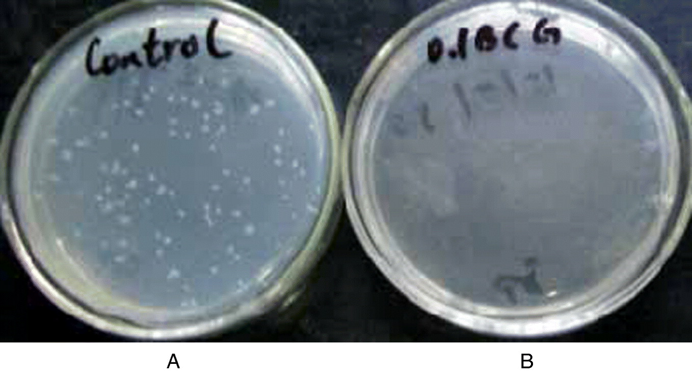
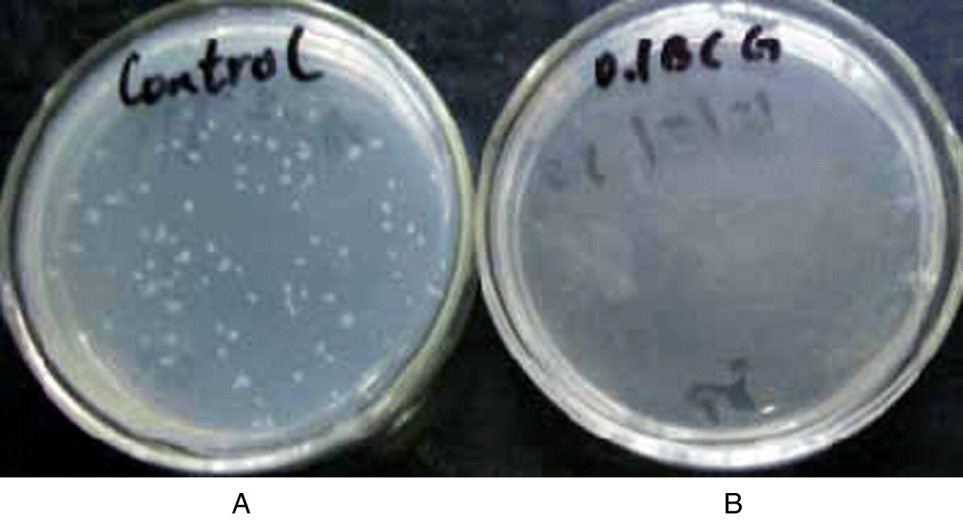
Seven-day-old mice were immunized with either 0.01 ml of BCG bacterial solution (containing about 2.0×10³ cfu living bacteria) or 0.2 ml BCG bacterial solution (containing about 4.0×10⁴ cfu living bacteria). Twenty-eight days later, mice were challenged by 0.5 ml of BCG bacterial solution (containing 105 cfu living bacteria), and the activity of anti-BCG in the control and immune groups was investigated. In the control group (not immune), a BCG colony (Fig. 1A) was observed after 2 days' culture of SPC suspension in vitro, and the number of colonies was stable after 8 days' culture. Typical colonies looked like cauliflowers. For mice in the high-dose group, no BCG colonies were seen after 2 days' culture of SPC in vitro, but a colony was observed in SPC of two mice after 8 days' culture. For the mice in the low-dose group, no colonies were observed after 12 days' culture (Fig. 1B).
Colonies of BCG in spleen cells of the inoculated and control mice after challenge with BCG, at 8 days' incubation in vitro. (A) Colonies of BCG from the control mouse spleen; (B) no colonies of BCG from the mouse inoculated with 0.01 ml BCG vaccine. (See online for a colour version of this figure.)
Impact of different doses of BCG and different ages of mice on BCG immunoprotection
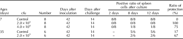
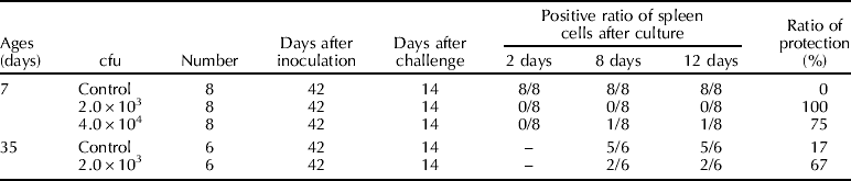
Seven-day-old neonate mice were immunized with either 0.01 ml of BCG bacterial solution (containing about 2.0×10³ cfu living bacteria) or 0.2 ml BCG bacterial solution (containing about 4.0×10⁴ cfu living bacteria). Twenty-eight days later, mice in the two groups were challenged by 0.5 ml of BCG bacterial solution (containing about 105 cfu living bacteria). The results of immunoprotection are listed in Table 1.
Detection of BCG in mouse spleens by incubation in vitro at 14 days after challenge with BCG
–, No detection.
As shown in the table, the low-dose group showed 100% immunoprotection, the high-dose group showed only 75% immunoprotection, and vaccine strain TB was detected after challenge in all SPC of mice not immunized by BCG. Table 1 also shows that 35-day-old mice showed 67% immunoprotection after being immunized with 0.01 ml BCG bacterial solution (containing about 2.0×10³ cfu live bacteria). All these results indicate that immunoprotection brought about by BCG was related to immune dosage of BCG and age of the immune animals.
Impact of different doses of BCG and different ages of mice on immune reaction type
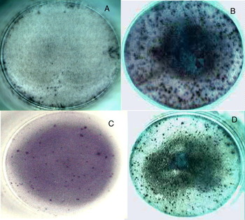
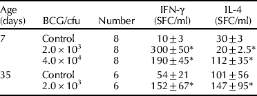
In this study, the ELISPOT assay was adopted to detect IFN-γ and IL-4 in SPC induced by BCG, and the results are shown in Fig. 2. The statistics shown in Table 2 indicate that the numbers of SPC that expressed IFN-γ increased significantly for neonate mice, both in the high-dose group and the low-dose group. However, the numbers of SPC of neonate mice that expressed IL-4 increased significantly in the high-dose group but decreased significantly in the low-dose group, which may be related to their immunoprotection ratio. For the 35-day-old immunized mice, the numbers of SPC that expressed IFN-γ and the numbers of SPC that expressed IL-4 both increased, and there existed marked differences in individual immune reactions, which may relate to the immune level of the organism before inoculation (depending on stimulation from outside, etc.).
The detection of IFN-γ (A and B) and IL-4 (C and D) in mice spleen cells, induced with 0.01 ml BCG (A and B) and 0.2 ml BCG (C and D) immunization. (A and C) Control mice; (B and D) immunized mice. One ELISPOT means one IFN-γ or IL-4 secreting cell. (See online for a colour version of this figure.)

Effect of different doses of BCG and age of mice on the IFN-γ/IL-4 induced by inoculation of BCG (SFC, mean±SD)
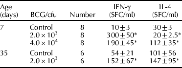
SFC, Spot forming cells. Each cell that secretes IL-4 or IFN-γ can combine their respective antibodies, forming one spot.
* Difference between groups is statistically significant at P<0.05 according to the t-test.
Discussion
Bacille Calmette–Guerin (BCG) is a live attenuated vaccine of TB and an inducer of a strong Th1 immune response. Its main component is BCG plasmonucleic acid; it has strong non-specific immunostimulation and it plays a role in immunoprotection via the above-mentioned Th1-type immune response (Vekemans et al., Reference Vekemans, Amedei and Ota2001; Hope et al., Reference Hope, Thom, Villarreal-Ramos, Vordermeier, Hewinson and Howard2005). This vaccine is widely used in the prevention of human TB and has a good protective effect (WHO, 2005), but its immunoprotective effect on cattle and sheep is controversial. Some research has indicated that vaccination failure of BCG was related to the change of immune response type induced by the vaccine itself (Suazo et al., Reference Suazo, Escalera and Torres2003; Lyashchenko et al., Reference Lyashchenko, Whelan and Greenwald2004; Denis et al., Reference Denis, Keen, Parlane, Storset and Buddle2007), especially a change of Th1-type immune response to Th2-type immune response (Buddle et al., Reference Buddle, Wedlock, Denis and Skinner2005).
The present study indicated that a Th1-type immune response induced by BCG led to a higher immunoprotection ratio, and weakened Th1-type immune response or Th2-type immune response led to a lower immunoprotection ratio. It also suggested that there are significant correlations between immunoprotection ratios and Th1/Th2-type cell immunoprotection reactions induced by BCG, indicating the need for a scientific method to analyse how BCG can induce animals such as cattle and sheep into immunoprotection, and to judge the immunoprotection ratio by detection of changes of Th1/Th2-type immune response in immunized cattle and sheep. In this study, the ELISPOT assay was adapted to analyse changes of expression of the Th1-type cytokine, IFN-γ and Th2-type cytokine, IL-4 (Power et al., Reference Power, Grand, Ismail, Peters, Yurkowski and Bretscher1999; Doherty et al., Reference Doherty, Demissie, Menzies, Andersen, Rook and Zumla2005). It has the characteristics of high sensitivity, high specificity, easy handling, etc., and it also offers a feasible scientific research method for the prevention and cure of bovine and ovine TB with BCG.
Immunoprotection induced by BCG is affected by many factors. Studies have indicated that a low dose of BCG showed larger immunoprotection in newly born individuals, and this kind of immunoprotection lasted a long time, but high doses of BCG showed weaker immunoprotection in newly born individuals, although it varied greatly between individuals. The reason for these individual differences is still unknown. It may be relevant to immune tolerance, thus Th1/Th2-type immune response are all involved, and this has been confirmed both in this study and similar earlier studies (Buddle et al., Reference Buddle, Wedlock, Parlane, Corner, De Lisle and Skinner2003, Reference Buddle, Wedlock, Denis and Skinner2005). This result indicates that effective immune dosing of BCG is one of the key factors in its immunoprotection, thus how to decide upon the effective immune dosage of BCG in different individuals should be the focal point of further study.
The age of animals at inoculation is another important factor that affects the immunoprotective effect of BCG (Marchant et al., Reference Marchant, Goetghebuer and Ota1999; Denis et al., Reference Denis, Wedlock and Buddle2005). In the present study, only 7-day-old neonate mice and 35-day-old mice were chosen for immunization with a low dose of BCG, and a significant difference in immunoprotection and immune response type was observed between them. Seven-day-old neonate mice had an immunoprotection ratio of 100% and more IFN-γ-secreting SPC, but the 35-day-old mice had an immunoprotection ratio of 67% and more SPC that secreting both IFN-γ and IL-4. This difference may be relevant to the immune state of the individual. Research has shown that factors such as the living environment of animals, infection by pathogenic microorganisms, etc., might intervene in the immunoprotection induced by BCG, thus affecting its immunoprotection ratio (Buddle et al., Reference Buddle, Wards, Aldwell, Collins and de Lisle2002; Brandt et al., Reference Brandt, Cunha and Olsen2002). Because of the complexity of this kind of interference, cattle and sheep should be inoculated early after birth for prophylactic immunization with BCG. It is therefore of great importance for us to study in more depth the early stages of immunization of cattle and sheep and methods to achieve adaptive immunity in fetuses.
Acknowledgement
This study was supported and funded by the Youth Innovation Foundation of Shandong Agricultural University and Youth Scholarship Foundation of Shandong Province.