Idiopathic dilatation of the right atrium is a rare anomaly with an unknown aetiology. It was first described in 1955Reference Binder, Rosenhek, Frank, Gwechenberger, Maurer and Baumgartner1–Reference Forbes, Kantoch, Divekar, Ross and Rebeyka3 and is included in the group of right atrium and coronary sinus malformations, which also include simple diverticulum, multiple diverticula, and diverticula of the coronary sinus.Reference Binder, Rosenhek, Frank, Gwechenberger, Maurer and Baumgartner1
Foetal diagnosis is extremely rare and requires the demonstration of diffuse enlargement of the right atrium in the absence of another cause.Reference Hofmann, Heilmann, Häusler, Dähnert, Kamin and Lachmann4 While associated cardiac malformations, such as hypertrophic cardiomyopathy, atrial septal defects,Reference Paladini, Tiesi, Buffi, Tuo and Marasini5 mitral valve prolapse, tricuspid dysplasia, and bronchial malformations, have been reported,Reference Binder, Rosenhek, Frank, Gwechenberger, Maurer and Baumgartner1 only few authors have tried to establish the aetiology.Reference Tenckhoff, Stamm and Beckwith6 However, the real pathogenesis has not been clarified. Early diagnosis in foetuses suggests a possible congenital aetiology.Reference Blaysat, Villain and Marcon2,Reference Tenckhoff, Stamm and Beckwith6,Reference Deora, Sharma and Garg7
The progression of this anomaly varies and may result in spontaneous regression, stabilisation of atrial size, and/or progressive enlargement.Reference Long8–Reference Huang10 The clinical presentation ranges from no symptoms to arrhythmias, heart failure, and even sudden death.Reference Srinivasa, Kharge, Patil, Rangan and Ramalingam11–Reference Özyurt, Baykan, Argun, Pamukçu, Mavili and Narin17 Treatment is controversial in asymptomatic patients, especially in children. Prophylactic antiplatelet treatment has been suggested to prevent the risk of thrombus formation,Reference Khare, Chandra, Agarwal and Dwivedi18 and surgical reduction has been performed in some symptomatic patients or in asymptomatic patients at high risk of progression, including initial severe right atrial dilatation, significant enlargement over a short period, compression of adjacent structures, or uncontrolled arrhythmias.Reference Deora, Sharma and Garg7
We present four cases of foetal diagnosis of idiopathic dilatation of the right atrium and discuss management from foetal to early postnatal life and follow-up until the patients reached the age of 5 to 6 years.
Materials and methods
Ours was a prospective clinical case study of four patients treated at Hospital Sant Joan de Déu (Paediatric Cardiology Department and BCNatal) in Barcelona, which is a tertiary referral centre for foetal and paediatric CHD.
All four patients were admitted to our hospital between December 2012 and April 2014. Foetal and sequential neonatal and paediatric evaluations including physical examination, blood analysis, and echocardiography were performed at the hospital. Three mothers had no disease, did not take medications, and did not experience complications during pregnancy. One mother was HIV-positive with undetectable viral load and was being treated with lamivudine, zidovudine, and ritonavir. Her pregnancy was also uncomplicated. The patients were born at term, with appropriate weight and no other cardiac or non-cardiac anomalies.
The study was approved by the local ethics committee, and written informed consent was obtained in all pregnancies from the parents or legal guardians of the patients included.
We recorded clinical characteristics such as gestational age at birth, delivery, Apgar score, umbilical artery pH, newborn anthropometric data (weight, body surface according to the Dubois formula), comorbidities, and gestational/neonatal complications.
Foetal echocardiographic data were recorded using a Siemens Sonoline Antares (Siemens Medical Systems, Malvern, PA, USA) with curved-array 2–6 and 2–10 MHz transducers. Newborn echocardiography data were recorded using an iE33 XMatrix device (Philips Ultrasound, Bothell, WA, USA) with X5–1 and/or S8–3 transducers. The CT angiography images were acquired using a PHILIPS Light Speed® device, and the MRI images were acquired using a GE Signa HD 1.5 T® device (Boston, MA, USA). The images recorded were obtained in the planes recommended by international guidelines for the assessment of cardiac parameters.
Results
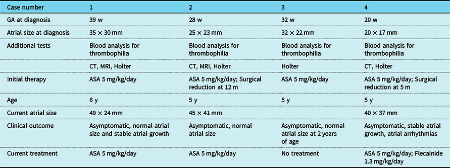
Case presentation (Table 1)
Summary of the characteristics and progression of the four cases of idiopathic dilatation of the right atrium

ASA: acetylsalicylic acid; GA: gestational age; kg: kilograms; m: months; mg: milligrams; mm: millimetres; w: weeks; y: years
Case 1
The first patient was a 28-year-old pregnant woman in whom foetal echocardiography revealed a significant increase in right atrium size at 39 weeks of gestation. The tricuspid valve was thin, with mobile leaflets, no regurgitation, and/or stenosis. The patient was in sinus rhythm. Postnatal echocardiography confirmed the diagnosis of idiopathic dilatation of the right atrium. Screening for thrombophilia was negative, and antiplatelet prophylaxis was started. The CT scan ruled out airway compression, and MRI helped to define the functioning of the right atrium, right ventricle, and tricuspid valve. Currently, 6 years later, the patient remains asymptomatic with a normal z-score for the right atrium area and normal growth in the right atrium. The electrocardiogram is normal, and the patient did not experience arrhythmias or thromboembolic events.
Case 2
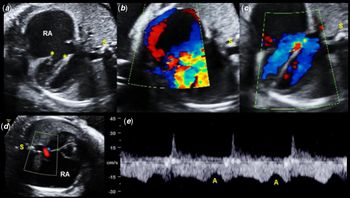
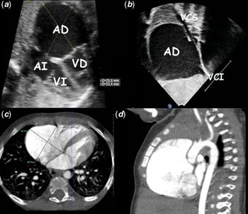
The second patient was a 33-year-old pregnant woman in whom ultrasound at 28 weeks of gestation detected cardiomegaly due to right atrium enlargement (Fig 1), with a normal tricuspid valve. This massive dilatation (35 × 34 mm), without thrombi (Fig 2a,b), was confirmed with postnatal echocardiography using spontaneous echo contrast. Slight tricuspid valve insufficiency was observed. Neither heart defects nor electrocardiographic anomalies were detected, although there was rapid growth of the right atrium (45 mm at 2 months and 54 mm at 3 months). A cardiac CT scan confirmed massive cavity dilatation without thrombi or airway compression (Fig 2c,d; Supplementary Fig S1a,b), MRI showed an absence of right atrium contractility, and late gadolinium enhancement ruled out fibrosis. At 7 months of age, the patient remained asymptomatic, and the right atrium had a diameter of 67 mm; therefore, reduction plasty with pericardial reinforcement was performed without cardiopulmonary bypass. The disease work-up did not reveal any specific lesions (Supplementary Fig S2). After surgery, the diameter of the atrium decreased dramatically to 37 mm, and follow-up was uncomplicated.
(a, b) Four-chamber view showing a markedly enlarged right atrium (RA) which is filled with blood at low-velocity flow. (*) Correct insertion of the tricuspid valve. (c) Normal tricuspid and mitral flow characteristics with equal-sized ventricles. Note the presence of a normal right-to-left shunt across the foramen ovale (*). (d) Predominant right-to-left shunt across the foramen ovale. (e) Normal pulmonary venous flow pattern. S: spine.

(a,b) Massive dilatation of the right atrium (RA) in a four-chamber and subcostal view via transthoracic contrast echocardiography at age of 5 months. (c,d) CT scan showing marked dilatation of the right atrium before atrial reduction plasty. SVC and IVC: superior and inferior vena cava.

Case 3
The third patient was a 32-year-old HIV-positive pregnant woman. Ultrasound screening at week 32 revealed abnormal growth of the right atrium. The diffuse growth was confirmed postnatally. No thrombi or associated heart defects were detected. In the postnatal follow-up, the abnormal growth of the right atrium had stopped, and at 2 years of age, the size was normal (right atrium area z-score of + 1.7).Reference Cantinotti, Scalese and Murzi19 At the age of five, the patient remained asymptomatic, with no rhythm abnormalities, and with normal growth of the right atrium; therefore, antiplatelet therapy was stopped.
Case 4
The fourth patient was a 28-year-old pregnant woman in whom echocardiography at 20 weeks of gestation detected cardiomegaly due to right atrium enlargement with a normal tricuspid valve. Postnatal echocardiography confirmed this massive dilatation, with no thrombi. Given the rapid and huge enlargement, CT angiography was performed and revealed compression of the other cardiac cavities and the pulmonary artery. In addition, episodes of atrial tachycardia that were difficult to control with antiarrhythmic treatment (flecainide, propranolol) were recorded; therefore, atrial reduction plasty was performed at the age of 5 months. The patient is now 5 years old, and the right atrial area has a z-score of 2 with stabilised atrial growth,Reference Cantinotti, Scalese and Murzi19 although atrial arrhythmias continue to be recorded on the electrocardiogram. Since the patient remains asymptomatic and no changes in left ventricle function or size have been observed, the therapeutic approach comprises radioablation in the cath lab only if the arrhythmias persist or worsen.
Discussion
Idiopathic dilatation of the right atrium is rarely described in children. Many of those diagnosed with this condition were initially classified as having Ebstein’s anomaly, tumours, or pericardial effusion.Reference Binder, Rosenhek, Frank, Gwechenberger, Maurer and Baumgartner1,Reference Özyurt, Baykan, Argun, Pamukçu, Mavili and Narin17,Reference Narain, Puri and Sethi20. The detection of four cases in a short period of time suggests that this anomaly could be underdiagnosed.
Reports of familial casesReference Jenni, Goebel, Schneider and Krayenbühl21,Reference Kurz, Oechslin, Kobza and Jenni22 and the association with Holt-Oram syndromeReference Paladini, Tiesi, Buffi, Tuo and Marasini5 reinforced the suspicion of a genetic origin, although the real pathogenesis was not demonstrated. Histopathological studies detected non-specific changes that did not make a significant contribution to the identification of the real cause of this condition. Some authors report non-specific findings such as tiny atrial tissue, as well as nonspecific muscular changes such as elongation of the fibres, lipomatous degeneration, lymphocytic infiltration, and patch fibrosis.Reference Özyurt, Baykan, Argun, Pamukçu, Mavili and Narin17,Reference Khare, Chandra, Agarwal and Dwivedi18,Reference Chatrath, Turek, Quivers, Driscoll, Edwards and Danielson23 The finding of inflammatory tissue in some samples suggested myocarditis as a possible cause,Reference Binder, Rosenhek, Frank, Gwechenberger, Maurer and Baumgartner1 although this was only detected in isolated cases. In our series, the patient in whom we performed a histopathological study showed non-specific elements, which, consistent with the literature, did not prove useful.
Many paediatric cases are asymptomatic, as were those we present, although other authors have reported palpitations, non-specific chest pain, dyspnoea due to airway compression, symptomatic atrial arrhythmias, syncope, and even sudden death.Reference Binder, Rosenhek, Frank, Gwechenberger, Maurer and Baumgartner1,Reference Tenckhoff, Stamm and Beckwith6,Reference Deora, Sharma and Garg7,Reference Özyurt, Baykan, Argun, Pamukçu, Mavili and Narin17 The possible risk of a thrombogenic event is known, and in idiopathic dilatation of the right atrium, initial antiplatelet treatment is accepted. Anticoagulation therapy is indicated in patients with atrial thrombosis or an added risk of thrombophilia. Arrhythmic treatment has been used successfully, and in refractory cases, ablation with radiofrequency or surgical resection with the Maze procedure has been performed.Reference Binder, Rosenhek, Frank, Gwechenberger, Maurer and Baumgartner1,Reference Tenckhoff, Stamm and Beckwith6,Reference Chatrath, Turek, Quivers, Driscoll, Edwards and Danielson23 In adults with rapid atrial enlargement, surgery is indicated if the atrium increases to over four times its normal size, although in children without symptoms, surgery is controversial. Given the relative ease of the intervention and the near absence of complications, this approach is justified, even in asymptomatic children, in order to avoid the risk of thrombosis and arrhythmias.Reference Deora, Sharma and Garg7 The technique varies from atrial resection to atrial reduction plasty without cardiopulmonary bypass.Reference Chatrath, Turek, Quivers, Driscoll, Edwards and Danielson23 We started antiplatelet prophylaxis with acetylsalicylic acid and maintained this approach indefinitely, since a review of the literature provided no indications with respect to the duration of prophylaxis. Treatment was stopped only in the patient with spontaneous normalisation of atrium size. We follow our patients periodically, and in the two patients in whom we detected progressive rapid atrial enlargement and uncontrolled arrhythmias, we opted for surgical reduction without the need for extracorporeal circulation.
Supplementary material
To view supplementary material for this article, please visit https://doi.org/10.1017/S1047951120001353
Acknowledgements
The authors are grateful to the following for their involvement and cooperation: the Foetal Cardiology Department of Hospital Clinic, Maternal Centre, Barcelona, Spain. Doctor Lorenzo Jiménez and the Department of Paediatric Cardiology, Hospital Miguel Servet, Zaragoza, Spain.
Financial support
This research received no specific grant from any funding agency or from the commercial or not-for-profit sectors.
Conflicts of interest
None.