Highlights
-
This study systematically evaluated risk factors, pathogen spectrum and antimicrobial resistance in secondary intracranial infections among patients with viral encephalitis.
-
Independent predictors included advanced age, comorbidities, prolonged corticosteroid therapy, multiple invasive procedures and elevated CSF protein levels.
-
A multivariable prediction model demonstrated strong discriminative performance (AUC = 0.86).
Background
Viral encephalitis remains a major public health challenge worldwide, with an estimated incidence of 5–8 cases per 100,000 population annually worldwide. Reference Seitz, Clarke and Tyler1–Reference Azab and Azzam3 Despite improvements in antiviral therapy and neurocritical care, the disease continues to be associated with high morbidity and mortality. Reference Wishart, Spiteri, Tan, Macia and King4–Reference Cheng, Wang and Xia7 The most common causative pathogens include herpes simplex virus and Japanese encephalitis virus, although other neurotropic viruses also contribute to the disease burden. Reference Zhao, Sun and Fang8,Reference Yeboah, Gorman and El-Duah9 Survivors frequently suffer from long-term neurological sequelae such as cognitive impairment, epilepsy and persistent functional disabilities, leading to considerable personal suffering and substantial societal and economic costs. Reference Wishart, Spiteri, Tan, Macia and King4,Reference Xu, Jia and Duan10,Reference Wurdack, Pruss and Finke11 Secondary intracranial infections represent one of the most frequent and severe complications during hospitalization for viral encephalitis. Reference Senel, Rapp and Mayer12–Reference Ratho, Sethi and Prasad14 Such infections substantially increase the risk of in-hospital mortality and disability, while also prolonging the duration of hospitalization and significantly escalating healthcare expenditures Reference Zhao, Sun and Fang8,Reference Depledge, Cudini and Kundu15 . Patients with severe disease, particularly those requiring intensive care or undergoing multiple invasive procedures, are at an especially high risk of developing these infections. Reference Yeboah, Gorman and El-Duah9,Reference Xu, Jia and Duan10,Reference Wakelin, Wolff and Angus-Leppan16 Given their impact on both short- and long-term outcomes, prevention and early identification of secondary intracranial infections are of critical importance in the management of viral encephalitis. Reference Sonneville, Lannuzel, Mathieu, Gonzalez-Dunia and Gaudin17
Several clinical risk factors for secondary intracranial infections following viral encephalitis have been reported, including advanced age, underlying comorbidities, immunosuppression, prolonged corticosteroid therapy and multiple invasive procedures, while certain laboratory parameters such as elevated CSF protein, C-reactive protein (CRP) and procalcitonin (PCT) have also been implicated as potential indicators of increased susceptibility. Reference Schlegel, Allartz, Wenzel, Faupel, Loester and Tappe6,Reference Pons-Espinal, Lopez-Perea, Masa-Calles, Munoz-Almagro, Tarrago and Launes18–Reference McMillen, Powell, Jani, Scipione, Seo and Babady20 However, evidence remains inconsistent due to limited sample sizes, heterogeneous infection definitions and the predominance of retrospective studies. Reference Gozdas21–Reference Dwibedi, Satapathy and Jain23 Previous reports further highlight that both Gram-positive cocci and Gram-negative bacilli contribute to these infections, with methicillin-resistant Staphylococcus aureus (MRSA) and multidrug-resistant Acinetobacter baumannii posing particular concern given their high prevalence and resistance profiles; rising carbapenem resistance in A. baumannii and extended-spectrum β-lactamase (ESBL)–producing Klebsiella pneumoniae exacerbate therapeutic challenges and restrict effective empirical antibiotic choices. Reference Cheng, Wang and Xia7,Reference Dharmagadda, Tambolkar, Mane and Singh24–Reference Ander, Fish, da Silva, Davenport, Parks and Morrison26
Comprehensive, multifactorial models tailored to viral encephalitis populations have not yet been established, leaving clinicians without validated tools for early risk stratification and timely preventive interventions. Reference Yeboah, Gorman and El-Duah9,Reference Xu, Jia and Duan10 This study aimed to systematically evaluate the risk factors, pathogen spectrum and antimicrobial resistance profiles associated with secondary intracranial infections in patients with viral encephalitis. In addition, we sought to develop a multifactorial prediction model based on clinical, therapeutic and laboratory variables to identify high-risk patients and enhance early recognition and prevention in clinical practice, which highlights the contributions of host factors, treatments and biological markers to secondary intracranial infections and adds evidence on neurocritical complications. By incorporating microbiological and resistance data, it provides useful information to guide the prevention and management of central nervous system (CNS) infections.
Methods
Study design and setting
This was a single-center cohort study conducted at Cangzhou Hospital of Integrated Traditional Chinese and Western Medicine Hospital, a tertiary care center in Hebei Province, China. Consecutive patients admitted between March 1, 2022, and October 31, 2024, with a confirmed diagnosis of viral encephalitis, were included. The study protocol was reviewed and approved by the institutional ethics committee. Given the retrospective nature of the study, the requirement for informed consent was waived. All data were anonymized prior to analysis to ensure patient confidentiality.
Inclusion criteria and exclusion criteria
Patients were eligible for inclusion if they were aged ≥18 years and fulfilled the established clinical diagnostic criteria for viral encephalitis. The diagnosis was based on a combination of acute onset of fever, altered mental status (including confusion, decreased level of consciousness or seizures) and supportive laboratory and imaging findings. Specifically, CSF analysis was required to show evidence of CNS inflammation (elevated white blood cell count, increased protein concentration and/or decreased CSF-to-blood glucose ratio), and neuroimaging (MRI or CT) was used to exclude alternative noninfectious causes and to support the diagnosis when encephalitic changes were evident. All included patients were free from documented intracranial infection at the time of admission and had complete demographic, clinical, laboratory and follow-up data available.
Exclusion criteria were as follows: (1) concurrent bacterial meningitis or fungal intracranial infection, confirmed by CSF culture or antigen testing; (2) patients with known HIV infection, AIDS or other severe immunocompromised states (such as those receiving long-term chemotherapy, radiotherapy or post-organ transplantation immunosuppressive therapy); (3) patients discharged or deceased within 48 hours of admission, which precluded adequate clinical evaluation and follow-up; and (4) patients with substantial missing data in key demographic, laboratory or clinical variables, preventing reliable analysis.
Time-to-event definition
For survival analyses, the time-to-event was defined from the date of hospital admission (time zero) to the date of the first confirmed diagnosis of secondary intracranial infection. Patients who did not develop intracranial infection were censored at the earliest of hospital discharge, in-hospital death without infection, transfer to another facility or the administrative end of follow-up (October 31, 2024).
Definition of secondary intracranial infection
Secondary intracranial infection was diagnosed based on a combination of clinical, laboratory and microbiological criteria. Clinically, patients were required to present with persistent fever, worsening disturbance of consciousness and/or signs of elevated intracranial pressure. Laboratory evidence included CSF abnormalities such as pleocytosis, elevated protein concentration and decreased CSF-to-blood glucose ratio. Microbiological confirmation was obtained through positive cultures from CSF or blood samples. Patients with noninfectious causes of fever, such as drug reactions or tumor-related fever, were carefully excluded.
Risk factor variables
Potential risk factors for secondary intracranial infection were categorized into three domains. Host-related variables included age, sex and comorbidities such as diabetes mellitus, hypertension and chronic pulmonary disease. Treatment-related variables comprised the duration of corticosteroid use and the number of invasive procedures performed during hospitalization, including tracheostomy, external ventricular drainage and central venous catheterization. Laboratory parameters included CSF indices (white blood cell count, protein concentration and CSF-to-blood glucose ratio) as well as systemic inflammatory markers, specifically CRP and PCT.
Data collection
Data were retrospectively extracted from the hospital’s electronic medical records, laboratory information system and infection control registry. The following information was collected for each eligible patient: demographic characteristics, including age and sex; comorbidities, such as hypertension, diabetes mellitus and chronic hepatic or renal disease; clinical presentation and severity, assessed by the Glasgow Coma Scale (GCS) score at admission; treatment-related factors, including the use of antiviral drugs, duration of corticosteroid therapy and the number and types of invasive procedures performed (e.g., tracheostomy, external ventricular drainage and central venous catheterization); laboratory and imaging data, including CSF analysis and hematological and biochemical parameters; and clinical outcomes, including the occurrence and timing of secondary intracranial infection, length of hospital stay and prognosis at discharge.
Microbiological analysis
Clinical specimens, including CSF, blood and sputum (when clinically indicated), were collected for microbiological examination. Pathogen isolation and identification were performed in the hospital’s central clinical laboratory using standard procedures, including automated systems such as VITEK 2 (bioMérieux, France) or matrix-assisted laser desorption ionization time-of-flight mass spectrometry. Antimicrobial susceptibility testing was conducted according to the guidelines of the Clinical and Laboratory Standards Institute, and resistance profiles were interpreted based on established breakpoints. For each major isolate, resistance rates to commonly used antimicrobial agents were recorded, and multidrug-resistant organisms (MDROs) were classified according to international consensus definitions.
Outcomes
The primary outcome of this study was the occurrence and time to onset of secondary intracranial infection, defined according to the clinical, laboratory and microbiological criteria described above. The secondary outcomes included (1) changes in length of hospital stay associated with the development of secondary intracranial infection and (2) the distribution of causative pathogens and their antimicrobial resistance profiles among infected patients.
Laboratory measurements
All laboratory tests were performed within 24 hours of admission and at key clinical time points during hospitalization. Routine hematological and biochemical parameters, including leukocyte count, hemoglobin, platelet count, serum glucose, electrolytes and liver and renal function, were measured using automated hematology analyzers and biochemical analyzers in the hospital’s central laboratory. Inflammatory biomarkers included CRP, determined by immunoturbidimetric assay, and PCT, assessed by electrochemiluminescence immunoassay. CSF analyses comprised cell counts, protein concentration, glucose and chloride levels, which were measured with automated cell counters and biochemical analyzers; the CSF-to-blood glucose ratio was calculated using simultaneous blood glucose values. All assays were conducted in accordance with national clinical laboratory standards, and both internal and external quality controls were routinely implemented to ensure reliability and accuracy.
Statistical analysis
Descriptive statistics were used to summarize baseline characteristics. Continuous variables were presented as mean ± standard deviation (SD) or median with interquartile range (IQR), depending on distribution, and categorical variables were expressed as frequencies and percentages. Group comparisons were performed using Student’s t test or the Mann–Whitney U test for continuous variables and the χ 2 test or Fisher’s exact test for categorical variables. Crude odds ratios (ORs) or hazard ratios (HRs) with 95% confidence intervals (CIs) were calculated in univariate analyses.
Variables with P < 0.10 in univariate analysis were entered into multivariate logistic regression models to identify independent risk factors, with results reported as adjusted ORs (95% CI). For time-to-event analyses, Kaplan–Meier curves were constructed to compare infection-free survival between groups stratified by key risk factors or model-based risk categories, and differences were assessed using the log-rank test. Multivariate Cox proportional hazards regression was further applied to estimate HRs (95% CI), and proportional hazards assumptions were evaluated using Schoenfeld residuals.
Logistic regression was used to estimate the overall risk of developing secondary intracranial infection during hospitalization, providing effect estimates in terms of ORs. In parallel, Cox proportional hazards regression was applied to evaluate the temporal dynamics of infection occurrence by incorporating time-to-event information and censoring, thereby identifying factors associated with earlier onset of infection. The combined use of these two models allowed complementary assessment of both risk magnitude and timing.
Model performance was assessed using receiver operating characteristic (ROC) curves, with the area under the curve (AUC) calculated and compared using DeLong’s test. Calibration was evaluated by calibration plots, and predictive performance was further assessed by sensitivity, specificity, positive predictive value (PPV), negative predictive value (NPV), Youden’s index and the Brier score.
Microbiological data were analyzed descriptively, including the distribution of isolated pathogens (illustrated by bar or donut charts), antimicrobial resistance patterns (depicted by heatmaps) and the prevalence of MDROs.
Subgroup analyses were performed according to age group, comorbidity status, corticosteroid use and number of invasive procedures. Sensitivity analyses were conducted by applying a strict definition of secondary intracranial infection (culture-confirmed cases only), excluding patients with extreme laboratory values and truncating follow-up at 7 days of hospitalization.
Results
Baseline characteristics
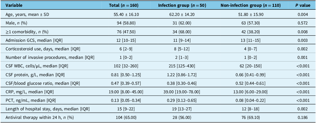
The median follow-up duration for the entire cohort was 16 days (IQR 10–23 days). During this period, 50 patients (31.3%) developed secondary intracranial infections. The overall incidence rate of infection was 2.9 events per 1000 patient-days, reflecting a substantial risk within the acute hospitalization period. A total of 160 patients diagnosed with viral encephalitis were included in the cohort, of whom 50 (31.3%) developed secondary intracranial infections during hospitalization. Baseline demographic and clinical characteristics stratified by infection status are summarized in Table 1.
Baseline characteristics of patients with viral encephalitis stratified by secondary intracranial infection status

Note: Continuous variables are expressed as mean ± SD or median [IQR] as appropriate. Categorical variables are expressed as n (%). Bold indicates statistically significant differences (P < 0.05).
CRP = C-reactive protein; GCS = Glasgow Coma Scale; IQR = interquartile range; PCT = procalcitonin; WBC = white blood cell count.
Patients in the infection group were significantly older than those without infection (62.20 ± 14.20 vs. 51.80 ± 15.90 years, P = 0.004). The prevalence of comorbidities was higher in the infection group (68.0% vs. 38.2%, P = 0.008). At admission, patients with infections presented with lower GCS scores (median 11 [IQR 9–14] vs. 13 [IQR 11–15], P = 0.003). Regarding treatment-related variables, the infection group had longer corticosteroid exposure (median 8 [IQR 5–12] vs. 4 [IQR 0–7] days, P = 0.002) and more invasive procedures (median 2 [IQR 1–3] vs. 1 [IQR 0–2], P = 0.001). Laboratory findings also revealed more severe abnormalities: higher CSF WBC counts (median 215 vs. 62 cells/µL, P < 0.001), higher CSF protein levels (median 1.22 vs. 0.66 g/L, P < 0.001), lower CSF/blood glucose ratios (median 0.38 vs. 0.52, P < 0.001) and elevated inflammatory biomarkers including CRP (median 39.00 vs. 13.00 mg/L, P < 0.001) and PCT (median 0.29 vs. 0.08 ng/mL, P < 0.001). In addition, patients who developed intracranial infections had longer hospital stays compared with noninfected patients (median 19 vs. 12 days, P = 0.002). No significant differences were observed in sex distribution (P = 0.572) or initiation of antiviral therapy within 24 hours of admission (P = 0.186).
Risk factor analysis
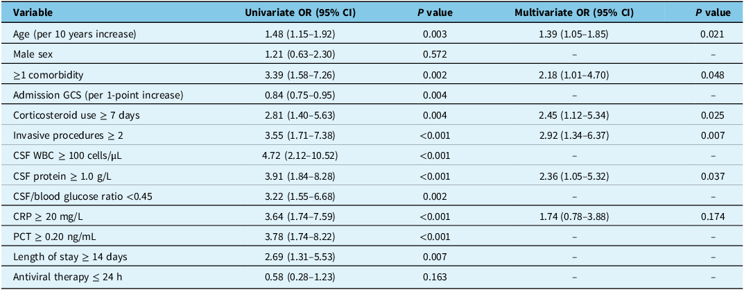
As shown in Table 2, univariate analysis identified older age, comorbidities, lower admission GCS scores, prolonged corticosteroid use, multiple invasive procedures, elevated CSF WBC and protein levels, reduced CSF/blood glucose ratios and increased CRP and PCT levels as significant predictors of secondary intracranial infections (all P < 0.05). In the multivariate logistic regression model, independent risk factors included older age (adjusted OR 1.39, 95% CI 1.05–1.85, P = 0.021), presence of comorbidities (adjusted OR 2.18, 95% CI 1.01–4.70, P = 0.048), corticosteroid use for ≥7 days (adjusted OR 2.45, 95% CI 1.12–5.34, P = 0.025), ≥2 invasive procedures (adjusted OR 2.92, 95% CI 1.34–6.37, P = 0.007) and elevated CSF protein levels (adjusted OR 2.36, 95% CI 1.05–5.32, P = 0.037). CRP, which was significant in univariate analysis, lost significance after adjustment.
Univariate and multivariate logistic regression analyses of risk factors for secondary intracranial infections in patients with viral encephalitis

Note: “–” indicates the variable was not retained in the final multivariate model.
CI = confidence interval; CRP = C-reactive protein; GCS = Glasgow Coma Scale; OR = odds ratio; PCT = procalcitonin; WBC = white blood cell count.
Survival analysis
To further assess the temporal dynamics of infection risk, Kaplan–Meier survival curves were generated with “time to secondary intracranial infection” as the outcome (Figure 1). Patients receiving corticosteroids for ≥7 days had a significantly shorter infection-free survival compared with those with shorter exposure (median infection-free time 12 vs. 20 days, log-rank P = 0.004). Similarly, patients undergoing ≥2 invasive procedures demonstrated a higher cumulative incidence of intracranial infections (median infection-free time 11 vs. 19 days, log-rank P < 0.001).
Kaplan–Meier survival curves for time to secondary intracranial infection in patients with viral encephalitis. (A) Patients stratified by corticosteroid exposure (≥7 vs. <7 days). Patients receiving prolonged corticosteroid therapy (≥7 days) had a significantly higher risk of intracranial infection and a shorter infection-free survival compared with those treated for <7 days (log-rank P = 0.011). (B) Patients stratified by the number of invasive procedures (≥2 vs. <2). Patients undergoing ≥2 invasive procedures experienced earlier onset of intracranial infections and a markedly lower probability of remaining infection-free compared with those with fewer procedures (log-rank P < 0.001). Legend: Kaplan–Meier curves show the cumulative probability of remaining free from secondary intracranial infection during hospitalization. The y-axis represents the infection-free survival probability (%), and the x-axis represents time since admission (days). Censored observations are indicated by tick marks. Log-rank tests were used to compare survival distributions between groups.

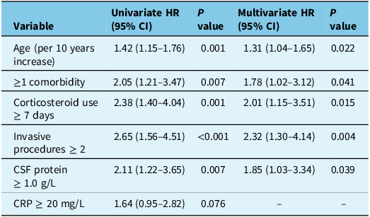
Univariate Cox regression identified older age, presence of comorbidities, prolonged corticosteroid use, multiple invasive procedures and elevated CSF protein as significant predictors of earlier infection onset (all P < 0.01; Table 3). In the multivariate model, independent risk factors included age (HR 1.31 per 10-year increase, 95% CI 1.04–1.65, P = 0.022), comorbidities (HR 1.78, 95% CI 1.02–3.12, P = 0.041), corticosteroid use ≥7 days (HR 2.01, 95% CI 1.15–3.51, P = 0.015), ≥2 invasive procedures (HR 2.32, 95% CI 1.30–4.14, P = 0.004) and CSF protein ≥1.0 g/L (HR 1.85, 95% CI 1.03–3.34, P = 0.039). CRP lost significance after adjustment.
Cox regression analysis of risk factors for time to secondary intracranial infection

Note: “–” indicates variable not retained in final model.
CI = confidence interval; CRP = C-reactive protein; HR = hazard ratio.
Predictive performance of biomarkers and multivariate model
ROC curve analysis demonstrated that single biomarkers, including CRP, PCT, CSF protein and the number of invasive procedures, showed moderate discriminative ability with AUCs ranging from 0.71 to 0.76 (Figure 2, Table 4). Among them, CSF protein achieved the highest AUC of 0.76 (95% CI 0.68–0.84).
Receiver operating characteristic (ROC) curves for predicting secondary intracranial infections. ROC curves are shown for four single predictors (C-reactive protein [CRP], procalcitonin [PCT], CSF protein and number of invasive procedures) and for the combined multivariate risk score. The multivariate model demonstrated superior discriminative ability compared with any individual biomarker. The x-axis represents the false positive rate (1 − specificity), and the y-axis represents the true positive rate (sensitivity).

Predictive performance of individual biomarkers and multivariate risk model

Note: Cut-off values for single predictors were determined using Youden’s index. The multivariate risk score was derived from the final logistic regression model.
AUC = area under the curve; CI = confidence interval; CRP = C-reactive protein; NPV = negative predictive value; PPV = positive predictive value.
The combined multivariate risk score substantially improved predictive performance, yielding an AUC of 0.86 (95% CI 0.80–0.92), with a sensitivity of 80.0% and a specificity of 81.8%. The Youden index (0.62) and Brier score (0.139) also indicated superior calibration and discrimination. DeLong’s test confirmed that the multivariate model significantly outperformed the best single biomarker (CRP) (P < 0.01).
Pathogen spectrum and antimicrobial resistance
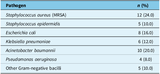
Among the 50 patients with secondary intracranial infections, positive cultures revealed a predominance of Gram-positive cocci (34.0%) and Gram-negative bacilli (66.0%) (Table 5, Figure 3). The most common isolates were MRSA (24.0%) and A. baumannii (20.0%), followed by Escherichia coli (16.0%) and K. pneumoniae (12.0%).
Pathogen distribution of secondary intracranial infections. A total of 50 positive cultures were obtained from patients with secondary intracranial infections. The predominant isolates were methicillin-resistant Staphylococcus aureus (MRSA, 24.0%) and Acinetobacter baumannii (20.0%), followed by Escherichia coli (16.0%) and Klebsiella pneumoniae (12.0%). Minor isolates included Staphylococcus epidermidis, Pseudomonas aeruginosa and other Gram-negative bacilli. Legend: The donut chart illustrates the relative proportions of isolated pathogens. MRSA and multidrug-resistant A. baumannii were the leading organisms, underscoring their clinical significance in hospital-acquired intracranial infections.

Distribution of pathogens isolated from patients with secondary intracranial infections (n = 50, positive cultures)

MRSA = methicillin-resistant Staphylococcus aureus.
Antimicrobial susceptibility testing demonstrated alarming resistance profiles across multiple pathogens (Table 6, Figure 4). MRSA exhibited near-universal resistance to penicillin (95%) and high resistance to fluoroquinolones (72%) and aminoglycosides (65%), although susceptibility to vancomycin was preserved. A. baumannii showed extensive multidrug resistance, with resistance rates of 85% to cephalosporins and 70% to carbapenems, underscoring the therapeutic challenges posed by this pathogen. Notably, extended-spectrum β-lactam resistance was also common among E. coli and K. pneumoniae.
Antimicrobial resistance patterns of major isolates (% resistant)

MRSA = methicillin-resistant Staphylococcus aureus.
Antimicrobial resistance profiles of major isolates. The heatmap summarizes resistance rates of the most frequently isolated pathogens against commonly used antibiotic classes. Methicillin-resistant Staphylococcus aureus (MRSA) exhibited extremely high resistance to penicillin (95%), cephalosporins (70%) and fluoroquinolones (72%), while remaining fully susceptible to vancomycin. A. baumannii displayed multidrug resistance, with resistance rates of 85% to cephalosporins and 70% to carbapenems, highlighting its therapeutic challenge. Legend: Rows represent pathogens, and columns represent antibiotic classes. Color intensity indicates the resistance percentage. The high prevalence of MRSA and carbapenem-resistant A. baumannii significantly limits empirical antibiotic options, stressing the importance of antimicrobial stewardship and precision therapy.

Subgroup analyses and sensitivity analyses
In predefined subgroups, the associations between risk factors and secondary intracranial infection were generally consistent (Supplementary Table S1). Older patients (≥60 years) and those with comorbidities exhibited higher infection risks (HR 1.72, 95% CI 1.05–2.81, P = 0.031; HR 1.88, 95% CI 1.12–3.14, P = 0.017, respectively). Prolonged corticosteroid use (≥7 days) and multiple invasive procedures (≥2) were also associated with earlier infection onset. No statistically significant interactions were observed between subgroups (all P interaction >0.05), suggesting robustness across patient strata. The findings remained stable in sensitivity analyses. When infection was strictly defined by CSF culture positivity, the association between risk exposures and infection persisted (HR 1.96, 95% CI 1.14–3.38, P = 0.015). Exclusion of patients with extreme laboratory values or truncating follow-up to those hospitalized for more than 7 days did not materially alter the effect estimates.
Discussion
In this study, we conducted a comprehensive evaluation of the risk factors, pathogen spectrum and antimicrobial resistance profiles associated with secondary intracranial infections among patients with viral encephalitis. We identified advanced age, the presence of comorbidities, prolonged corticosteroid therapy (≥7 days), multiple invasive procedures (≥2) and elevated CSF protein levels as independent predictors of infection. Importantly, the multifactorial prediction models developed using logistic and Cox regression analyses demonstrated superior discriminative performance compared with individual clinical or laboratory indicators, with an area under the ROC curve of 0.86, indicating robust predictive capability. Furthermore, microbiological analyses revealed that MRSA and A. baumannii were the predominant pathogens, both of which exhibited high rates of multidrug resistance, underscoring the substantial therapeutic challenges in this population.
By integrating logistic and time-to-event analyses, our study provides a comprehensive characterization of both the risk and temporal patterns of secondary intracranial infections in patients with viral encephalitis. Our findings are generally consistent with prior research on intracranial infections in neurocritical care populations. Reference Cheng, Wang and Xia7,Reference Ye, Ding, Shen, Wang, Wu and Chen27–Reference Hu, Huang, Zhu, Sun and Zhang30 Advanced age and the presence of comorbidities have repeatedly been identified as major risk factors, Reference Goel, Chakravarti, Mantan, Kumar and Ashraf31 reflecting the impact of immune-senescence and impaired host defenses on infection susceptibility. Reference Yang, Li, Tu, Li and Wei32–Reference Sun, Ni, Liu and Liu34 Similarly, prolonged corticosteroid exposure has been reported in both domestic and international studies as a double-edged sword: while corticosteroids may attenuate cerebral inflammation, excessive or extended use suppresses immunity and predisposes patients to secondary infections. Reference Singh, Singh, Mishra, Singh and Kumar35–Reference Maharaj, Bosco-Lauth and Langevin37 The association between multiple invasive procedures and infection risk is also in line with evidence from neurosurgical and intensive care unit settings, underscoring the iatrogenic contribution of device-related infections. Reference Ye, Ding, Shen, Wang, Wu and Chen27,Reference Yang and Wei28,Reference Bakovic, Bhalla, Alem, Campbell, Zhou and Narayanan38 Notably, elevated CSF protein emerged as a novel predictor in our study, suggesting that disruption of the blood–brain barrier (BBB) and increased permeability may play a role in facilitating pathogen entry into the CNS. Reference Olsen, Morrey, Smee and Sidwell39–Reference Bonney, Seitz and Ryan43
Importantly, the multivariable model developed in this study is intended for risk stratification rather than for diagnostic or prognostic decision-making. Its primary purpose is to identify patients at higher risk of developing secondary intracranial infection during hospitalization, thereby facilitating early preventive measures and closer clinical monitoring. Given the observational design and lack of external validation, the model should not be interpreted as a standalone diagnostic or prognostic tool.
With regard to microbiology and antimicrobial resistance, the predominance of MRSA and A. baumannii in our cohort mirrors the pathogen spectrum commonly reported in neurocritical care units. Reference Zhang, Zhang and Han44 The high carbapenem resistance rate observed in A. baumannii is consistent with data from multicenter antimicrobial surveillance studies in China, highlighting its significance as a major multidrug-resistant pathogen. In contrast, international studies have reported a relatively higher prevalence of ESBL-producing Enterobacteriaceae, whereas MRSA and carbapenem-resistant A. baumannii appear more prominent in our cohort, indicating potential regional differences in resistance patterns and emphasizing the importance of local epidemiological data to guide empirical therapy. Reference Li, Cheng and Wei45–Reference Corral-Rodriguez, Moreno-Contreras and Sierra-Campos48 It should be noted that the antimicrobial resistance patterns observed in this study likely reflect local epidemiology and institutional antimicrobial practices at our center. Therefore, these findings may not be directly generalizable to other regions or healthcare settings with different pathogen distributions and resistance profiles.
Several plausible mechanisms may underlie the observed associations. From a host perspective, advanced age and underlying comorbidities likely increase vulnerability through immune-senescence and diminished organ function, which together reduce the capacity to mount effective immune responses. Reference Senel, Rapp and Mayer12,Reference Yang, Xiong and Liu49–Reference Rempel, Murray, Meisner and Buchmeier51 Corticosteroid therapy, while commonly employed to mitigate cerebral inflammation in viral encephalitis, suppresses cellular immunity and delays clearance of pathogens, thereby creating conditions conducive to secondary infection. Reference Reiss, Chesler, Hodges, Ireland and Chen52–Reference Poluektova, Gorantla, Faraci, Birusingh, Dou and Gendelman54 From a treatment-related standpoint, repeated invasive procedures disrupt natural anatomical barriers of the CNS, providing entry routes for colonizing microorganisms and facilitating nosocomial transmission. Reference Melamed, Avraham and Rothbard55,Reference Mancini, Caignard and Charbonneau56 Finally, laboratory markers such as elevated CSF protein may reflect BBB disruption and increased permeability, which can enhance microbial invasion into the CNS. Reference Li, Wang and Yu41,Reference Bonney, Seitz and Ryan43 Elevated CSF protein may reflect underlying BBB dysfunction or neuroinflammatory activity and therefore serve as a surrogate marker of increased vulnerability to secondary intracranial infection. Importantly, CSF protein should be interpreted as a risk indicator or pathophysiological correlate rather than a direct causal mechanism. This finding highlights a potential pathophysiological link between BBB dysfunction and susceptibility to intracranial infection. Reference Li, Wang and Yu41
The findings of this study carry important implications for clinical practice. Risk stratification using the proposed multivariable model may enable early identification of high-risk patients and guide individualized preventive strategies. Infection control should focus on reducing unnecessary invasive procedures and using corticosteroids judiciously to avoid prolonged or high-dose regimens that increase susceptibility to infection. In addition, empirical antimicrobial therapy needs to provide adequate coverage for MRSA and carbapenem-resistant A. baumannii, while antimicrobial stewardship remains essential to ensure precision prescribing and mitigate the spread of resistance.
Limitations and future directions
This study has several limitations. First, it was a single-center retrospective analysis, which may have introduced selection bias and information bias. Second, the sample size was relatively limited, and the statistical power of subgroup analyses was insufficient. Third, microbiological testing carries an inherent risk of false negatives, and the interactions between viral pathogens, secondary bacterial invasion and host immune responses could not be fully captured. Fourth, although secondary intracranial infection was defined using a combination of clinical features, CSF abnormalities and microbiological evidence, not all cases were culture-confirmed. Culture-negative infections may occur due to prior antimicrobial exposure or the limited sensitivity of microbiological testing in CNS infections, which may introduce a degree of misclassification bias. To address this issue, we performed sensitivity analyses restricted to culture-confirmed cases, and the main associations remained largely consistent. However, residual misclassification cannot be completely excluded and should be considered when interpreting the results. Finally, long-term outcomes, such as cognitive function and overall survival, were not assessed, restricting the evaluation of the broader prognostic impact of secondary intracranial infections. Future research should aim to validate these findings in multicenter, large-sample, prospective cohorts or randomized controlled trials. Integrative multi-omics approaches, including genomics, metabolomics and immunomics, may provide deeper insights into host susceptibility and mechanisms of secondary infection. Development of dynamic risk prediction tools that combine imaging, biomarkers and artificial intelligence could further enhance early identification of high-risk patients. In addition, individualized prevention and antimicrobial treatment strategies should be designed and tested for their ability to improve patient outcomes and optimize healthcare resource utilization.
Conclusion
In summary, this study evaluated the risk factors, pathogen spectrum and antimicrobial resistance profiles of secondary intracranial infections in patients with viral encephalitis. Advanced age, comorbidities, prolonged corticosteroid use, multiple invasive procedures and elevated CSF protein were identified as independent predictors. The multivariable prediction model demonstrated strong discriminative performance, offering a practical tool for early risk stratification. The predominance of MRSA and carbapenem-resistant A. baumannii underscores the urgent need for tailored infection control and antimicrobial strategies. These findings provide important evidence to guide individualized prevention and management of secondary intracranial infections in neurocritical care.
Supplementary material
The supplementary material for this article can be found at https://doi.org/10.1017/cjn.2026.10532.
Data availability statement
The datasets generated and/or analyzed during the current study are available from the corresponding author upon reasonable request.
Acknowledgments
The authors sincerely thank all study participants for their invaluable contributions.
Author contributions
Yuman Zhang contributed to the study conception and design, performed the data analysis and drafted the manuscript. Baodong Li and Jingfeng Liu assisted in study design, data collection and interpretation of the results. Yanxiao He, Qin Li and Jing Ma participated in data acquisition, case management and literature review. Jingjun Cui provided critical revision of the manuscript for important intellectual content and supervised the overall study as the corresponding author. All authors read and approved the final version of the manuscript.
Funding statement
This study was supported by the Cangzhou Key R&D Program (Self-funded Project) under Grant No. 20251102075 (Project Leader: Yuman Zhang; Project Title: Risk Factors and Precision Prevention and Control Strategies of Secondary Intracranial Infections Following Viral Encephalitis).
Competing interests
The authors declare that they have no competing interests.
Ethical standard
The study protocol was reviewed and approved by the Research Ethics Committee of Cangzhou Hospital of Integrated Traditional Chinese and Western Medicine. All procedures were conducted in accordance with the ethical principles outlined in the Declaration of Helsinki. Research data were securely stored in compliance with institutional privacy and data protection regulations.
Consent for publication
Not applicable.
Informed consent
Written informed consent was obtained from all participants prior to enrollment in the study.