Double chambered right ventricle is a rare CHD characterised by hypertrophied anomalous muscle bundle in right ventricle. The muscle bundle causes obstruction to the ventricular outflow and divides the ventricle into a proximal high-pressure and a distal low-pressure chamber. The most common associated lesion is ventricular septal defect. In this brief report, we describe an uncommon association of coarctation of aorta with double chambered right ventricle in a child with bi-ventricular failure. This case is being reported for its rarity and challenges in management.
Case
A 10-year-old girl was brought to the emergency with history of progressively worsening dyspnoea and easy fatigability for the past 2 years. This was associated with palpitations, pedal oedema, abdominal distention, and episodes of exertional presyncope in the last 5 months. On examination, the child had a pulse rate of 120 beats per minute with slow rising femoral pulses and a systolic blood pressure difference of 40 mmHg between right arm and leg. Child was in congestive heart failure as evidenced by cardiomegaly and elevated jugular venous pulsation.
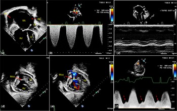
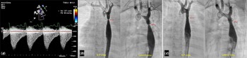
Transthoracic echocardiogram confirmed the presence of dilated right atria and right ventricle. The right ventricular systolic function was reduced as estimated by the decreased tricuspid annular plane systolic excursion (Fig 1a–c). A hypertrophied anomalous muscle bundle was visualised in the right ventricular cavity that was leading to severe obstruction with the gradient estimated to be about 130 mmHg (Fig 1d–f). Interrogation of aortic arch revealed a discrete narrowing with a peak gradient of 55 mmHg and a holo-diastolic spill (Fig 2a).
( a ) Transthoracic echocardiographic image in apical 4 chamber view demonstrating dilated RA and RV. Moderate pericardial effusion is also seen. ( b ) TR jet estimated right ventricular systolic pressures was 120 mmHg over and above the RA pressure. ( c ) Tricuspid annular plane systolic excursion is decreased suggesting severe RV dysfunction. ( d and e ) Subcostal short axis view demonstrating a discrete muscle bundle (Mus) in RV cavity resulting in severe obstruction. ( f ) Continuous wave Doppler trace showing forward flow extending even into the diastole being contributed by the right atrial “a” wave (red dashed arrows) suggestive of severe ventricular dysfunction as a result of the obstruction.

( a ) Continuous wave Doppler tracing across the juxta-ductal aorta showing a peak velocity of 3.7 cm/s and holo-diastolic spill of flow suggestive of tight coarctation of aorta. ( b and c ) Aortogram images profile the coarctation segment before and after balloon aortoplasty.

An urgent cardiac surgical consultation was sought, and a plan for transcatheter therapy for coarctation followed by surgical repair of double chambered right ventricle was drafted. Aortogram profiled a tight narrowing in the juxta-ductal aorta with an invasive pressure gradient of 40 mmHg across the narrowing. A balloon aortoplasty was performed using a Tyshak II 8 mm × 3 cm balloon (NuMed Inc., Hopkinton, NY, United States of America). The procedure was successful with a fall in gradient to 15 mmHg and a good angiographic result (Fig 2b and c). Subsequently, the child was taken up for urgent cardiac surgery.
After median sternotomy and aortobicaval cannulation, cardiopulmonary bypass was established with core-cooling to 32°C. A thick muscular diaphragm was visualised in the right ventricular cavity which was excised. No evidence of a ventricular septal defect was found during surgery. The child had an uneventful postoperative recovery with improvement in right ventricular function and heart failure symptoms.
Discussion
Double chambered right ventricle is a rare CHD characterised by hypertrophied anomalous muscle bundle in the right ventricular cavity. This causes obstruction of ventricular outflow as a result of which the right ventricle is divided into a proximal high-pressure and a distal low-pressure chamber. Various subtypes of double chambered right ventricle have been described based on the location of the muscle bundles within the right ventricular cavity.Reference Loukas, Housman, Blaak, Kralovic, Tubbs and Anderson1
The embryological basis of double chambered right ventricle is unclear. Possible theories include incomplete resorption of right ventricular trabeculations, hypertrophy of bulbar musculature, and arrested incorporation of the bulbus cordis into right ventricular body.Reference Wang, Wu and Chang2 The irregular expansion of bulboventricular junction results in an incomplete fusion of bulbar and endocardial cushions, thus explaining the frequent association of ventricular septal defect with double chambered right ventricle, which can be seen in up to 90% of these patients.Reference Hoffman3 Another commonly associated lesion is a discrete sub-aortic membrane. Association between sub-aortic membrane and double chambered right ventricle is reported to be nine times greater than expected. Sub-aortic membrane is found in up to 20% of cases with double chambered right ventricle.Reference Baumstark, Fellows and Rosenthal4 One series has reported sub-aortic membrane in up to 72% patients who had double chambered right ventricle along with a malalignment type of ventricular septal defect.Reference Wang, Wu and Chang2 Other less commonly associated lesions include tetralogy of Fallot, double outlet right ventricle, atrial septal defect, ruptured sinus of Valsalva aneurysm, etc.Reference Loukas, Housman, Blaak, Kralovic, Tubbs and Anderson1
Coarctation of aorta is almost never associated with a right-sided obstructive lesion, especially double chambered right ventricle. Review of available literature revealed only four patients with this combination.Reference Hoffman3,Reference Kahr, Alonso-Gonzalez and Kempny5 Abnormality of a common embryologic pathway may fail to explain the association between double chambered right ventricle and coarctation of aorta; rather, an aberration of two different pathways could have resulted in this extremely uncommon association. Double chambered right ventricle repair requires a midline sternotomy approach. In view of the anticipated difficulty in repairing coarctation of aorta from the midline, a hybrid approach was resorted to in the index patient.
Conclusions
Double chambered right ventricle is a rare cause of right ventricular outflow obstruction. The most common associated lesions are ventricular septal defect followed by sub-aortic membrane. Coarctation of aorta is almost never seen in association with a right-sided obstructive lesion. We hereby report an extremely rare association of coarctation of aorta with double chambered right ventricle causing severe heart failure in a child who was successfully managed adopting a hybrid approach.
Acknowledgement
None.
Authors’ contributions
BA, SR, and VD were involved in the management of case. BA wrote the first manuscript draft which was finalised by SR and VD.
Financial support
This research received no specific grant from any funding agency, commercial, or not-for-profit sectors.
Conflict of interest
None.
Ethical standards
The authors assert that all procedures contributing to this work comply with the ethical standards as mentioned in the Helsinki Declaration of 1975, as revised in 2008.
Declaration of patient consent
The authors certify that they have obtained all appropriate patient consent forms. In the form, the patient(s) has/have given his/her/their consent for his/her/their images and other clinical information to be reported in the journal. The patients understand that their names and initials will not be published and due efforts will be made to conceal their identity, but anonymity cannot be guaranteed.