





Introduction
The genus Opegaster Ozaki, 1925 (Opecoelidae Ozaki, 1925) comprises a diverse group of digenean trematodes parasitising marine and freshwater fishes. Opecoelidae is one of the largest families of marine digeneans, currently comprising more than 1000 described species distributed among approximately 100 genera worldwide. Members of Opegaster have been reported primarily from marine and freshwater fishes across multiple marine ecoregions (Martin, Reference Martin, Gardner and Gardner2024). A comprehensive revision of the marine representatives of Opegaster was provided by Bray and Justine (Reference Bray and Justine2013), who recognized 27 valid marine species and compiled detailed morphometric and ratio-based characters to facilitate interspecific comparisons. Since that revision, additional species have been described, including Opegaster mezcal Santillán-Pérez, García-Varela & Aguilar-Aguilar, 2025 (Santillán-Pérez et al., Reference Santillán-Pérez, García-Varela and Aguilar-Aguilar2025) and Opegaster gorgonioi Aguilar-Aguilar, 2025 (Aguilar-Aguilar, Reference Aguilar-Aguilar2025) from marine fishes in Mexico. Earlier, Opegaster elongata Yamaguti, 1959, described from Ostorhinchus semilineatus (Apogonidae) in the Pacific Ocean (Yamaguti, Reference Yamaguti1959), and Opegaster oplegnathi Wang et al., 1992, described from the reef-associated fish Oplegnathus fasciatus Temminck & Schlegel, 1844 (Wang et al., Reference Wang, Wang and Zhang1992), were not included in the compilation of Bray and Justine (Reference Bray and Justine2013). Despite the relatively large number of described species, knowledge of the genus remains limited because most species are known only from their original descriptions and have not subsequently been reported. Furthermore, molecular data are available for only a few taxa. A partial 28S rDNA sequence was generated from an unidentified species of Opegaster parasitizing Tomicodon sp. (Gobiesocidae) from the Pacific coast of Mexico (Pérez-Ponce de León and Hernández-Mena, Reference Pérez-ponce de León and Hernández-Mena2019) and was subsequently assigned to O. mezcal (Santillán-Pérez et al., Reference Santillán-Pérez, García-Varela and Aguilar-Aguilar2025). Given the high diversity and morphological complexity within Opecoelidae, integrative taxonomic approaches combining morphological and molecular data are essential for reliable species delimitation.
Mudskippers (Oxudercinae) represent a distinctive ecological group of gobioid fishes inhabiting soft-bottom intertidal zones and mangrove ecosystems throughout the Indo–West Pacific region, with a single representative occurring in tropical West Africa. Their distribution extends from the western coast of Africa eastwards to Samoa. Members of Oxudercinae exhibit remarkable morphological and physiological adaptations to amphibious life, including modified pectoral fins for terrestrial locomotion and specialized respiratory mechanisms involving vascularized epithelia and modified gill structures (Murdy, Reference Murdy, Patzner, Van Tassell, Kovacic and Kapoor2011). The subfamily currently comprises 43 species in 10 genera (Parenti, Reference Parenti2021). The giant mudskipper, Periophthalmodon schlosseri (Pallas, 1770), is widely distributed in India, Indonesia, Malaysia, Singapore, Thailand and Vietnam (Hoese et al., Reference Hoese, Larson and Bleher2024). In Thailand, 9 species of mudskippers belonging to the genera Boleophthalmus Valenciennes, 1837, Periophthalmus Bloch & Schneider, 1801 and Scartelaos Swainson, 1839 have been recorded, including P. schlosseri (Larson, Reference Larson2019; Hoese et al., Reference Hoese, Larson and Bleher2024). Despite their ecological importance and distinctive life history, helminthological data on mudskippers in Thailand are virtually non-existent. Globally, the helminth fauna of Oxudercinae remains poorly known. Only a few adult digeneans have been documented, including Lecithaster ghanensis Fischthal & Thomas, 1971 (Lecithasteridae) from Periophthalmus barbarus (Linnaeus, 1766) in Ghana and Opegaster ouemoensis Bray & Justine, 2013 from Periophthalmus argentilineatus Valenciennes, 1837 in New Caledonia (Bray and Justine, Reference Bray and Justine2013). Subsequently, Hidayaturrahmah et al. (Reference Hidayaturrahmah, Santoso, Nurapriana, Badruzsaufari and Muhamat2019) reported the presence of Opecoeloides sp. in P. schlosseri from Borneo Island, Indonesia, but without detailed morphological or molecular characterization sufficient to confirm its taxonomic placement.
During a survey of helminths infecting mudskippers in Chumphon Province, Thailand, specimens of an undescribed species of Opegaster were recovered from P. schlosseri. Detailed morphological examination and comparison with previously described congeners support its recognition as a new species, which is described herein.
Materials and methods
Host sampling
Mudskippers were collected in April 2025 from 3 localities in Chumphon Province, Thailand: Hat Sai Ri Subdistrict, Mueang Chumphon District (10.37381° N, 99.24138° E; n = 3 fishes examined); Pak Nam Chumphon Subdistrict, Mueang Chumphon District (10.38339° N, 99.22828° E; n = 4 fishes examined) and Pak Khlong Subdistrict, Pathio District (10.87374° N, 99.45584° E; n = 1 fish examined). All 3 sites comprise mangrove habitats subject to anthropogenic influence and are exploited daily by local artisanal fisheries.
Fish were identified as P. schlosseri using the morphological criteria of Hoese et al. (Reference Hoese, Larson and Bleher2024). Live specimens were transported to the laboratory and euthanized by immersion in an overdose of tricaine methanesulphonate (MS-222). The gastrointestinal tract was removed and examined under a stereomicroscope for helminths.
Parasite recovery and fixation
Digeneans recovered from hosts collected at Hat Sai Ri and Pak Khlong were processed as follows. Specimens intended for morphological study were relaxed and fixed in hot saline, transferred to 70% ethanol, stained with Semichon’s acetocarmine, dehydrated through a graded ethanol series, cleared in xylene and mounted in Canada balsam. Specimens intended for scanning electron microscopy (SEM) were fixed in 2.5% glutaraldehyde in cold 0.1 M sodium cacodylate buffer (pH 7.4).
Specimens recovered from hosts collected at Pak Nam Chumphon were processed differently. Following euthanasia, hosts were frozen and dissected subsequently. Recovered digeneans were stained with Semichon’s acetocarmine to confirm their conspecificity with specimens from the other localities. These specimens were used exclusively for the estimation of epidemiological parameters and were not included in detailed morphological analyses because freezing of the hosts may have affected trematode morphology. Prevalence was calculated as the percentage of infected hosts among those examined and mean intensity as the average number of parasites per infected host, following Bush et al. (Reference Bush, Lafferty, Lotz and Shostak1997).
Light microscopy study
Stained specimens were examined using a Leica DMLB light microscope (Leica Microsystems, Wetzlar, Germany). Identification to the generic level followed Cribb (Reference Cribb, Jones, Bray and Gibson2005). Measurements were taken using an ocular micrometre. Illustrations were prepared with the aid of a drawing tube attached to the microscope and were subsequently digitized and refined using Adobe Illustrator (Adobe Systems, San Jose, CA, USA). Measurements are given in micrometres (µm) and are presented as range followed by the mean in parentheses, when appropriate.
Scanning electron microscopy
One specimen was fixed in 2.5% glutaraldehyde in a 0.1 M sodium cacodylate buffer (pH 7.4) at 4 °C, rinsed in this buffer, post-fixed in 1% osmium tetroxide containing 0.9% potassium ferricyanide in the same buffer at 4 °C for 1 h, and finally rinsed in Milli-Q water. The fixed digenean was dehydrated through a graded ethanol series (80%, 90%, 96% and absolute), followed by critical-point drying with carbon dioxide in an Emitech K850X (Quorum Technologies Ltd, Laughton, East Sussex, UK). The dried worm was then mounted on a stub using conductive adhesive tape, coated with carbon in an Emitech K950X evaporator (Quorum Technologies Ltd.) and examined with a JSM-7001F field emission SEM (JEOL Ltd) operated at 10 kV at the Scientific and Technological Centres (CCiTUB), University of Barcelona, Spain.
Molecular analyses
Total genomic DNA was extracted from 2 specimens using the Geneaid gDNA Mini Kit (Geneaid Biotech Ltd, Taipei, Taiwan) following the manufacturer’s instructions. Polymerase chain reaction (PCR) was used to amplify fragments of the nuclear 18S ribosomal RNA (rRNA) gene, nuclear 28S rRNA gene and mitochondrial cytochrome c oxidase subunit I (cox1) gene. Each PCR reaction was performed in a final volume of 30 µL, containing 15 µL of 2x i-Taq™ Master Mix (iNtRON Biotechnology, Gyeonggi, South Korea), 10–50 µM of each primer and 1 ng µL−1 of DNA template. Amplification was conducted in a T100™ thermocycler (Bio-Rad, Hercules, CA, USA). Primer sequences and thermocycling conditions followed those described by Bowles et al. (Reference Bowles, Blair and McManus1992), Tkach et al. (Reference Tkach, Littlewood, Olson, Kinsella and Swiderski2003) and Routtu et al. (Reference Routtu, Grunberg, Izhar, Dagan, Ucko and Ben-ami2014). PCR products of approximately 800, 1200 and 440 bp for the 18S, 28S and cox1 genes, respectively, were visualized on a 1% agarose gel stained with SYBR™ Safe DNA Gel Stain (Invitrogen, Waltham, MA, USA). Amplicons were sequenced commercially using Fast Next Generation Sequencing technology (Tsingke Biotech, Beijing, China).
Electropherograms were inspected and edited in BioEdit 7.0 (Hall, Reference Hall1999). Multiple sequence alignments for each marker were generated in ClustalX 2.1 (Thompson et al., Reference Thompson, Gibson and Higgins2002) using reference sequences of Opecoelidae obtained from GenBank (Table 1). Preptetos caballeroi Pritchard, 1960 and Enenterum aureum Linton, 1910 were selected as outgroup taxa.
Taxa included in the phylogenetic analyses and their corresponding GenBank accession numbers

Table 1 Long description
Rows list species names and columns give GenBank accession numbers for three markers: 18S rRNA, 28S rRNA, and Cox1, with some cells marked by a dash to indicate no sequence used. Opegaster plateen, this study, includes 18S rRNA PZ487612 and PZ487613, 28S rRNA PZ487610 and PZ487611, and Cox1 PZ485872 and PZ485873. Several taxa have accessions in only one marker, such as Opegaster sp. with Cox1 PV576489, Opecoeloides pedicathedrae with 18S rRNA OQ101906, and Opecoelus sp. with Cox1 ON661076. Some taxa have accessions in two markers, such as Anomalotrema koiae with 18S rRNA KU320582 and 28S rRNA MH161429 and KU320595, and Helicometra fasciata with 18S rRNA KU320584, 28S rRNA KU320597, and Cox1 KJ996006. Multiple Podocotyle entries list Cox1 accessions only, including Podocotyle atomon PP333594, Podocotyle odhneri PP333611, Podocotyle reflexa PP333609, and Podocotyle sp. PP333598, while Podocotyle atomon also lists 28S rRNA MH161437. Outgroup rows at the end include Preptetos zebravaranus with Cox1 MZ695197, Preptetos caballeroi with 18S rRNA AJ287563 and 28S rRNA AY222236, and Enenterum aureum with 18S rRNA AY222124 and 28S rRNA AY222232.
Sequences generated in the present study are indicated in bold. A ‘—’ indicates sequences were not used.
Phylogenetic analyses were performed using the maximum likelihood (ML) and neighbour-joining (NJ) methods in MEGA X (Kumar et al., Reference Kumar, Stecher, Li, Knyaz and Tamura2018). The best-fit nucleotide substitution model was selected for the ML analyses, and nodal support was assessed using 1000 bootstrap replicates. Phylogenetic trees were visualized and edited in FigTree 1.3.1 (Rambaut, Reference Rambaut2009). Pairwise interspecific genetic distances were calculated in MEGA X using the p-distance model (Kumar et al., Reference Kumar, Stecher, Li, Knyaz and Tamura2018).
Results
Taxonomy
Phylum: Platyhelminthes Minot, 1876
Class: Trematoda Rudolphi, 1808
Order: Plagiorchiida La Rue, 1957
Family: Opecoelidae Ozaki, 1925
Genus: Opegaster Ozaki, 1925
Species: Opegaster plateen Poonlaphdecha & Ribas (Figure 1)
Opegaster plateen n. sp., adult specimen. Scale bar = 200 µm.

Type host: Periophthalmus schlosseri (Pallas, 1770) (Gobiiformes: Oxudercinae), giant mudskipper.
Type locality: Hat Sai Ri Subdistrict, Mueang Chumphon District (10.37381° N, 99.24138° E, Thailand).
Site in host: Intestine.
Type specimens: Holotype MZB 2026-0183 and paratypes deposited in the Museum of Natural Sciences of Barcelona, Spain (Voucher nos: MZB 2026-0184 to MZB 2026-0192).
Collection date: 24 April 2025.
Prevalence and intensity: 62.5% prevalence, 4–16 trematodes from 5 positive hosts.
Representative DNA sequences: GenBank accession numbers: 18S (PZ487612 and PZ487613), 28S (PZ487610 and PZ487611) and cox1 (PZ485872 and PZ485873).
Etymology: The species epithet plateen is derived from the Thai vernacular name of the host fish, ‘pla teen’ (pla = fish; teen = foot), referring to its use of the pectoral fins to locomote on mud. The name is treated as a noun in apposition.
Description
Description based on 10 whole-mounted specimens (Figures 1 and 2) with additional observations from specimens examined by SEM (Figure 3). All measurements are provided in micrometres; ranges followed by the mean in parentheses.
Conformation of the terminal part of the male genitalia of Opegaster plateen n. sp. Scale bar 50 μm. 1: genital pore, 2: cirrus sac, 3: internal seminal vesicle, 4: external seminal vesicle, 5: metraterm.

Scanning electron micrograph focusing of the acetabulum (asterisks indicate 3 of the tegumental papillae of the acetabulum of Opegaster plateen n. sp.). Not all 6 papillae in each lip can be observed simultaneously in this SEM image.

Body elongated, fusiform, 1098–1506 (1290) long; 264–378 (335) wide, usually at level of the ventral sucker or the gonads. Tegument smooth, without spines. Forebody 364–505 (420) long, representing approximately 32.6% of the total body length. Oral sucker subterminal, 117–159 (139) long, 101–151 (126) wide. Prepharynx short, observable by light microscopy in 2 of 10 specimens, indistinguishable in 8. Pharynx muscular, 67–113 (88) long, 79–106 (93) wide. Oesophagus relatively long, 79–151 (101) long. Intestinal bifurcation situated anterior to the ventral sucker; distance from the bifurcation to the anterior margin of the ventral sucker 65–99 (87). Caeca extending posteriorly to near the posterior extremity of the body. Ventral sucker well developed, 175–234 (202) long, 180–258 (220) wide; oral to ventral sucker ratio approximately 1:1.4–1.6. Tegumental papillae present on the ventral sucker, 5–6 on each margin (Figure 3). Distance from the ventral sucker to the anterior extent of the vitellarium 37–94 (73).
Testes 2, lobulated, tandem in the posterior half of the body. Anterior testis 79–132 (112) long, 86–171 (133) wide. Posterior testis 76–151 (123) long, 106–159 (131) wide. Intertesticular distance 0–37 (11). Post-testicular region 186–409 (307) long. Ovary lobulated, pretesticular, 58–90 (73) long, 76–118 (93) wide; situated immediately anterior to the anterior testis; distance between the ovary and the anterior testis 0–5 (2). Distance from the ventral sucker to the ovary 7–137 (68). Seminal vesicle elongate, 124–170 (152) long, 38–65 (54) wide, posterior end sometimes C-shaped. Anterior end situated anterior to the ventral sucker. Cirrus sac membranous and, short, containing internal seminal vesicle, ejaculatory duct and cirrus. Vitellarium follicular, lateral, extending from slightly posterior to the level of the ventral sucker to near the posterior extremity. Uterus occupying the intercaecal space between the ovary and the ventral sucker. Eggs operculated, 47–69 (61) long, 24–33 (29) wide (n = 45). Uroproct present connected to excretory vesicle.
Remarks
The only species previously described from a mudskipper host is O. ouemoensis from New Caledonia. The present specimens are clearly distinguished by having a larger ventral sucker (175–234 µm in length and 180–258 µm in width) compared with O. ouemoensis (62–148 µm in length and 66–154 µm in width) (Bray and Justine, Reference Bray and Justine2013). In addition, the ventral sucker in O. plateen n. sp. is larger than the oral sucker, whereas in O. ouemoensis it is only slightly larger.
Opegaster plateen n. sp. can also be differentiated from other congeners parasitizing marine fishes based on morphometric characters, although its intertidal ecology makes it improbable that its life cycle is completed in fully marine environments.
Among congeners, differences in the papillae of the ventral sucker provide a useful means of discrimination. Opegaster apogonichthydis Yamaguti, 1938, O. elongata Yamaguti, 1959, O. gobii Yamaguti, 1952, O. macrorchis Yamaguti, 1938, O. syngnathi Yamaguti, 1934 and O. synodi Manter, 1947 each possess only 3 papillae or papilliform structures on each lip of the ventral sucker (Yamaguti, Reference Yamaguti1938, Reference Yamaguti1942, Reference Yamaguti1952, Reference Yamaguti1959; Manter, Reference Manter1947). Opegaster hippocampi Shen, 1982 differs in having 4 papillae on each lip, whereas O. longivesicula Yamaguti, 1952 possesses 3 strongly flattened papillae on each lip (Yamaguti, Reference Yamaguti1952; Shen, Reference Shen1982). In O. plotosi Yamaguti, 1940, the ventral sucker bears 3 papilliform protuberances on each lip (Yamaguti, Reference Yamaguti1940).
The relative position of the ovary and ventral sucker also distinguishes several congeners from O. plateen n. sp. In O. dendrochiri Yamaguti, 1970, O. ditrematis Yamaguti, 1942, O. hawaiiensis Yamaguti, 1970 and O. queenslandica Aken’Ova, 2007, the distance between the posterior margin of the ventral sucker and the ovary is considerably greater than in O. plateen n. sp. (Yamaguti, Reference Yamaguti1970; Bray and Cribb, Reference Bray and Cribb1989; Bray and Justine, Reference Bray and Justine2013).
Differences in testicular morphology, position and relative size further separate O. plateen n. sp. from several species. In O. dactylopteri Yamaguti, 1970, the testes are separated by a few vitelline follicles, a condition not observed in O. plateen n. sp. In O. apogonichthydis Yamaguti, 1938, the anterior testis is situated to the left of the median line (Yamaguti, Reference Yamaguti1938). Opegaster ovata Ozaki, 1928 has obliquely arranged testes, with one situated posterior to the other (Ozaki, Reference Ozaki1928). In O. gobii Yamaguti, 1952 and O. macrorchis Yamaguti, 1938, the testes are larger than the relevant sucker, whereas, in O. parapristipomatis Yamaguti, 1934 and O. paramacrorchis Hafeezullah, 1971, the testes are wider than the ventral sucker; in O. paramacrorchis, they are also separated from each other (Yamaguti, Reference Yamaguti1938, Reference Yamaguti1952; Hafeezullah, Reference Hafeezullah1971). In contrast, the testes of O. plateen n. sp. are narrower than the ventral sucker. Opegaster plotosi Yamaguti, 1940 differs further in having deeply lobulated testes (Yamaguti, Reference Yamaguti1940), while O. lutiani Bravo-Hollis & Manter, 1957 has bilobed testes and a trilobate ovary, as well as smaller eggs (Bravo-Hollis and Manter, Reference Bravo-Hollis and Manter1957).
The relative size of the suckers provides additional distinguishing characters. In O. hippocampi Shen, 1982 and O. oplegnathi Wang, Wang & Zhang, 1992, the oral sucker is smaller than the ventral sucker (Shen, Reference Shen1982; Wang et al., Reference Wang, Wang and Zhang1992). In O. iniistii Yamaguti, 1970, the ventral sucker is positioned considerably farther from the oral sucker (Yamaguti, Reference Yamaguti1970).
The vitellarium and anterior digestive region also show species-specific differences. In O. acuta Manter, 1940, the vitelline fields are situated anterior to the ventral sucker and may overlap the testes dorsally, while the genital pore is located at the posterior margin of the oral sucker (Manter, Reference Manter1940). In O. bothi Yamaguti, 1970, the vitellarium extends to the level of the pharynx (Yamaguti, Reference Yamaguti1970). Opegaster cryptocentri Yamaguti, 1958 has an expanded prepharynx, whereas O. brevifistula Ozaki, 1928 lacks an oesophagus (Ozaki, Reference Ozaki1928; Yamaguti, Reference Yamaguti1958).
Other congeners differ in genital and reproductive structures. In O. dermatogenyos Yamaguti, 1970, the genital pore reaches the posterior level of the pharynx (Yamaguti, Reference Yamaguti1970). Opegaster dendrochiri Yamaguti, 1970 and O. longivesicula Yamaguti, 1952 possess a very long seminal vesicle (Yamaguti, Reference Yamaguti1952, Reference Yamaguti1970).
The present species is also distinguished from species lacking ventral sucker papillae, including O. gorgonioi Aguilar-Aguilar, 2025, O. mezcal Santillán-Pérez, Garcia-Varela & Aguilar-Aguilar, 2025, O. pritchardae Overstreet, 1969, O. recta Ozaki, 1928 and O. tamori Yamaguti 1938 (Aguilar-Aguilar, Reference Aguilar-Aguilar2025; Santillán-Pérez et al., Reference Santillán-Pérez, García-Varela and Aguilar-Aguilar2025).
Molecular analyses
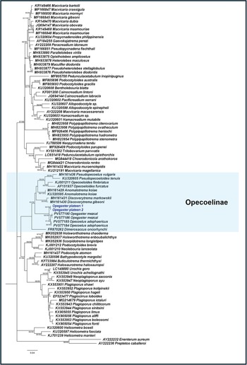
The phylogenies inferred from the nuclear 18S and 28S rRNA genes and the mitochondrial cox1 gene supported the placement of O. plateen n. sp. within the subfamily Opecoelinae (Figures 4, 5 and 6). In the 28S phylogeny, O. plateen n. sp. formed a monophyletic clade with O. mezcal and Opecoelus adsphaericus, occupying a sister position to the clade comprising O. mezcal and O. adsphaericus. Pairwise interspecific genetic distances among O. plateen n. sp., O. mezcal and O. adsphaericus ranged from 1.2 to 1.5%. No intraspecific variation was observed among the O. plateen n. sp. sequences for either nuclear rRNA genes.
Maximum likelihood phylogeny of Opecoelidae inferred from the nuclear 18S rRNA gene (Kimura 2-parameter + G + I). Numbers at nodes indicate ML/NJ bootstrap support values; only values greater than 70 are shown. Sequences of Opegaster plateen n. sp. generated in the present study are shown in blue, and members of the subfamily Opecoelinae are enclosed within blue boxes.

Figure 4 Long description
Phylogenetic tree depicting the evolutionary relationships within the Opecoelidae family, inferred from nuclear 18S rRNA gene sequences. The tree is oriented vertically, with branches extending from top to bottom. Each branch represents a different species or group, labeled with accession numbers and species names. Bootstrap support values are indicated at nodes, with only values greater than 70 shown. The tree includes sequences generated in the present study, highlighted in blue and members of the subfamily Opecoelinae are enclosed within blue boxes. The tree shows two main clades labeled as Opecoelinae, with species such as Opecoelus plateen n. sp., Opecoelus conorchis and Opecoelus pedunculatus forming distinct groups. The tree illustrates the placement of Opecoelus plateen n. sp. within the subfamily Opecoelinae, forming a monophyletic clade with Opecoelus mezcal and Opecoelus adsphaericus. The diagram provides a detailed view of the genetic relationships and evolutionary lineage within the family.
Maximum likelihood phylogeny of Opecoelidae inferred from the nuclear 28S rRNA gene (GTR + G + I). Numbers at nodes indicate ML/NJ bootstrap support values; only values greater than 70 are shown. Sequences of Opegaster plateen n. sp. generated in the present study are shown in blue, and members of the subfamily Opecoelinae are enclosed within blue boxes.

Figure 5 Long description
The phylogenetic tree is vertically oriented, depicting the evolutionary relationships within the family Opecoelidae, specifically highlighting the subfamily Opecoelinae. The tree is structured with branches extending from a central trunk, each labeled with species names and accession numbers. The subfamily Opecoelinae is enclosed within a blue shaded box, indicating its members. Branches within this box show various species, including Opecoelus adsphaericus and O. plateen n. sp., forming a monophyletic clade. The tree includes bootstrap support values at nodes, with only values greater than 70 shown. The layout progresses from top to bottom, illustrating the hierarchical relationships among the species. The tree also includes sequences generated in the present study, marked in blue, emphasizing their placement within the subfamily. The diagram visually represents genetic distances and evolutionary lineage connections among the species listed.
Maximum likelihood phylogeny of Opecoelidae inferred from the mitochondrial cox1 gene (HKY + G). Numbers at nodes indicate ML/NJ bootstrap support values; only values greater than 70 are shown. Sequences of Opegaster plateen n. sp. generated in the present study are shown in blue, and members of the subfamily Opecoelinae are enclosed within blue boxes.

Although no reference sequences of Opegaster or Opecoelus were available for the 18S phylogeny, pairwise genetic distances between O. plateen n. sp. and other genera within Opecoelinae ranged from 3.6 to 4.7%. These values exceeded those observed among the other sampled genera within the subfamily, for which pairwise genetic distances ranged from 2.1 to 3.2%.
In the cox1 phylogeny, O. plateen n. sp., O. mezcal and O. adsphaericus formed a monophyletic clade. Pairwise interspecific genetic distances among these taxa ranged from 12.5 to 12.9%. An intraspecific genetic distance of 0.3% was observed among the O. plateen n. sp. sequences.
Discussion
Despite the wide geographical distribution of the giant mudskipper and the recognized importance of mangrove ecosystems for biodiversity and ecosystem services (Otieno et al., Reference Otieno, Owuor, Pacheco, Coppo, Monteiro Santos, Ferreira and Bernardino2026), parasitological studies on this host remain scarce. Although the life cycle of Opegaster remains unknown, studies of other opecoelids with known life cycles, such as that of Opecoelus variabilis Cribb, 1985, suggest the involvement of an aquatic snail as the first intermediate host and a crustacean as the second intermediate host (Cribb, Reference Cribb1985). Periophthalmodon schlosseri feeds predominantly on crabs but also consumes other locally abundant prey items (Hoese et al., Reference Hoese, Larson and Bleher2024). In Pattani Province, relatively close to the present sampling localities, stomach-content analyses of 11 specimens revealed the presence of crabs, fishes and shrimps (Swennen et al., Reference Swennen, Ruttanadakul, Haver, Piummongkol, Prasertsongskum, Intanai, Chaipakdi, Yeesin, Horpet and Detsathit1995). Consequently, a crab is a plausible candidate as the second intermediate host of O. plateen n. sp.
The host specificity of this trematode remains uncertain. Although mudskipper species often occur sympatrically, parasite sharing among hosts is poorly documented owing to the limited number of studies available. Furthermore, differences in habitat use, diet and distribution along the mudflat gradient (Swennen et al., Reference Swennen, Ruttanadakul, Haver, Piummongkol, Prasertsongskum, Intanai, Chaipakdi, Yeesin, Horpet and Detsathit1995; Su and S. SL, Reference Su and S. SL2016) may act as barriers to parasite transmission. Nevertheless, host sharing has been demonstrated in marine environments, where O. mezcal has been reported from fishes belonging to 2 different families, Labrisomidae and Gobiesocidae (Santillán-Pérez et al., Reference Santillán-Pérez, García-Varela and Aguilar-Aguilar2025).
The molecular data generated from both nuclear and mitochondrial markers support the placement of O. plateen n. sp. within the Opecoelinae. This subfamily currently comprises more than 20 genera, including Opegaster. Using concatenated 18S and 28S rRNA gene sequences, Bray et al. (Reference Bray, Cribb, Timothy Littlewood and Waeschenbach2016) demonstrated the monophyly of Opecoelinae, including representatives of Dimerosaccus Shimazu, 1988, Anomalotrema Zhukov, 1957, Pseudopecoeloides Yamaguti, 1940 and Opecoeloides Odhner, 1928. Likewise, Opecoelinae was recovered as monophyletic in the present 28S and cox1 phylogenies. O. plateen n. sp. showed a close genetic relationship with O. mezcal and O. adsphaericus, as evidenced by both the 28S and cox1 phylogenies. A close relationship between O. mezcal and O. adsphaericus was also recovered by Martínez-Aquino et al. (Reference Martínez-Aquino, Rincon-sandoval, Flores-lopez, Ruiz-campos, Aguirre-macedo and Ceccarelli2025), in which O. adsphaericus, a parasite of the intertidal fish Clinocottus analis, was recovered as the sister taxon of the Opegaster clade. The sequences generated herein represent only the second molecular data available for a species of Opegaster and contribute to the limited molecular resources currently available for the genus.
In conclusion, O. plateen n. sp. is described from the giant mudskipper on the basis of both morphological and molecular evidence. As most described species of Opegaster parasitize marine fishes, the present findings contribute to knowledge of trematode diversity in intertidal environments. The scarcity of studies on mudskipper parasites may reflect the limited commercial importance of these fishes; however, their distinctive ecology and habitat make them particularly valuable hosts for investigations of trematode diversity, host-parasite associations and digenean taxonomy.
Data availability statement
All data generated or analysed during this study are included in this published article. The 18S rRNA, 28S rRNA and cox1 gene sequences generated in the present study have been deposited in GenBank under accession numbers PZ487612–PZ487613, PZ487610–PZ487611 and PZ485872–PZ485873, respectively.
Acknowledgements
We are grateful to the staff and students from the Department of Agricultural Technology, Program in Fisheries Science and Aquatic Resources, King Mongkut’s Institute of Technology Ladkrabang, Prince of Chumphon Campus, Chumphon, Thailand, for their logistical support in sample collection and for providing laboratory facilities for this project. The authors also thank the staff of the Scientific and Technological Centres of the University of Barcelona (CCiTUB) for their assistance in the preparation of scanning electron microscopy (SEM).
Author contributions
S.P., N.S. and A.R. conceived and designed the study. S.P., J.M. and A.R. performed morphological analyses. S.P., A.C., U.T. and V.C. performed molecular analyses. S.P., A.C., U.T. and A.R. conducted data analyses and wrote the paper. S.P., N.S., A.C., U.T., J.M., V.C. and A.R. edited the paper.
Financial support
The authors declare that no financial support was received for the research and/or publication of this article.
Competing interests
The authors declare that there are no conflicts of interest.
Ethical standards
The animal study was approved by the Animal Care and Use Committee of King Mongkut’s Institute of Technology Ladkrabang, Thailand (Approval No. ACUC-KMITL-RES/2025/009). The study was conducted in accordance with local legislation and institutional requirements.




 Open access

