Introduction
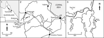
The Quinton Formation is part of the Silurian to Lower Devonian Graveyard Creek Group in the Broken River Province, a Paleozoic tectonostratigraphic terrane located approximately 200 km west of Townsville in northeast Queensland, Australia (Fig. 1.1, 1.2). Spanning most of the Silurian (Fig. 2), the formation is a thick (up to 5,000 m), predominantly turbiditic sequence of siltstones, sandstones, and mudstones with some conglomerates, interpreted as deepwater deposits of a submarine fan system adjacent to an active fault zone along the margin of the Precambrian Georgetown Inlier lying to the west, the probable source of the sediments (Withnall, Reference Withnall1993; Sloan et al., Reference Sloan1995; Henderson and Withnall, Reference Henderson and Withnall2013). In the eastern part of its outcrop area, in the vicinity of Gray Creek (Fig. 1.3), the lower part of the formation also contains a number of allochthonous limestones, ranging in size from pebble-sized clasts in conglomerates to large blocks some hundreds of meters long considered to have been derived from a contemporaneous carbonate shelf situated to the north or northwest. These limestones contain diverse middle Silurian fossil faunas of which only the corals have as yet been extensively documented (Munson and Jell, Reference Munson and Jell1999). Other faunal elements include stromatoporoids, brachiopods, molluscs, trilobites, and conodonts. The largest of these limestone bodies, informally named the Tomcat Creek limestone, contains the most diverse and best preserved trilobite fauna known from the Broken River Province, including members of the Illaeninae, Bumastinae (these two collectively referred to as illaenimorphs), Scutelluidae, Harpetidae, Proetidae, Aulacopleuridae, Brachymetopidae, Cheiruridae, Encrinuridae, Calymenidae, Lichidae, and Odontopleuridae. We here document the illaenimorphs and scutelluids; other groups will be dealt with in later contributions.
Locality maps for the Tomcat Creek limestone in north Queensland: (1) northeastern Australia (approximate area of 2 indicated by rectangle); (2) Townsville–Greenvale region (approximate area of 3 indicated by square); (3) approximate location of Tomcat Creek limestone outcrops (TCL).

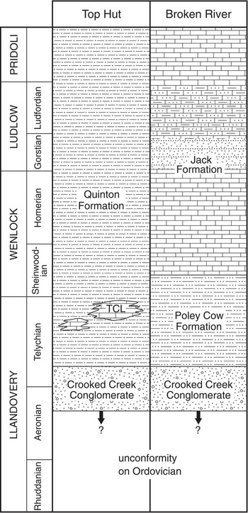
Stratigraphic correlation of the Silurian sequences in the Top Hut and Broken River regions of the Broken River Province, north Queensland; TCL=Tomcat Creek limestone.

The only trilobites previously described from the Quinton Formation are a small fauna dominated by the illaenimorph Rhaxeros and also including fragmentary remains of Sphaeroxochus, a possible warburgelline, an encrinurid, and a calymenid from redeposited limestone clasts in a conglomerate at a locality on the bank of Gray Creek about 3 km north of the Tomcat Creek limestone (Lane and Thomas, Reference Lane and Thomas1978a, Reference Lane and Thomas1980). From shales of the Quinton Formation in the same area, Öpik (in White, Reference White1965, p. 43) identified Encrinurus, two species of scutelluids, Proetus? and Sphaerexochus? or Onycopyge?, but the specimens were not illustrated and, as reported by Holloway (Reference Holloway1994), their present whereabouts are unknown, so the identifications cannot be verified. A diverse Silurian trilobite fauna including representatives of the Scutelluidae, Proetidae, Aulacopleuridae, Scharyiidae, Cheiruridae, Encrinuridae, Calymenidae, and Odontopleuridae was described by Holloway (Reference Holloway1994) from siltstones of the Poley Cow Formation in the vicinity of the Broken River, 20 to 30 km south of the Tomcat Creek limestone. This formation is a lateral equivalent of the lower part of the Quinton Formation (Fig. 2), and some authors consider that the latter name should be applied to it and use of the term Poley Cow Formation discontinued (Sloan et al., Reference Sloan1995; Talent et al., Reference Talent, Mawson, Simpson and Brock2002).
Locality, material, and age
The Tomcat Creek limestone is exposed on a low hill just to the east of Tomcat Creek, a tributary of Gray Creek, about 4 km southeast of the ‘Top Hut’ cattle yards (known locally as ‘Benson’s Hut’) and about 28 km south of Greenvale township (Fig. 1.2, 1.3). The limestone is predominantly massive, up to about 60 m thick (Sloan et al., Reference Sloan1995), and discontinuously exposed for about 400 m along strike. It appears to represent at least two separate lenses, and Sloan et al. (Reference Sloan1995) suggested that there were three. Most of the trilobites collected from the limestone came from Museum Victoria invertebrate fossil locality PL3309 about halfway along the combined length of the exposures (latitude -19.2412, longitude 144.8976). Smaller collections came from PL3312 and PL3314, each about 200 m from PL3309 at the southernmost and northernmost ends, respectively, of the limestone exposures. The specimens are preserved in pale to dark gray, rarely reddish, micrite and biosparite, mostly as disarticulated exoskeletal elements, which in some cases have been broken prior to fossilization; some partly articulated specimens have also been recovered. During mechanical preparation, the matrix usually parts readily from the exoskeleton, but in some places the two have become firmly attached as a result of recrystallization and cannot be separated.
The Tomcat Creek limestone is dated as Telychian to possibly earliest Sheinwoodian on the basis of conodonts of the Pterospathodus celloni and P. amorphognathoides biozones (Simpson, Reference Simpson1999). Conodonts of the celloni Biozone have also been recovered from other limestones in the lower part of the Quinton Formation in the vicinity of Top Hut (Lane and Thomas, Reference Lane and Thomas1978a; Simpson, Reference Simpson1999). A Telychian age is consistent with graptolites of the Monoclimacis griestoniensis and M. crenulata biozones identified by Rickards and Jell (Reference Rickards and Jell2002) from clastics of the Quinton Formation in Gray Creek near Top Hut.
Faunal comparisons
The trilobite fauna of the Tomcat Creek limestone includes representatives of at least 11 families, but effaced Illaenina (Illaeninae and Bumastinae) and members of the Scutelluidae predominate. The predominance of these forms is typical of trilobite faunas from lithologically similar relatively pure limestones of Silurian age deposited in carbonate shelf or carbonate mound environments in many parts of the world (e.g. ‘Illaenid-Scutelluid-Lichid Association’ of Mikulic, Reference Mikulic1999; ‘Styginid-Cheirurid-Harpetid Association’ of Thomas and Lane, Reference Thomas and Lane1999; ‘Scutelluid-Illaenimorph Association’ of Hughes and Thomas, Reference Hughes and Thomas2011). Faunas of this type occur elsewhere in eastern Australia in the Wenlock to Ludlow Borenore Limestone and allochthonous limestones within the stratigraphically equivalent Mirrabooka Formation in the Orange district of central western New South Wales (Holloway and Lane, Reference Holloway and Lane1998, Reference Holloway and Lane2012). Similarities in the composition of those faunas and that of the Tomcat Creek limestone reflect the similarity in lithofacies. Of the eight scutelluid genera present in the Tomcat Creek limestone, four (Australoscutellum, Illaenoscutellum, Japonoscutellum, Kosovopeltis) are shared with the stratigraphically younger faunas from Orange, but the other four (Dolabrapex, Iotoryx, Perizostra, Quintonia) are new and not known to occur elsewhere. Australoscutellum also occurs in mudstones of Wenlock age in the Walker Volcanics near Canberra and together with Japonoscutellum and Kosovopeltis in the late Ludlow Yarwood Siltstone Member of the Yass Basin (Chatterton and Campbell, Reference Chatterton and Campbell1980; Holloway and Lane, Reference Holloway and Lane2012), but these genera are not common in either stratigraphic unit. None of the illaenimorph genera in the Tomcat Creek limestone (Cybantyx?, Stenopareia, Opsypharus) are shared with the limestone faunas from Orange. However, Rhaxeros occurs both at Orange and in the small trilobite fauna of Pterospathodus celloni Biozone age described by Lane and Thomas (Reference Lane and Thomas1978a, Reference Lane and Thomas1980) from limestone clasts in the Quinton Formation at a locality near Tomcat Creek, where it is the only member of the Illaenina present. Given the similarity in age of this locality and the Tomcat Creek limestone, the absence of Rhaxeros in the latter is surprising and suggests that these allochthonous limestones may have been derived from different source areas.
The Tomcat Creek limestone fauna is not similar to other Telychian trilobite faunas known from eastern Australia, most of which occur in siliciclastics. The fauna from siltstones of the Poley Cow Formation (Holloway, Reference Holloway1994), equivalent to the lower part of the Quinton Formation in the south of the Broken River Province, is similar in age to the Tomcat Creek limestone fauna or slightly older (Spirograptus turriculatus–Monoclimacis griestoniensis biozones; Rickards and Jell, Reference Rickards and Jell2002). It lacks illaenimorphs, and the only scutelluid recorded is a fragmentary cranidium that Holloway (Reference Holloway1994) assigned to Kosovopeltis but which may belong to Japonoscutellum mawsonae n. sp. Illaenina are absent from the faunas of the Richea Siltstone of southwestern Tasmania (Holloway and Sandford, Reference Holloway and Sandford1993) and the Springfield Sandstone and Chintin Formation of central Victoria (Sandford and Holloway, Reference Sandford and Holloway2006), all of which also differ from the Tomcat Creek limestone fauna in the presence of phacopids and/or dalmanitids as well as homalonotids. Fine siltstones of the Cotton Formation (early Telychian, Spirograptus turriculatus Biozone) of central western New South Wales contain a low diversity trilobite fauna that was interpreted by Edgecombe and Sherwin (Reference Edgecombe and Sherwin2001) as inhabiting a deeper-water, more offshore environment. The fauna includes only Aulacopleura, Odontopleura (Sinespinaspis), and Raphiophorus, none of which are present in the Tomcat Creek limestone. Tuffaceous limestones of the late Telychian to early Sheinwoodian (Pterospathodus amorphognathoides to Kockelella ranuliformis biozones; Cockle, Reference Cockle1999) Boree Creek Formation of central western New South Wales contain a diverse trilobite fauna that is mostly undescribed, but genera represented were listed by Holloway (in Pickett et al., Reference Pickett2000, p. 163). The fauna is dominated by the phacopid Ananaspis, the tropidocoryphid Decoroproetus, and the odontopleurid Dicranurus rather than by members of the Illaenina, the only representatives of the latter shared with the Tomcat Creek limestone being the ubiquitous Japonoscutellum and Kosovopeltis.
Systematic paleontology
Morphological terminology follows Whittington and Kelly (Reference Whittington and Kelly1997), Holloway and Lane (Reference Holloway and Lane1998, pp. 863–865), and Holloway and Lane (Reference Holloway and Lane2012, p. 420); the last authors clarified the homology of the cephalic borders and border furrows in scutelluids. The new term ‘librigenal preocular boss’ is employed for the small swelling adjacent to the facial suture at the anteromedial extremity of the eye in some species of Australoscutellum, and which also occurs rarely in some Devonian scutelluids (see discussion in Holloway and Lane, Reference Holloway and Lane2012, p. 423). The very narrow, prominent band on the anterior and lateral cephalic margins of Cybantyx? ergodes and some other illaenimorph trilobites is referred to as the ‘rim’ rather than the ‘border.’ On the librigena, the rim does not correspond in width to that of the doublure and thus is not homologous with the cephalic border in other trilobites.
All specimens are housed in the invertebrate paleontology collections of Museum Victoria (NMV).
Suborder Illaenina Jaanusson, Reference Jaanusson1959
Family Illaenidae Hawle and Corda, Reference Hawle and Corda1847
Subfamily Illaeninae Hawle and Corda, Reference Hawle and Corda1847
Genus Stenopareia Holm, Reference Holm1886
Type species
Illaenus Linnarssonii (sic) Holm, Reference Holm1883, from the Boda Limestone Formation (Upper Ordovician, Hirnantian) of Dalarna, Sweden, by original designation.
Remarks
The year of publication of Holm’s work in which Stenopareia linnarssonii was originally proposed has generally been cited as 1882, as shown on the title page. However, on p. 148 it is stated that printing was concluded in January 1883, and that is the year of publication cited by Holm (Reference Holm1886) for his earlier work.
Stenopareia aff. thomsoni (Salter, Reference Salter1867)
Material
A large, weathered, and mostly exfoliated pygidium NMV P313646, from PL3309, and a small pygidium NMV P313576, from PL3312.
Remarks
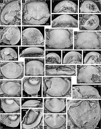
The large pygidium is approximately 59 mm wide and 52 mm long. In size and proportions it resembles pygidia of S. thomsoni as illustrated by Howells (Reference Howells1982, pl. 3) but differs in that the axis is a little narrower anteriorly and the pleural margin correspondingly a little wider adaxial to the fulcrum; the articulating facets are possibly not directed as strongly backward; and the doublure expands much less strongly backward. It is not possible to determine whether the inner margin of the doublure is deflected forward medially, into either a double cusp as in S. thomsoni or a single cusp as in some other Stenopareia species, but from what is preserved of the external mold of the doublure, a double cusp seems unlikely. The appearance of a forward deflection in the doublure margin just to the right of the sagittal line in Figure 5.30 is an artifact caused by erosion and weathering of the specimen.
The small pygidium, 8 mm wide and 4.7 mm long (Fig. 5.29), is proportionally much wider and shorter than the large specimen. The difference in proportions may be explained by the disparity in size between the specimens, as progressive elongation of the pygidium during ontogeny has been documented in other members of the Illaenina (e.g., see Chatterton and Ludvigsen, Reference Chatterton and Ludvigsen2004, p. 22; Holloway and Lane, Reference Holloway and Lane2012, p. 476). The subconical axis is a little narrower than that of the large pygidium (approximately 33% of the maximum pygidial width as opposed to almost 40%), is bounded laterally but not posteriorly by a weak axial furrow, and has five rings defined mainly by terrace ridges. There is a prominent sagittal ridge on the posterior half of the pygidium.
Family Uncertain
Subfamily Bumastinae Raymond, Reference Raymond1916
Remarks
Despite the currently intractable problems in elucidating evolutionary relationships among highly effaced forms of the Illaenina (see Lane and Thomas, Reference Lane and Thomas1978b, p. 9; Reference Lane and Thomas1983; Holloway and Lane, Reference Holloway and Lane1998, p. 865; Chatterton and Ludvigsen, Reference Chatterton and Ludvigsen2004, p. 19), we agree with Adrain et al. (Reference Adrain, Chatterton and Blodgett1995, p. 726) that the Bumastinae will likely prove to be monophyletic (although Adrain, Reference Adrain2013, p. 300, has recently advanced a different view). The Bumastinae was traditionally included within the Illaenidae (e.g., Jaanusson, Reference Jaanusson1959) but was placed by Ludvigsen and Tripp (Reference Ludvigsen and Tripp1990) and Adrain et al. (Reference Adrain, Chatterton and Blodgett1995) in the ‘Styginidae’, which in their concept also encompassed the Scutelluidae. Whittington (Reference Whittington1997) did not recognize the Bumastinae as a distinct subfamily but stated that he considered it “most probable that Bumastus is an illaenid, and not an effaced scutelluid” (p. 889), and he unequivocally assigned to the Illaenidae the genus Cybantyx, which is generally regarded as closely related to Bumastus. Hughes and Thomas (Reference Hughes and Thomas2014), however, included the Bumastinae in the Scutelluidae. We are unable to provide convincing arguments for assigning the Bumastinae to one or the other of the Illaenidae or Scutelluidae, and hence regard it at present of uncertain family affinity.
Adrain et al. (Reference Adrain, Chatterton and Blodgett1995, p. 729) stated that virtually all bumastines for which information is available have a rostral plate “with a posteromedially convex lobe that is produced into a dorsally re-entrant flange, along the posterior edge of which the hypostomal suture runs.” Adrain et al. (1995) contrasted this morphology with that of their species ‘Paracybantyx’ occidentalis in which the rostral plate lacks an upturned flange and its posterior margin is indented medially by the hypostomal suture, a condition they regarded as derived within bumastines. In fact, the only bumastine having a rostral plate with a vertically upturned posterior flange is Bumastus itself, in which the dorsal margin of the flange is also indented medially by the rostral suture (Lane and Thomas, Reference Lane and Thomas1978b, pl. 2, fig. 1b, c). Bumastines in which the rostral plate lacks a flange include Cybantyx Lane and Thomas (Reference Lane and Thomas1978b, pl. 5, figs. 6, 7; see also Whittington, Reference Whittington1997, fig. 5.6, 5.8), Liolalax Holloway and Lane (Reference Holloway and Lane1998, pl. 5, fig. 7; see also Šnajdr, Reference Šnajdr1957, text-fig. 2, pl. 10, fig. 8, pl. 11, figs. 6, 10; Lane and Thomas, Reference Lane and Thomas1978b, pl. 3, figs. 7, 8, pl. 4, fig. 1), and Opsypharus Howells (Reference Howells1982, pl. 2, fig. 9). We therefore consider that the presence of the rostral flange, rather than its absence, is the derived condition in bumastines.
Genus Cybantyx Lane and Thomas, Reference Lane and Thomas1978b
Type species
Cybantyx anaglyptos (sic) Lane and Thomas, Reference Lane and Thomas1978b, from the Much Wenlock Limestone Formation (upper Wenlock, Homerian), Dudley, West Midlands, UK, by original designation.
Remarks
The generic name is of feminine gender, rather than masculine as stated by Lane and Thomas (Reference Lane and Thomas1978b), so the correct form of the name of the type species is C. anaglypte.
Cybantyx? ergodes new species
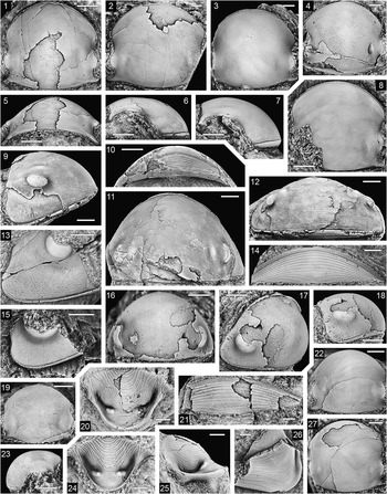
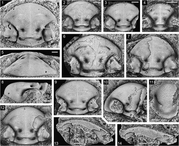
Cybantyx? ergodes n. sp., Tomcat Creek limestone, locality PL3309. (1, 5, 6) Cranidium, NMV P313583: (1) plan view, (5) posterodorsal view, (6) lateral view; (2) cranidium, NMV P313587, plan view; (3) cranidium, NMV P313588, plan view; (4) exfoliated cranidium, NMV P313589, plan view; (7, 8) cranidium, NMV P313584: (7) lateral view, (8) plan view; (9–12) holotype, cephalon, NMV P313578: (9) lateral view, (10) ventral view, (11) dorsal view, (12) anterodorsal view; (13) librigena, NMV P313597, oblique view; (14) rostral plate, NMV P313605, ventral view; (15) librigena, NMV P313603, oblique view; (16–18) small cephalon, NMV P313579: (16) dorsal view, (17) oblique view, (18) lateral view; (19, 23) smallest cranidium, NMV P313594: (19) plan view, (23) lateral view; (20, 25) hypostome, NMV P313607: (20) ventral view, (25) lateral view; (21) rostral plate, NMV P313606, ventral view; (22) small cranidium, NMV P313592, dorsal view; (24) small hypostome, NMV P313610, ventral view; (26) fragmentary librigena, NMV P313599, ventral oblique view; (27) cranidium, NMV P313591, plan view. (1, 5–13) Scale bars=5 mm; (2–4, 14–18, 20–22, 25–27) scale bars=2 mm; (19, 23, 24) scale bars=1 mm.

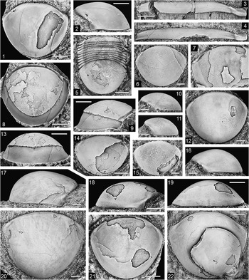
Cybantyx? ergodes n. sp., Tomcat Creek limestone, locality PL3309. (1, 2) Pygidium, NMV P313645: (1) dorsal view, (2) lateral view; (3) thoracic segment, NMV P313611, dorsal view; (4) thoracic segment, NMV P313612, dorsal view; (5) small thoracopygon, NMV P313641, dorsal view; (6, 10) small pygidium, NMV P313639: (6) dorsal view, (10) lateral view; (7) pygidium, NMV P313616, detail of internal mold in dorsal view; (8, 9, 13) pygidium, NMV P313621, internal mold with external mold of doublure: (8) dorsal view, (9) lateral view, (13) posterodorsal view; (11, 15) smallest pygidium, NMV P313640: (11) lateral view, (15) dorsal view; (12, 16) pygidium, NMV P313631: (12) dorsal view, (16) lateral view; (14) pygidium, NMV P313638, dorsal view; (17, 20) pygidium, NMV P313614: (17) lateral view, (20) dorsal view; (18, 19, 21) pygidium, NMV P313619: (18) lateral view, (19) posterior view, (21) dorsal view; (22) cranidium, NMV P313595, stacked on pygidium, NMV P313596, dorsal view of pygidium. (1–4, 7–9, 13, 17–22) Scale bars=5 mm; (5, 12, 14, 16) scale bars=2 mm; (6, 10, 11, 15) scale bars=1 mm.

Types
Holotype, cephalon NMV P313578, from PL3309. Paratypes, small cephalon NMV P313579, cranidia NMV P313580–P313595, librigenae NMV P313597–P313603, rostral plates NMV P313605, P313606, hypostomes NMV P313607–P313610, thoracic segments NMV P313611–P313613, pygidia NMV P313544, P313551, P313614–P313640, P313645, small pygidium with 10 thoracic segments NMV P313641, and small pygidium with two thoracic segments NMV P313642, from PL3309; rostral plate NMV P313643 and hypostome NMV P313644, from PL3312.
Diagnosis
Cephalon with axial furrow faint or not impressed behind lunette and absent in front of it; lunette situated level with δ–δ, poorly defined externally. Eye rather small, situated almost its own length from posterior cephalic margin. Librigena with broad, shallow subgenal notch and broadly rounded genal angle. Terrace ridges present only on front of cranidium and anterolateral part of librigena; librigena abaxial to subocular depression coarsely pitted. Rostral plate gently convex sagittally in anterior two-thirds and weakly concave in posterior third; posterior edge weakly notched medially by hypostomal suture; connective sutures almost transversely aligned. Pygidium with first pleural furrow weakly impressed.
Description
Cephalon strongly convex, in dorsal view approximately 160% as wide as long (sag.) and semielliptical in outline; in lateral view sagittal profile describing an arc of about 100° from posterior cephalic margin to posterior edge of very narrow, projecting marginal rim. Marginal rim becomes less prominent and bounding furrow becomes less sharply impressed medially on cranidium and posteriorly on librigena; on cranidium, outer face of rim slopes steeply downward to rostral suture. Cranidium with width at δ–δ slightly greater than at β–β and slightly less than at ω–ω; width at γ–γ a little greater than at ε–ε and about equal to sagittal length of cranidium in plan view; posterior margin nonfulcrate, describing an almost uniform arc in transverse profile (Fig. 3.5). Glabella comprising about 75% cranidial width posteriorly where its lateral extent is indicated by a gentle change in curvature of cranidial margin in plan view (Fig. 3.1–3.3, 3.8); glabella about 70% as wide at lunette as posteriorly. Omphalus and anterolateral internal pit present (Fig. 3.4), the latter situated closer to anterior rim than to omphalus and slightly closer than omphalus to sagittal line; median internal pit present on posterior part of glabella but not expressed externally as a node. G0 and G1 expressed on interior of exoskeleton as conjoined area of reticular appearance, extending farther back than posteromedian internal pit and at least as far forward as cranidial midlength (Fig. 3.1). Palpebral area sloping laterally from lunette at about same angle as adjacent part of glabella, decreasing in slope onto palpebral lobe, which is gently downturned along margin. Anterior branch of facial suture diverging at about 15° to sagittal line from γ to β; posterior branch curving backward and strongly outward. Eye situated with anterior edge well behind cephalic midlength (sag.); visual surface surrounded abaxially by broad, shallow depression. Cephalic doublure rather flattened posteriorly, increasing greatly in convexity anteriorly; transverse vincular furrow present just in front of posterior edge (Fig. 3.26). Rostral plate lenticular in outline, length (sag.) about 25% maximum width, which is transversely opposite median part of hypostomal suture or slightly farther forward; connective suture almost straight; rostral portion of hypostomal suture comprising about 25% maximum width of rostral plate. Hypostome about 160% as wide across anterior wings as long (sag.), anterior margin broadly rounded in ventral view, lateral and posterior margins parabolic in outline, without shoulders. Anterior lobe of middle body elliptical in outline, comprising 65%–70% hypostomal length (sag.), strongly convex transversely and moderately convex sagittally; posterior lobe moderately convex transversely and gently concave longitudinally between inflated and transversely elongated maculae. Lateral border furrow deep adjacent to anterior lobe of middle body, rapidly shallowing backward adjacent to posterior lobe and merging with even shallower posterior border furrow; lateral and posterior borders narrow except anteriorly where lateral border expands to merge with anterior wing; outer face of lateral border rather high; outer face of posterior border decreases in height medially between posterior wings.
Thorax probably of 10 segments (Fig. 4.5); axis and pleurae forming an almost uniform arch in transverse profile. Axis gradually widening from segments 1 to 6 and narrowing from segments 7 to 10, comprising almost 75% of segmental width throughout thorax; articulating furrow defined only on lateral-most part of axial ring but very weak (Fig. 4.3, 4.4); axial furrow shallow. Pleurae with fulcrum situated at or very close to axial furrow; articulating facet with small process on anterior edge close to its inner end (Fig. 4.3); pleural tips pointed on anterior segments and truncated on posterior ones.
Pygidium strongly convex, 115%–125% as wide as long (sag.), outline in dorsal view broadly rounded anteriorly and semielliptical laterally and posteriorly, maximum width level with about one-third sagittal length. Axis undefined but its width anteriorly as indicated by adaxial extent of first pleural furrow is about 66% to almost 75% of maximum pygidial width. Very shallow pleural furrow fades abaxially behind articulating facet, anterior edge of which has a small process at midwidth (Fig. 4.18), as on thoracic segments. Very faint holcos distinguishable on some pygidia, not extending farther back than about two-thirds sagittal length of pygidium; narrow, shallow sagittal furrow present posteriorly (Fig. 4.19), fading anteriorly above doublure and replaced in front of doublure by weak sagittal ridge that is more distinct on internal molds (Fig. 4.8, 4.14, 4.20); doublure convex in outer half and weakly concave in inner half, almost twice as wide sagittally as anterolaterally but deceasing in slope anterolaterally so that in ventral view it appears to be of uniform width, about 15% sagittal pygidial length.
Sculpture of terrace ridges on front of cranidium and anterolateral part of librigena; entire dorsal exoskeleton of cephalon, except lunette and visual surface, with sparsely distributed pits, coarsest and most distinct on librigena and very weak on posteromedian part of cranidium; interspersed with pits on outer part of librigena is a pattern of very fine, anastomosing, incised lines running roughly parallel to cephalic margins and extending along posterior edge of cranidium; interior of cephalon with caecal pattern on fixigena and subocular depression of librigena, reflected also on external surface of some specimens; coarse terrace ridges present on cephalic doublure and hypostome, excluding posterior lobe of middle body and a narrow band around front of rostral plate. Thorax with terrace ridges on articulating facets, and some segments with a few extending across middle (sag., exsag.) of axial ring; pits present on pleurae behind articulating facets and on posterior half of axial rings. Terrace ridges on dorsal surface of pygidium mainly restricted to anterolateral region including articulating facets, but a few extend adaxially in a narrow band just behind anterior margin; sparse pits present on entire dorsal surface excluding articulating facets but weak anteromedially; terrace ridges on doublure become more closely spaced toward outer edge; inner surface of exoskeleton inside doublure with caecal pattern (Fig. 4.7, 4.14).
Etymology
Greek, troublesome.
Remarks
The specimens vary considerably in size, with maximum lengths ranging from 3.2 to 40 mm for cranidia/cephala, from ~7 to ~24 mm for librigenae, and from 4.3 to 39 mm for pygidia. Significant changes in morphology that occur with increasing specimen size include decrease in the cranidial convexity (sag.), increase in the prominence of the anterior rim on the cranidium, decrease in the relative size of the eye (compare Fig. 3.9 and 3.18, 3.13 and 3.15), and an increase in the backward deflection of the genal angle (Fig. 3.13, 3.15). Variation in the width-to-length ratio of the pygidium is partly related to specimen size, the smallest pygidia being relatively wider than large ones though there is some variation unrelated to size among large pygidia.
Cybantyx? ergodes is similar to the type species in most aspects of morphology but differs markedly in the form of the rostral plate. In C. anaglypte, the rostral plate is subrhomboidal in outline as a result of the strongly oblique orientation of the connective suture, and the posteromedian part of the plate forms a gently convex (sag., exsag.), lobate projection (Lane and Thomas, Reference Lane and Thomas1978b, pl. 5, figs. 6, 7; Whittington, Reference Whittington1997, fig. 5.6, 5.8). In C.? ergodes, however, the rostral plate is lenticular in outline and lacks a posteromedian projection, the connective sutures being oriented almost transversely (Fig. 3.14, 3.21). Because of this difference from the type species in a feature thought to be of considerable taxonomic significance in illaenimorph trilobites (e.g., Holloway and Lane, Reference Holloway and Lane1998, p. 863), we assign ergodes to Cybantyx with question. In both C. anaglypte and C.? ergodes, there is on the front of the rostral plate a narrow band devoid of terrace ridges, running parallel and adjacent to the rostral suture. Similar smooth anterior bands are present on the rostral plates of Liolalax olibros (Holloway and Lane, Reference Holloway and Lane1998, pl. 5, fig. 6), L. bouchardi (see Šnajdr, Reference Šnajdr1957, pl. 11, figs. 6, 10) and Opsypharus magnificus (Chatterton and Ludvigsen, Reference Chatterton and Ludvigsen2004, pl. 6, figs. 4, 7).
Apart from the form of the rostral plate, C.? ergodes also differs from C. anaglypte in that the cephalic axial furrow is even weaker; the eye is smaller in the largest cephala; the lunette is weaker on the exterior of the exoskeleton and is situated farther forward; the librigena has a shallow subgenal notch and a more rounded genal angle (compare Fig. 3.9 with Lane and Thomas, Reference Lane and Thomas1978b, pl. 5, fig. 3c); there are weaker terrace ridges and larger pits on the librigenal field (compare Fig. 3.13 with Whittington, Reference Whittington1997, fig. 5.4); and the first pygidial pleural furrow is slightly impressed.
Cybantyx? nebulosa Hughes and Thomas (Reference Hughes and Thomas2014, figs. 14, 15A–15J) from the Telychian of North Greenland is questionably assigned by us to the genus because it has a rostral plate that is similar to that of C.? ergodes rather than to that of the type species. Cybantyx? nebulosa differs from C.? ergodes in that the cephalon is less convex (sag.) with a more prominent anterior rim; the axial furrow is more distinct on internal molds; the median internal pit on the posterior of the glabella is larger; the eye is longer in cephala of similar size, with its posterior edge closer to the posterior cephalic margin; the librigena lacks a subgenal notch; and the holcos on the pygidium is more distinct.
Genus Opsypharus Howells, Reference Howells1982
Type species
Illaenus (Bumastus) Maccallumi (sic) Salter, Reference Salter1867, from the Mulloch Hill Formation (lower Llandovery, Rhuddanian) of the Girvan district, Scotland, by original designation.
Other species
Opsypharus ambiguus (Foerste, Reference Foerste1885), Brassfield Formation (upper Rhuddanian–Aeronian), Ohio; O. arenaceus (Whittard, Reference Whittard1938), Hughley Shales and Pentamerus Beds (Aeronian–Telychian), England (see Curtis and Lane, Reference Curtis and Lane1997); O. asulcatus (Ludvigsen and Tripp, Reference Ludvigsen and Tripp1990), upper Road River Group (Ludlow), Yukon Territory, Canada; O. commodus (Apollonov, Reference Apollonov1980), upper Chokpar Formation, Osju limestone (Hirnantian), southern Kazakhstan; O. convexus (Kiaer, Reference Kiaer1908) (=‘Bumastus’ sulcatus var. kiaeri Whittard, Reference Whittard1939), Steinsfjorden Formation (Homerian), Norway; O. depressus (Kiaer, Reference Kiaer1908) (= O. maccallumi according to Helbert et al., Reference Helbert, Lane, Owens, Siveter and Thomas1982, p. 132; ?=pygidium type E of Whittard, Reference Whittard1939) and O. longicaudatus (Kiaer, Reference Kiaer1908), lower Rytteråker Formation (upper Aeronian), Norway (see Whittard, Reference Whittard1939); O. magnificus (Chatterton and Ludvigsen, Reference Chatterton and Ludvigsen2004), Becscie Formation (Rhuddanian), Anticosti Island, Quebec, Canada; O. niagarensis (Whitfield, Reference Whitfield1880), Racine Formation (Sheinwoodian–lower Homerian), Wisconsin and Illinois (see Whittington, Reference Whittington1992, pl. 113); O. nudus (Angelin, Reference Angelin1854), upper Boda Limestone (Hirnantian), Sweden (see Warburg, Reference Warburg1925; Suzuki and Bergström, Reference Suzuki and Bergström1999, fig. 5E, F); O. orbicaudatus (Billings, Reference Billings1859), Ellis Bay Formation (Hirnantian), Anticosti Island, Canada (see Bolton, Reference Bolton1981, pl. 7, fig. 4); O. sulcatus (Lindström, Reference Lindström1885), Slite Group (Sheinwoodian–Homerian), Gotland, Sweden; O. taimyricus (Balashova, Reference Balashova1959), Andreevskaya Formation (Aeronian), Eastern Taimyr, Russia (see Balashova, Reference Balashova1960); O. tegartensis (Norford, Reference Norford1994), Tegart Formation (Telychian), British Columbia, Canada; O. wangi (Chatterton and Ludvigsen, Reference Chatterton and Ludvigsen2004), upper Gun River to lower Chicotte formations (Aeronian–Telychian), Anticosti Island, Quebec, Canada; Opsypharus sp. of Lane (Reference Lane1984) (= Ligiscus sp. in pl. 1, fig. 1 of the same paper), Offley Island and/or Hauge Bjerge formations (Telychian), North Greenland.
Diagnosis
Cephalon and pygidium of low to moderate convexity. Omphalus and/or anterolateral internal pit present; axial furrow commonly extending forward to omphalus on internal molds. Shallow border furrow may be present on librigena; eye socle absent; genal angle broadly rounded to acute. Rostral plate lenticular to rhombic in outline; rostral portion of hypostomal suture transverse or gently convex forward. Thorax with very broad axis comprising 65%–75% segmental width and with ‘nonfulcrate’ pleurae. Pygidium with first pleural furrow impressed, meeting distinct holcos distally.
Remarks
Opsypharus was regarded by Morris (Reference Morris1988) as a junior synonym of Failleana Chatterton and Ludvigsen, Reference Chatterton and Ludvigsen1976, a view accepted by Lane and Siveter (Reference Lane and Siveter1991), who considered both generic names to be synonyms of Meitanillaenus Zhang, Reference Zhang1974, Jell and Adrain (Reference Jell and Adrain2003), and Chatterton and Ludvigsen (Reference Chatterton and Ludvigsen2004). There are certainly similarities between Opsypharus and Failleana in many characters, including the low to moderate convexity of the cephalon and pygidium, the course of the cephalic axial furrows, the presence of the omphalus, the form of the rostral plate, and the thorax with very broad axis and ‘nonfulcrate’ pleurae. However, we consider that Opsypharus and Failleana are distinct genera, and we restrict Failleana to the species (all Ordovician) listed by Chatterton and Ludvigsen (Reference Chatterton and Ludvigsen1976, p. 38) as well as F. limbata (Raymond, Reference Raymond1910) of Shaw and Bolton (Reference Shaw and Bolton2011, p. 433, fig. 11.10–11.18, 11.23) from the Mingan Formation of Quebec, F. memorabilis (Reed, Reference Reed1906) from the lower Balclatchie Group of Scotland (see Morris and Tripp, Reference Morris and Tripp1986, p. 165, pl. 2, fig. 8), and Failleana sp. of Tremblay and Westrop (Reference Tremblay and Westrop1991, p. 808, fig. 9.15, 9.16) from the upper Sunblood Formation of Canada. A distinctive difference between Opsypharus and Failleana is that in the latter the cephalic axial furrow is continuous posteriorly with a transverse furrow in front of a posterior fixigenal articulating flange that is longest (exsag.) toward its distal end where it overlies a fulcral socket (see Ludvigsen and Chatterton, Reference Ludvigsen and Chatterton1980, pl. 1, figs. c–g). This articulating flange is similar in structure to that on the posterior cephalic margin of scutelluids, as described by Whittington (Reference Whittington1999, p. 415, who erroneously referred to the flange as the posterior border) and Holloway and Lane (Reference Holloway and Lane2012, pp. 419–420). By contrast, the axial furrow in Opsypharus may be continuous posteriorly with a weak border furrow that may extend onto the librigena (see Whittington, Reference Whittington1992, pl. 113). Failleana also differs from Opsypharus in that the posterior branch of the facial suture is curved in a convex-forward arc instead of a convex-backward arc; there is on the front of the cranidium a band of very strong terrace ridges that are much more prominent than those on the front of the cranidium in Opsypharus; genal spines in Failleana, when present as on small librigenae of the type species F. calva Chatterton and Ludvigsen, Reference Chatterton and Ludvigsen1976 (see Ludvigsen and Chatterton, Reference Ludvigsen and Chatterton1980, pl. 1, figs. s–v) or on large librigenae of F. limbata (see Shaw, Reference Shaw1968, pl. 19, fig. 5), are broad, flattened, and blade-like, whereas the genal spines on an immature cephalon of O. pandanensis n. sp. are slender and spike-like (Fig. 5.4, 5.6); and the first pygidial pleural furrow is not impressed in mature pygidia of Failleana. Failleana has a stratigraphic range of Darriwilian to Katian, whereas the range of Opsypharus is Hirnantian to Ludlow. Although Lane and Siveter (Reference Lane and Siveter1991) regarded the poorly known Meitanillaenus as a senior synonym of Opsypharus and Failleana, we consider that it is distinct from both genera, as the type species, M. binodosus Zhang (Reference Zhang1974, pl. 81, figs. 5–7, pl. 82, figs. 11, 12) from the upper Rhuddanian to Aeronian of Guizhou, China, differs in that it lacks the omphalus; the rostral plate is subtrapezoidal in outline rather than lenticular or rhombic; and the thorax has a narrower axis and fulcrate pleurae.
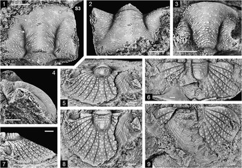
Opsypharus and Stenopareia from Tomcat Creek limestone, all except (16) and (29) from locality PL3309. (1–22, 24–28) Opsypharus pandanensis n. sp.: (1) cranidium, NMV P313556, plan view; (2, 3) mostly exfoliated cranidium, NMV P313558: (2) plan view, (3) lateral view; (4, 6) ?meraspid cephalon, NMV P313571: (4) dorsal view, (6) oblique view; (5, 9, 10, 14) holotype, cranidium, NMV P313560: (5) posterodorsal view, (9) plan view, (10) dorsal view, (14) lateral view; (7, 8, 11, 12) small, mostly exfoliated cephalon, NMV P313557: (7) oblique view, (8) dorsal view, (11) lateral view, (12) ventral view; (13, 17) small cranidium, NMV P313562: (13) dorsal view, (17) lateral view; (15, 19) cranidium, NMV P313561: (15) lateral view, (19) plan view; (16) pygidium, NMV P313575, dorsal view, locality PL3314; (18) incomplete thoracic segment, NMV P313577, dorsal view; (20) small pygidium, NMV P313570, dorsal view; (21) incomplete librigena, NMV P313563, oblique view; (22, 25) pygidium, NMV P313567: (22) dorsal view, (25) lateral view; (24) incomplete librigena, NMV P313564, oblique view; (26) small pygidium, NMV P313569, dorsal view; (27) small librigena, NMV P313565, oblique view; (28) incomplete pygidium, NMV P313568, latex cast of natural mold, ventral view. (23, 29, 30) Stenopareia aff. thomsoni (Salter, Reference Salter1867); (23, 30) large exfoliated and corroded pygidium, NMV P313646: (23) lateral view, (30) plan view; (29) small pygidium, NMV P313576, dorsal view, locality PL3312. (1–3, 16, 18, 21, 22, 24, 25, 28) Scale bars=2 mm; (4–15, 17, 19, 20, 26, 27, 29) scale bars=1 mm; (23, 30) scale bars=5 mm.

Ludvigsen and Tripp (Reference Ludvigsen and Tripp1990, p. 8, pl. 1, figs. 1–15, pl. 11, figs. 1–5) proposed the genus Paracybantyx for their species P. asulcatus (sic) from Ludlow strata of the Road River Formation of the northern Yukon Territory. The diagnosis provided for the new genus was directed mainly toward distinguishing it from Cybantyx by the absence of an anterior rim on the cranidium. The other characters mentioned are rather generalized and present in many illaenimorphs. We are unable to find any characters differentiating Paracybantyx from Opsypharus and consequently regard it as a junior synonym of that genus.
Opsypharus pandanensis new species
Types
Holotype, cranidium NMV P313560, from PL3309. Paratypes, incomplete cephala NMV P313556, P313557, cranidia NMV P313558, P313559, P313561, P313562, librigenae NMV P313563–P313565, fragmentary thoracic segment NMV P313577, pygidia NMV P313566–P313570, and probable meraspid cephalon NMV P313571, from PL3309; cranidium NMV P313572 and librigenae NMV P313573, P313574, from PL3312; pygidium NMV P313575, from PL3314.
Diagnosis
Cranidium moderately convex but anterior outline only gently curved; width at γ–γ approximately equal to sagittal length in plan view; eye relatively large, situated half its own length from posterior margin; genal angle broadly rounded; axial furrow only distinguishable immediately in front of posterior cranidial margin; anterolateral internal pit present but omphalus absent; lunette very weakly defined, situated opposite posterior half of palpebral lobe in plan view. Pygidium less convex than cephalon, 1.4–1.5 times as wide as long (sag.); anterior width of axis a little less than half maximum pygidial width; transverse, adaxial portion of anterior pleural margin approximately as wide as articulating facet. Dorsal surface of exoskeleton densely covered with tiny, shallow pits.
Description
Cranidium with sagittal profile in lateral view describing an arc of about 130° in largest specimen (Fig. 5.3); transverse profile across δ–δ with greatest curvature sagittally (Fig. 5.5). Cranidial widths at β–β, δ–δ, and ω–ω approximately in ratio 0.9:1:1.1, width at ε–ε slightly greater than at γ–γ. Glabella comprises a little more than half total cranidial width at posterior margin, width at lunette approximately 75% posterior width; on internal mold of largest cranidium, a forwardly diverging row of very weak impressions extending from front of lunette approximately halfway toward anterolateral pit (Fig. 5.2, most distinct on left side) either coincides with position of axial furrow or runs subparallel and just adaxial to it. Inner surface of glabella on largest cranidium also with weak sagittal furrow (ridge on internal mold); G1 muscle scars large, subcircular or ovate, situated opposite palpebral lobe in plan view and closer to lunette than to sagittal line; other paired muscle scars indistinguishable. Palpebral lobe gently curved in outline, length (exsag.) about one-third sagittal cranidial length in plan view, situated about half its length from posterior cranidial margin in dorsal view and with anterior edge opposite cranidial midlength in plan view; palpebral furrow generally not defined but weakly expressed in an incomplete cephalon (Fig. 5.1), running subparallel with palpebral margin and very close to it. Anterior branch of facial suture weakly diverging forward at about 10° to sagittal line from γ to β; posterior branch initially diverging moderately backward from ε before curving strongly outward close to posterior margin. Posterior cranidial margin with narrow, horizontal, articulated portion adaxial to fulcrum, beyond which fixigena is downturned (Fig. 5.5). Librigena with posterior margin directed anterolaterally to genal angle; visual surface bounded below by broad, shallow furrow. Doublure known only in a small cephalon (Fig. 5.12), broadly rounded (sag., exsag.) in section anteromedially; rostral plate apparently lenticular in outline (see remarks that follow).
Thorax known only from an incomplete segment, probably from the anterior part of the thorax judging from the oblique orientation of the axial furrow (Fig. 5.18). Segment more or less uniformly arched (tr.) overall, without distinct fulcrum. Axial furrow very weakly defined, directed posterolaterally from a shallow notch on anterior edge of segment; axial ring and undifferentiated articulating half ring gently convex (sag., exsag.). Pleura curved gently backward abaxially to about midwidth and recurved forward thereafter, with truncated tip; broad (tr.) articulating facet extends inward to anterior end of axial furrow; anterior edge of facet with a narrow (tr.) articulating flange proximally, ending distally in a small process.
Pygidium evenly curved in outline laterally and posteriorly. Axis defined only by forward deflection of anterior margin adaxial to transverse, articulated portion of pleural margin and by inward extent of first pleural furrow; articulating facet deflected backward at about 50° to sagittal line. First pleural furrow long (exsag.) and shallow, distally joining even shallower border furrow that dies out posteriorly about level with two-thirds sagittal pygidial length. Doublure flattened and steeply inclined sagittally where it comprises about 20% of pygidial length, increasing in convexity and decreasing in slope anterolaterally, narrowing slightly toward its anterior end.
Fine, dense pitting of dorsal exoskeleton not present on articulating facets of thoracic segment and pygidia. Terrace ridges present on front of cranidium and outer part of librigena, on articulating facet and anterior part of pleural band of thoracic segment, on articulating facet and outer part of pygidium, and on cephalic and pygidial doublures; terrace ridges on outer margins of cranidium and librigena are coarse and prominent as on cephalic doublure and increase in number anteromedially on librigena, from one at genal angle to four or more at front of librigena (Fig 5.24, 5.27).
Etymology
After ‘Pandanus’ cattle station, on which the Tomcat Creek limestone occurs.
Remarks
The very fine, dense pitting on the dorsal surface distinguishes the various exoskeletal elements of this species from those of Cybantyx? ergodes, in which the pits are larger and more sparsely distributed. The nature of the exoskeletal pits is the reason that the thoracic segment in Figure 5.18 is assigned to Opsypharus pandanensis rather than to C.? ergodes, segments of which are otherwise very similar. Cranidia of O. pandanensis are also somewhat similar to those of C.? ergodes but are readily distinguished by the distinct geniculation of the posterior fixigenal margin (compare Figs. 3.5 and 5.5), the less strongly curved anterior outline, and the longer (exsag.) palpebral lobe.
As a result of significant morphological changes that occur during growth of O. pandanensis, smaller specimens differ in several respects from the largest ones on which the preceding description is based. Smaller cephala/cranidia are more convex than larger ones (compare Fig. 5.3 and 5.13) and the eye is relatively larger (Fig. 5.1, 5.8). A very small cephalon about 2.8 mm long in dorsal view has a slender, spike-like genal spine (Fig. 5.4, 5.6), and the spine is still present in a cephalon about 5.2 mm long (maximum length of librigena about 4 mm; Fig 5.7, 5.8, 5.11) though the spine is broken and its length is unknown. In a very small cranidium about 3.5 mm long in dorsal view, the axial furrow is distinguishable though very faint as far forward as about one-quarter of the sagittal cranidial length from the anterior (Fig. 5.17). The occipital furrow is very weakly impressed medially, and muscle scars G0–G3 are distinguishable on the exterior of the exoskeleton, G1 being most distinct, G2 and G3 smaller, very faint, and situated successively farther from the sagittal line. Small pygidia are relatively a little wider than large ones.
The rostral plate is known only in a small cephalon (Fig. 5.7, 5.8, 5.11, 5.12) but is not well delineated. The connective suture is very indistinct on the left side in Figure 5.12, converging backward at about 65° to an exsagittal line and apparently meeting the hypostomal suture rather close to the sagittal axis. The rostral portion of the hypostomal suture is also indistinct but appears to be transverse. The right-hand edge of the doublure in Figure 5.12 is at least partly broken and does not indicate that the connective suture curves backward posteriorly.
Opsypharus pandanensis is easily distinguished from other species of the genus by the gentle curvature of the anterior cranidial margin in dorsal view, the almost nonexistent cephalic axial furrows, the absence of the omphalus, and the less elongated outline of the pygidium. In pygidial morphology, O. pandanensis is exceedingly similar to the type species of Bumastella, B. spicula (Kobayashi and Hamada, Reference Kobayashi and Hamada1974) (see Holloway and Lane, Reference Holloway and Lane1998, pl. 1, figs. 1–21, pl. 2, figs. 1–15, 17, 18). Differences in the cephalic morphology of B. spicula, in particular its much greater convexity, the posterior margin that in dorsal view is deflected backward rather than forward abaxial to the fulcrum, the smaller eye situated farther forward, the anterior branch of the facial suture that converges rather than diverges forward, the weaker backward divergence of the posterior branch of the suture, and the more elongated (sag.) rostral plate, suggest that the two species are not closely related, even at the generic level.
Family Scutelluidae Richter and Richter, Reference Richter and Richter1955
Remarks
In pygidia of some Ordovician and Silurian scutelluids, the interpleural furrows originating toward the posterolateral part of the axis become very shallow or effaced at their proximal ends, the furrows affected usually being the third to the fifth, but they may include others. Commonly associated with the weakening of these furrows is a slight inflation of the pleural region immediately adjacent to the axis, both aspects of the feature being more distinct in larger pygidia than in smaller ones. Among the scutelluids from the Tomcat Creek limestone, the feature is well shown in Japonoscutellum mawsonae (Fig. 12) and is evident in Kosovopeltis avita n. sp. (Fig. Reference Cockle14.3). The feature is not always easy to distinguish, but other scutelluids showing it to various extents include Eokosovopeltis atava (Webby, Reference Webby1974, pl. 28, fig. 16), Japonoscutellum allaarti (Lane, Reference Lane1984, pl. 1, fig. 16), J. gephyricum Holloway and Lane (Reference Holloway and Lane2012, fig. 17O, R), Kosovopeltis trepida Holloway and Lane (Reference Holloway and Lane2012, fig. 22A, D), Planiscutellum planum (Hawle and Corda, Reference Hawle and Corda1847) (see Šnajdr, Reference Šnajdr1960, pl. 1, fig. 1), P. andersoni (Etheridge and Nicholson in Nicholson and Etheridge, Reference Nicholson and Etheridge1879) (see Howells, Reference Howells1982, pl. 2, figs. 3, 5–7), Securifrons borealis (Poulson, Reference Poulsen1934) (see Ludvigsen and Tripp, Reference Ludvigsen and Tripp1990, pl. 2, fig. 11, pl. 3, fig. 2), and three species of Jungosulcus described by Hughes and Thomas (Reference Hughes and Thomas2014). Hughes and Thomas regarded the feature as diagnostic of Jungosulcus, but its relatively widespread but inconsistent presence in scutelluids suggests that it is not diagnostic at the generic level. In Illaenoscutellum psephos n. sp. from the Tomcat Creek limestone the feature is conspicuously developed, and because of the effacement of the axial furrow in this species, the smooth and inflated adaxial parts of the pleural region appear to be part of the axis, though they extend farther posteriorly than the axial terminus (Fig. 9.1, 9.2, 9.9, 9.10). The function of these extra-axial inflated areas is unclear. It is possible that they represent muscle attachment sites, perhaps for the dorsal longitudinal muscles; however, we are unable to find any evidence on the interior of the exoskeleton of a concentration of muscle insertions in these areas. In addition, a function as muscle attachment sites seems incompatible with the inconsistent development of the feature among some species of a few genera of scutelluids. The feature is not apparent in some other species of the genera mentioned in the preceding—for example in Kosovopeltis ennadifi and K. maroccanica of Alberti (Reference Alberti1970, pl. 1, figs. 2, 4), Japonoscutellum diascetum Holloway and Lane (Reference Holloway and Lane2012, fig. 16I–O), and Illaenoscutellum tryo Holloway and Lane (Reference Holloway and Lane2012, fig. 11N, Q, R)—nor is it present in most other Silurian scutelluid genera or any of the large number of Devonian genera. However, if these inflated areas do indeed represent the sites of muscle attachment, they may be homologous with the pair of large, elliptical, extra-axial muscle scars that have been documented in the pygidia of some illaenimorph genera such as Bumastella (Holloway and Lane, Reference Holloway and Lane1998, pl. 2, fig. 8), Bumastus (Lane and Thomas, Reference Lane and Thomas1978b, pl. 1, fig. 7), Liolalax (Šnajdr, Reference Šnajdr1957, pl. 11, fig, 11, text-fig. 34F; Lane and Thomas, Reference Lane and Thomas1978b, pl. 3, fig. 10), Opsypharus (Lane, Reference Lane1984, pl. 1, fig. 5) and Stenopareia (Zhou et al., Reference Zhou, Yin and Zhou2014, fig. 21H).
Genus Australoscutellum Chatterton and Campbell, Reference Chatterton and Campbell1980
Type species
Bronteus longispinifex Mitchell, Reference Mitchell1887, from the Yarwood Siltstone Member, Black Bog Shale (upper Ludlow, Ludfordian), Yass Basin, New South Wales, by original designation.
Other species
Australoscutellum iota, A. microps, A. pulex, A. trica, and Australoscutellum? sp., all of Holloway and Lane (Reference Holloway and Lane2012); A. talenti n. sp.; Australoscutellum n. sp. A of Chatterton and Campbell (Reference Chatterton and Campbell1980).
Diagnosis
See Holloway and Lane (Reference Holloway and Lane2012, p. 422).
Occurrence
Upper Llandovery (Telychian) to upper Ludlow (Ludfordian); eastern Australia (New South Wales, Australian Capital Territory, Queensland).
Australoscutellum talenti new species
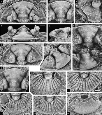
Australoscutellum talenti n. sp., Tomcat Creek limestone, all except (3) from locality PL3309; dorsal views unless otherwise stated. (1, 4, 5) Holotype, cephalon, NMV P313420: (1) dorsal view, (4) anterodorsal view, (5) lateral view; (2) cranidium, NMV P313427; (3) librigena, NMV P313435, oblique lateral view, locality PL3312; (6) cranidia, NMV P313425 (above) and P313426 (below); (7, 8) cephalon, NMV P313421: (7) dorsal view, (8) oblique view; (9) cranidium, NMV P313422; (10) pygidium, NMV P313429; (11) pygidium, NMV P313434; (12) pygidium, NMV P313432; (13) pygidium, NMV P313433; (14) pygidium, NMV P313431, ventral view. Scale bars=1 mm.

Types
Holotype, cephalon NMV P313420, from PL3309. Paratypes, cephalon NMV P313421, six cranidia NMV P313422–P313427, and six pygidia NMV P313429–P313434, from PL3309; a librigena NMV P313435, and two pygidia NMV P313436, P313437, from PL3312.
Diagnosis
Glabella moderately convex, expanding strongly forward in front of S1, more than twice as wide across frontal lobe as at L1; S1 and S2 separate but connected by poorly defined exsagittal depression, S1 relatively shallow, S2 even shallower; S3 wide (tr.), rather shallow, not distinctly connected to axial furrow; preglabellar furrow shallow abaxially, faint or nonexistent medially; preocular librigenal boss large, prominent; eyes large with bulbous visual surface raised high on cheek; anterior branch of facial suture initially converging weakly forward from γ and diverging across anterior part of genal field and border, meeting anterior cephalic margin a little farther from the sagittal line than γ; lateral border furrow rather sharply impressed on fixigena; genal angle backwardly deflected, subgenal notch not present. Pygidium elliptical in outline, with seven pairs of pleural ribs and lacking marginal spines. Dorsal sculpture of variably sized tubercles and granules interspersed with and rarely superimposed on terrace ridges.
Description
Cephalon lenticular in outline in dorsal view, about twice as wide across genal angles as long (sag.). Glabella narrowing strongly forward across occipital ring, subparallel-sided or expanding very weakly forward across L1, thereafter expanding rather uniformly except for slight constriction level with outer end of S3; widths at posterior margin, L1, and frontal lobe approximately in ratio 1.5:1:2.2; width across frontal lobe equal to cephalic length. Occipital ring lenticular in outline, strongly arched in transverse profile with greatest curvature at sagittal line and flattened on flanks, with short, stout median spine on posterior edge (Fig. 6.6); occipital furrow broad (sag., exsag.), shallow, more sharply defined anteriorly than posteriorly, containing weak, subcircular muscle scar abaxially. L1 uniformly arched transversely, as long abaxially as sagittal length of occipital ring excluding median spine; S1 directed anteromedially from axial furrow; bolus not defined; S2 shallow, subcircular to subtriangular, situated at or just in front of glabellar midlength and exsagittally in line with inner edge of S1; S3 oblique, elliptical to slit-like, deepest abaxially, inner end situated in line (exsag.) with axial furrow adjacent to L1. Axial furrow well impressed, deepest adjacent to lunette and at intersection with lateral border furrow, shallowest adjacent to occipital ring and frontal lobe; lunette elongated, extending well in front of posterolateral edge of S1. Eye situated opposite occipital furrow; visual surface strongly overhanging cheek anterolaterally, laterally, and posteriorly, with anteromedial and posteromedial edges in line (exsag.) with lateral extremity of frontal lobe; palpebral lobe steeply upturned and convex (tr.), approximately equal in height to median part of occipital ring, its posterior half with short, stout spine directed posterodorsally and slightly adaxially; palpebral furrow well impressed, exsagittally aligned; very weak eye ridge extends from front of palpebral lobe toward axial furrow adjacent to L3. Anterior branch of facial suture describing concave outward curve, running on low, narrow ridge that interrupts lateral border furrow; posterior branch directed laterally and slightly forward from ε, in front of posterior articulating flange furrow on fixigena, and curving backward distally to meet posterior cephalic margin a little less than halfway between axial furrow and genal angle. Librigenal field decreasing in slope and increasing in width anteriorly; lateral border furrow shallowing posteriorly on librigena, fading as it closely approaches base of eye (Fig. 6.3); lateral border concave anteriorly, posteriorly becoming less concave in its outer half and weakly convex in its inner half; genal angle a little less than 90°. Terrace ridges present on entire dorsal surface of cephalon except axial and glabellar furrows but including occipital furrow; ridges scarce and weak on cheek in front of eye but prominent on preocular librigenal boss; vague pitting present between tubercles on genal field; prominent pair of larger tubercles commonly present medially on glabella opposite front of S1.
Pygidium approximately 1.6 times as wide as long, maximum width lying just behind level of axial terminus. Axis comprising about one-third maximum pygidial width anteriorly and one-third sagittal length, subpentagonal in outline except for lateral remnant of first ring, which projects well beyond general outline and dies out medially; remainder of axis longitudinally trilobate, median lobe with strongest inflation posteromedially and in some specimens possibly with indistinct segmentation defined by alignment of tubercles (Fig. 6.10, 6.13). Pleural region gently vaulted, concave in outer part; interpleural furrows expanding very slightly abaxially, terminating distally a short distance from margin; furrows 1 and 2 successively more weakly concave backward, furrows 3 and 4 straight, furrows 5 to 7 successively more strongly concave forward and successively shallower proximally; furrow 6 developed only adaxially in one pygidium (Fig. 6.13). Postaxial rib wider proximally than adjacent paired rib, subparallel-sided or expanding weakly backward in anterior half and expanding more strongly in posterior half; in one pygidium, postaxial rib is wider than in others, is subdivided proximally by a pair of very short longitudinal furrows, and has a weak sagittal keel farther back (Fig. 6.10). Doublure comprising about 55% sagittal length of pygidium. Tubercles dense on most of dorsal surface but sparse and weak on lateral lobe of axis and remnant of first ring; coarser tubercles absent in marginal zone of pleural region; terrace ridges present on articulating half ring, median lobe of axis, and outer part of pleural region.
Etymology
After Emeritus Professor J. A. Talent of Macquarie University, New South Wales.
Remarks
All of the specimens assigned to species of Australoscutellum from the known areas of occurrence are small for members of the Scutelluidae (Holloway and Lane, Reference Holloway and Lane2012, p. 422). With lengths ranging from 3.2 to 5.5 mm for cephala and cranidia (excluding the occipital spine) and from 3.9 to 5.7 mm for pygidia, specimens of A. talenti are similar in size to those of the type species A. longispinifex and A. iota but a little larger than those of A. microps, A. pulex, and A. trica. Australoscutellum talenti differs from all of these stratigraphically younger (Wenlock to Ludlow) species in that the glabella is less convex; S2 is distinctly separated from S1 instead of being superimposed on it anteromedially; the preocular librigenal boss is larger; a subgenal notch is not present on the librigena; and the pygidium has seven pairs of pleural ribs rather than five or six. We regard these differences as representing less derived character states. The species that is most similar to A. talenti is A. pulex but, in addition to the differences listed above, in A. talenti S1 and S2 are shallower; S3 is wider (tr.) and not distinctly joined to the axial furrow; the facial suture meets the anterior cephalic margin farther from the sagittal line; the lateral border furrow is deeper anteriorly; the maximum width of the pygidium lies farther forward; the lateral remnant of the first pygidial axial ring is more reduced; the first two interpleural furrows on the pygidium do not curve as strongly backward abaxially; and terrace ridges are much more extensive on the dorsal exoskeleton.
Genus Dolabrapex new genus
Type species
Dolabrapex acomus n. gen. n. sp.
Diagnosis
Glabella weakly convex, narrowing forward across occipital ring and occipital furrow, subparallel-sided across L1 and L2, thereafter expanding very strongly; axial furrow not impressed adjacent to frontal lobe of glabella, which merges with lateral border; preglabellar furrow absent. S1–S3 very weak and poorly defined; S1 subrectangular, bolus not distinct; S2 and S3 isolated from axial furrow, S2 subtriangular to kidney shaped, S3 elliptical to weakly arcuate, slightly obliquely oriented. Fixigena broad, width opposite δ almost equal to that of glabella; lateral border furrow and/or epiborder furrow very weak on front of fixigena, not clearly differentiated. Pygidial axis of low convexity, bell shaped, not distinctly trilobate longitudinally; pleurae with six pairs of ribs of low relief; interpleural furrows shallow. Terrace ridges present on front of cranidium and on pygidium; remainder of cranidium smooth.
Etymology
Latin dolabra, an ax, and apex, top, referring to the shape of the glabella; gender masculine.
Remarks
Dolabrapex is known only from the type species. The genus resembles Kosovopeltis, especially species such as K. trepida Holloway and Lane, Reference Holloway and Lane2012 and K. avita n. sp., in the gently convex glabella, the strongly laterally expanded frontal lobe that tends to merge abaxially with the cephalic border, the absence of the preglabellar furrow, the weak S1–S3 and their shape, the fixigena that is about as wide as the glabella opposite δ, and the poorly differentiated lateral and/or epiborder furrows on the anterior fixigena. Kosovopeltis differs from Dolabrapex in that there is a median swelling on the posterior edge of the occipital ring; the glabella expands forward from the occipital furrow or the front of L1 instead of from the front of L2; many species have a median swelling on the glabella opposite S2; the pygidial axis is subtriangular to parabolic in outline rather than bell shaped, is longitudinally trilobate, and includes a small lateral remnant of the first ring; and there are seven rather than six pairs of pygidial pleural ribs.
Apart from Dolabrapex, the only other rather effaced Silurian scutelluid genera with six pairs of pleural ribs in the pygidium are Ciliscutellum and Sangzhiscutellum, both proposed by Lin (Reference Lin1987) and known only from their respective type species from the Aeronian of Hunan, China. Ciliscutellum appears rather similar to Dolabrapex in cranidial morphology except that the occipital furrow is much weaker or effaced and a shallow preglabellar furrow is present at least abaxially. Differences between the pygidia of Ciliscutellum and Dolabrapex are more significant: in Ciliscutellum the axis is subtriangular rather than bell shaped and is more poorly defined; the axis is said to have four indistinct rings, each with a pair of tubercles, though the tubercles are not distinguishable in the published photographs; the pleurae are more convex and have a gently concave border; and the pleural furrows are much weaker, the more posterior ones being almost or completely effaced. The type species of Sangzhiscutellum, S. flabelliforme (Liu, Reference Liu1982), is known only from a cranidium and a pygidium that differ from Dolabrapex in that the occipital furrow appears to be much weaker; the glabella is not subparallel-sided across L1 and L2 but widens anteriorly from the front of the lunette; the frontal lobe is not as expanded (tr.); a shallow preglabellar furrow is present; the fixigena is much narrower, especially opposite the palpebral lobe; the pygidium appears to be more convex; the pygidial axis is semielliptical in outline rather than bell shaped, has two rings, and is indistinctly trilobate posteriorly; the postaxial rib is subparallel-sided; and the interpleural furrows do not extend as close to the margin.
Dolabrapex can be readily distinguished from Eobronteus Reed, Reference Reed1928 and Protobronteus Šnajdr, Reference Šnajdr1960, to which are assigned Late Ordovician species having six pairs of pygidial pleural ribs. Eobronteus (see Whittington, Reference Whittington2000) differs from Dolabrapex in having a well-defined preglabellar furrow joining the axial furrow abaxially so that the frontal lobe of the glabella is clearly differentiated from the border; the anterior fixigena is longer and wider; the lateral border furrow is well impressed and differentiated from the epiborder furrow on the front of the fixigena; the posterior border furrow is distinct on the fixigena and curves forward adaxially to reach the posterior edge of the lunette; and the pygidial axis is parabolic in outline rather than bell shaped. In the poorly known Protobronteus, with type and only assigned species P. reedi (Sinclair, Reference Sinclair1949) from the Trenton Group in Quebec, the preglabellar furrow is effaced as in Dolabrapex, but the axial furrow is impressed though shallow adjacent to the frontal lobe of the glabella; the lateral and epiborder furrows are clearly differentiated on the front of the fixigena; the pygidial axis is less well defined, especially posteriorly; and the postaxial rib is broad and subparallel-sided. Another genus that has been regarded as a scutelluid with six pleural ribs in the pygidium is Craigheadia Hupé, Reference Hupé1955, with type and only assigned species ‘Bronteus’ grayi Reed, Reference Reed1904, known only from a mold of a very large pygidium from the Upper Ordovician of Scotland. However, this pygidium does not belong to the Scutelluidae but to the Lichidae, its pleural region having three pairs of furrowed segments as do many lichids. The apparent absence of marginal spines and the proportionally very large axis suggest that the pygidium may belong to Leiolichas Schmidt, Reference Schmidt1885 (revised by Thomas and Holloway, Reference Thomas and Holloway1988), and Craigheadia is probably a junior synonym of that genus.
‘Kosovopeltis’ obsoletus [sic] Wu, Reference Wu1977, from the Telychian of southeastern Sichuan, China, is based on a rather effaced cranidium that shows similarities with Dolabrapex. The species was excluded from Kosovopeltis by Holloway and Lane (Reference Holloway and Lane2012, p. 472), but in the absence of other exoskeletal parts, especially the pygidium, it cannot be assigned to a genus with confidence.
Dolabrapex acomus new species
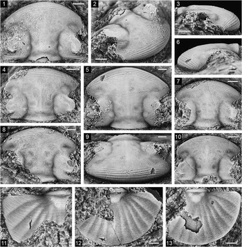
Dolabrapex acomus n. gen. n. sp., Tomcat Creek limestone, all except (5, 6, 9) from locality PL3309; dorsal views unless otherwise stated. (1–3) Holotype, cranidium, NMV P313497: (1) dorsal view, (2) oblique view, (3) lateral view; (4) cranidium, NMV P313498; (5, 6, 9) cranidium, NMV P313505: (5) dorsal view, (6) lateral view, (9) anterodorsal view, locality PL3314; (7) cranidium, NMV P313499; (8) cranidium, NMV P313428; (10) cranidium, NMV P313500; (11) pygidium, NMV P313502; (12) pygidium, NMV P313503; (13) pygidium, NMV P313504. Scale bars=1 mm.

Types
Holotype, cranidium NMV P313497, from PL3309. Paratypes, cranidia NMV P313428, P313498–P313500, pygidia NMV P313502–P313504, P313514, from PL3309; cranidium NMV P313505, from PL3314.
Diagnosis
As for genus.
Description
Cranidium slightly wider at ω than at β, width at β generally greater than at δ but may be equal to it (Fig. 7.10); anterior outline uniformly arcuate or slightly more strongly curved medially than laterally. Glabella mushroom shaped, approximately three-quarters as wide across L1 and L2 as at posterior margin, expanding to about twice this width at level of S3, in front of which it widens abruptly though its lateral extent is unclear. Occipital ring weakly convex sagittally, more strongly arched transversely than remainder of glabella, with small and faint median tubercle; occipital furrow long (sag.) and shallow, lateral third containing a poorly defined muscle impression that indents front of occipital ring. S1 situated about level with two-thirds cranidial length from anterior; S2 and S3 situated about level with half and one-third cranidial length, respectively, S3 with inner edge more or less in line (exsag.) with outer edge of S2 (Fig. 7.10). Indistinct sagittal glabellar ridge is evident in some cranidia, extending from occipital furrow forward to about level of S3 (Fig. 7.4). Axial furrow slightly deeper adjacent to occipital ring than adjacent to L1, L2, and posterior part of L3, effaced at inner end of eye ridge but reappearing behind lateral part of frontal lobe where it merges imperceptibly in a broad curve with a long (exsag.), shallow, and posterolaterally directed depression that probably includes both epiborder and lateral border furrows. Lunette almost indistinguishable in small cranidia, in larger ones more distinct and elliptical to subcircular in outline (Fig. 7.1, 7.5). Palpebral lobe extending from opposite front of occipital furrow to opposite posterior edge of S2, very weakly convex (tr.) and only weakly upturned in relation to slope of palpebral area, lower than medial parts of L1 and L2; palpebral margin more strongly curved in posterior half than in anterior half; palpebral furrow barely distinguishable, its curvature apparently variable but much less than that of palpebral margin (Fig. 7.7, 7.10); eye ridge weak, fading anteromedially, its anterior end lying opposite midlength of L3. Anterior branch of facial suture diverging forward from γ at 40°–50° to sagittal line before curving forward in a broad arc to β; posterior branch curving backward and strongly outward from ε to run subparallel to posterior cranidial margin before curving backward to ω. Articulating flange furrow on posterior fixigena very shallow.
Pygidium approximately 1.6 times as wide as long, evenly curved in outline laterally and posteriorly. Axis of low convexity, comprising about one-quarter maximum pygidial width anteriorly and one-quarter sagittal length or a little more; articulating furrow expanded backward medially in lenticular depression; remainder of axis most inflated posteromedially. Inner part (about two-thirds) of pleural region gently convex behind axis, decreasing in convexity anterolaterally; outer part weakly concave; paired pleural ribs with long, gentle anterior slopes and very short, steeper posterior slopes, ribs 1 and 2 expanding more strongly abaxially than remainder, rib 4 excluded from axial furrow proximally (Fig. 7.11); postaxial rib expanding uniformly backward, a little more than twice as wide distally as proximally, with greatest convexity at about midlength and with very weak sagittal furrow distally (Fig. 7.13); interpleural furrows fading proximally and distally; doublure comprising a little more than half postaxial length of pygidium. Terrace ridges widely spaced on most of dorsal surface of pygidium but closely spaced on articulating half ring and adjacent to lateral and posterior margins.
Etymology
Greek akomos, bald, alluding to the paucity of sculpture on the cephalon.
Remarks
Though the bolus is not distinct in any of the specimens, it is discernible in the holotype as a faint swelling within S1 (Fig. 7.1, particularly left side), and it causes a weak outward deflection of the axial furrow adjacent to it. This weak deflection of the axial furrow is present also in some other cranidia in which there is no other indication of the bolus (e.g., Fig. 7.5). In two of the four pygidia available, the anterolateral part of the axis is slightly inflated and lobate in appearance (Figs. 7.13, 14.7); this part of the axis is possibly faintly swollen in another pygidium (Fig. 7.11) but in the fourth pygidium has no trace of independent convexity (Fig. 7.12).
Genus Illaenoscutellum Kobayashi and Hamada, Reference Kobayashi and Hamada1974
Type species
Illaenoscutellum platiceps Kobayashi and Hamada, Reference Kobayashi and Hamada1974, from the Fukata Formation (upper Wenlock to lower Ludlow; Umeda, Reference Umeda1998) of Gomi, Yokokurayama, Kôchi Prefecture, Shikoku, Japan, by original designation.
Other species
Illaenoscutellum psephos n. sp.; I. tryo Holloway and Lane, Reference Holloway and Lane2012.
Diagnosis
See Holloway and Lane (Reference Holloway and Lane2012, p. 444).
Occurrence
Upper Llandovery (Telychian) to Ludlow; eastern Australia (New South Wales, Queensland), Japan.
Remarks
Kobayashi and Hamada (Reference Kobayashi and Hamada1974, pp. 59, 68) assigned Illaenoscutellum to their new subfamily Meroperixinae, but Holloway and Lane (Reference Holloway and Lane2012, pp. 444–445) noted that although there is some resemblance to Meroperix, mainly in cephalic features, there is little similarity with the other genera originally included in that subfamily. More similar to Illaenoscutellum is Eokosovopeltis Přibyl and Vaněk, Reference Přibyl and Vaněk1971, with type species E. romanovskii (Weber, Reference Weber1948) from the Sandbian of southern Kazakhstan (also see Koroleva, Reference Koroleva1984, pl. 8, figs. 1–6) and represented in the Katian of southeastern Australia by E. atava (Webby, Reference Webby1974), E. currajongensis Edgecombe and Webby, Reference Edgecombe and Webby2007, and E. grandicurvata Edgecombe et al., Reference Edgecombe, Banks and Banks2004. The similarities between E. grandicurvata and Illaenoscutellum are particularly striking. They include (1) the low convexity of the cephalon; (2) the effacement of S1–S3 and the anterior part of the cephalic axial furrow; (3) the absence of the preglabellar furrow (at least in mature specimens of I. psephos; it is present but shallow in juveniles of I. psephos and in the stratigraphically younger I. platiceps and I. tryo); (4) the wide preocular fixigena; (5) the absence of the lateral border and epiborder furrows on the librigena; (6) the very short and broad genal spine; (7) the thoracic segments with a broad axis having a weak transverse depression close to the posterior edge of the ring, and with truncated rather than spinose pleural tips (compare Fig. 8.11 with Edgecombe et al., Reference Edgecombe, Banks and Banks2004, figs. 5F, 6); (8), the wide, rather weakly delimited pygidial axis; (9) the gently convex pygidial pleural region with gently rounded ribs and with shallow interpleural furrows that do not expand significantly abaxially. Because of these similarities, we consider that Illaenoscutellum was probably derived from Eokosovopeltis, most likely from a species such as E. grandicurvata.
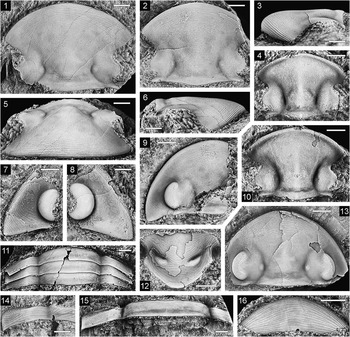
Illaenoscutellum psephos n. sp., Tomcat Creek limestone, all except (8, 12) from locality PL3309; dorsal views unless otherwise stated. (1, 5, 6) Holotype, cranidium, NMV P313515: (1) dorsal view, (5) anterodorsal view, (6) lateral view; (2, 3) cranidium, NMV P313520: (2) dorsal view, (3) lateral view; (4) smallest cranidium, NMV P313523; (7) librigena, NMV P313525, dorsolateral view; (8) librigena, NMV P313540, dorsolateral view, locality PL3314; (9) incomplete cephalon, NMV P313516; (10) small cranidium, NMV P313522; (11) three articulated thoracic segments, NMV P313549; (12) hypostome, NMV P313541, ventral view, locality PL3312; (13) cephalon, NMV P313553; (14) incomplete thoracic segment, NMV P313528, detail of left pleura and part of axis; (15) thoracic segment, NMV P313550; (16) rostral plate, NMV P313554, ventral view. (1–3, 5–9, 11–16) Scale bars=2 mm; (4, 10) scale bars=1 mm.

Illaenoscutellum psephos new species
Illaenoscutellum psephos n. sp., Tomcat Creek limestone, locality PL3309; dorsal views unless otherwise stated. (1) Cranidium, NMV P313517, and pygidium, NMV P313518; (2) pygidium, NMV P313533; (3) small pygidium, NMV P313538; (4, 6, 7) pygidium, NMV P313545: (4) posterodorsal view, (6) dorsal view, (7) enlargement of axial region showing muscle scars on internal mold; (5) small pygidium, NMV P313537; (8) pygidium, NMV P313535; (9) pygidium, NMV P313542; (10) pygidium, NMV P313532; (11) pygidium, NMV P313530, latex cast of natural mold, ventral view. (1, 2, 4, 6–11) Scale bars=2 mm; (3, 5) scale bars=1 mm.

Types
Holotype, cranidium NMV P313515, from PL3309. Paratypes, cephala NMV P313516, P313553, cranidia P313517, P313519–P313523, librigenae NMV P313525–P313527, rostral plates NMV P313554, P313555, thoracic segments NMV P313528, P313549, P313550, pygidia NMV P313518, P313529–P313539, P313542, P313545, from PL3309; librigena NMV P313540, from PL3314; hypostome NMV P313541, from PL3312.
Diagnosis
Cranidium wider at β than at δ; preglabellar furrow absent in mature specimens; pygidial axis poorly differentiated posterolaterally from unfurrowed and inflated proximal part of pleural region; interpleural furrows very shallow; pleural ribs weakly convex; postaxial rib indistinctly separated from seventh interpleural furrow in its posterior half; posterior margin only weakly dorsally arched medially.
Description
Cephalon of low convexity; maximum width situated slightly in front of genal angle and about level with posterior edge of eye, about twice sagittal length or a little less; anterior outline a little more strongly curved medially than laterally; posterior margin deflected backward in weakly sigmoidal curve from fulcrum to genal angle. Cranidium approximately equal in width at ω and δ, about 1.2 times wider at β. Axial furrow deepest at lunette; shallow but distinct behind lunette and converging forward at 40°–50° to sagittal line; in front of lunette very weak, initially diverging gently to a point almost opposite γ and thereafter diverging more strongly (Fig. 8.1, 8.13), dying out anteriorly opposite cranidial midlength. Glabella gently convex transversely, more flattened in sagittal profile except anteriorly, comprising two-thirds cranidial width posteriorly, width at lunette about two-thirds posterior width. Occipital ring weakly convex (sag., exsag.), slightly longer (sag., exsag.) at outer end of occipital furrow than medially; occipital furrow long (sag; exsag), very shallow, lateral section indistinct due to presence of poorly differentiated G0 that weakly indents front of occipital ring. Other glabellar muscle scars almost indistinguishable; G1 of indeterminate shape, its anterior edge more or less opposite γ; G2 subcircular, situated opposite β or slightly farther back; G3 elliptical, oblique, much smaller than G2, situated close to it and farther from the sagittal line. Lunette weakly inflated, poorly differentiated abaxially. Palpebral lobe about twice as long as lunette and about half its own length from posterior cephalic margin; palpebral furrow indistinct, not as strongly curved as palpebral margin; anterior branch of facial suture sigmoidally curved, diverging strongly forward at 45°–55° to sagittal line; posterior branch also sigmoidal, strongly diverging backward. Palpebral area more or less horizontal in transverse profile; posterior fixigena crossed by very shallow, transverse, abaxially expanding articulating flange furrow that meets axial furrow close to posterior edge of occipital ring and does not extend on to librigena distally. Librigena with broad, shallow, and poorly defined epiborder furrow that extends inward almost to base of visual surface and dies out opposite posterior edge of eye; dorsal surface of librigena lateral to epiborder furrow gently rounded; lateral border furrow not defined; genal angle drawn out into broad, tongue-like projection. Terrace ridges on dorsal surface of cephalon strongest on anterior and lateral margins; less continuous and more sinuous on frontal lobe, median part of glabella behind S3 including median part of occipital ring, and on lateral and posterolateral part of librigena; sparse or absent elsewhere.
Rostral plate lenticular in outline in ventral view, gently convex (sag., exsag.), a little more so abaxially than medially, maximum width a little more than three times sagittal length; connective suture converging strongly backward at 65°–70° to sagittal line; hypostomal suture transversely arched, curving slightly backward abaxially in ventral view; at intersection of hypostomal and connective sutures is a very short, obtuse, dorsally curved projection on rostral plate. Hypostome approximately 1.5 times as wide across anterior wings as long (sag.); shoulder prominent, situated opposite hypostomal midlength; posterolateral margin converges at about 45° to sagittal line from shoulder to strongly rounded posterior termination. Anterior lobe of middle body elliptical in outline; posterior lobe crescentic, indistinctly separated from posterior border by long (sag., exsag.), very shallow, and poorly defined posterior border furrow; macula inflated, oblique, its inner end level with shoulder; middle furrow deep in front of macula but not impressed medially; lateral border furrow very shallow from opposite posterior edge of anterior wing to widest part of macula; border weakly convex at and behind shoulder. Terrace ridges on rostral plate and hypostome stronger than on most of dorsal surface.
Thoracic segments with gently arched axis comprising about half total width or a little less; axial ring and undifferentiated articulating half ring crossed by weak transverse depression dividing it into a gently convex (sag., exsag.) anterior band of almost uniform length and a shorter, flattened posterior band that expands medially; anterior band has a very short, sharply differentiated, raised rim extending across most of its anterior margin. Axial furrow shallow. Pleura with fulcrum situated at about one-quarter distance from axial furrow, flattened adaxial to fulcrum, gently expanding abaxially beyond fulcrum and with short (exsag.) articulating facet; pleural tip obliquely truncated or with very short backward projection. Terrace ridges run in convex-forward arcs on axis and subparallel to somite margins on most of pleural region but obliquely on articulating facet.
Pygidium 1.35–1.5 times as wide as long, evenly curved in lateral and posterior outline, widest at or just behind posterior edge of articulating facet, which is indistinctly differentiated distally from first pleural rib. Axis moderately convex, about twice as wide as long, comprising 40%–45% maximum pygidial width anteriorly and about 25%–30% sagittal pygidial length; axial furrow indistinct anteriorly and posteromedially, not impressed posterolaterally where extent of axis is unclear due to incorporation into it of inflated proximal part of pleural region (see preceding remarks on Scutelluidae); articulating furrow faint or effaced laterally, very shallow medially where it is expanded (sag.) and gently convex backward; articulating half ring with very short, raised anterior rim as present on thoracic segments (Fig. 9.1); longitudinal trilobation of axis weakly indicated in some specimens by slight independent convexity of median lobe posteriorly (Fig. 9.1, 9.10). Pleural region with seven pairs of shallow and successively weaker interpleural furrows dying out distally in weakly concave zone close to margin; first rib with marked geniculation at fulcrum (Fig. 9.4), wider than other paired ribs and expanding more strongly than them abaxially; remainder of pleural region downturned from ‘axis’; postaxial rib much wider than paired ribs, subparallel-sided or expanding weakly backward but poorly defined in posterior half, with a low, rounded median longitudinal ridge. Doublure comprising about 55% sagittal pygidial length, narrowing anterolaterally, with broad, low, rounded ridge sagittally and narrow, faint radial ridges below pleural ribs. Terrace ridges on dorsal surface run in convex forward arcs on axis and inflated adaxial portion of pleural region, elsewhere roughly transversely except on first and second ribs, where they run more or less parallel with ribs, and on articulating facet, where they are directed anterolaterally; terrace ridges on doublure running obliquely to inner margin anteromedially and obliquely to outer margin anterolaterally, those on outer part of doublure more closely spaced than on inner part. Interior of exoskeleton with slightly raised, subelliptical to irregularly shaped patches, concentrated below interpleural furrows but not restricted to them, and extending onto axis (Fig. 9.7).
Etymology
Greek, pebble, referring to the highly effaced dorsal surface.
Remarks
Pronounced morphological differences between very small cranidia and pygidia and the large specimens on which the preceding description is based are the result of ontogenetic changes, similar to those documented by Holloway and Lane (Reference Holloway and Lane2012, p. 448) in I. tryo from the Wenlock to Ludlow of New South Wales. The two smallest cranidia of I. psephos, with sagittal lengths of ~3.5 mm and ~4 mm (Fig. 8.4, 8.5), differ from larger ones in that the preglabellar furrow is present though it is shallower and shorter medially than laterally; the axial furrow is deeper and extends farther forward to meet the preglabellar furrow though it is very shallow adjacent to the frontal lobe; the glabella is more convex (tr.) and does not narrow as strongly forward from the posterior margin to the lunette; the occipital furrow is a little deeper, and the median occipital node is much larger and more prominent; S1 is more distinct, especially in the smallest cranidium; the articulating flange furrow on the posterior fixigena is deeper; and the palpebral lobe is relatively larger with its anterior edge situated farther forward, the palpebral furrow is more distinct, and the palpebral area is more convex. The two smallest pygidia, ~4.3 mm and ~4.6 mm (Fig. 9.3) in width, are relatively wider than larger pygidia (width-to-length ratio of about 1.75 compared to about 1.35 in the largest pygidia); the axis is better defined, about equal in width to the pleural region rather than much wider than it, and is distinctly trilobate, the median lobe having traces of segmentation in the arrangement of widely spaced (tr.) pairs of tubercles; the pleural ribs and interpleural furrows are more distinct; and the postaxial rib is more convex. These very small cranidia and pygidia also differ from larger specimens in having fine, sparse granules interspersed with and superimposed on the terrace ridges on the dorsal surface.
The type species I. platiceps from Japan is known only from a single large and poorly preserved cranidium (Holloway and Lane, Reference Holloway and Lane2012, fig. 10M, N, Q), which can, however, be distinguished from I. psephos by the presence of the preglabellar furrow and the more rounded outline at β, where the cranidium is about as wide as at δ rather than being considerably wider. Illaenoscutellum tryo differs from I. psephos in that the preglabellar furrow is present in large as well as small cranidia and cephala; the anterior branch of the facial suture is abaxially concave in its anterior half instead of weakly abaxially convex; the posterior hypostomal border does not expand in length adaxially and is not as strongly curved in outline medially; the terrace ridges on the thoracic pleurae are oriented obliquely instead of running subparallel to the somite margins; the pygidial axis is better defined posteromedially; the pleural region of the pygidium is more concave abaxially, the interpleural furrows are deeper, the pleural ribs more convex, and the postaxial rib is better delineated in its posterior half; and the pygidial margin is more strongly arched upward posteromedially.
Genus Iotoryx new genus
Type species
Iotoryx clarksoni n. gen. n. sp.
Diagnosis
Glabella narrow and subparallel-sided from occipital furrow to front of bolus, thereafter expanding very strongly forward, strongly convex (tr.) across L1 and adaxial to exsagittally elongated S2, gently convex at combined L2+L3 and across frontal lobe. L1 long (sag, exsag.); S1 deep and expanded (tr.) adaxial to bolus, meeting axial furrow only weakly behind and in front of bolus; bolus rather large, gently inflated. S2 very deep, confluent with S1 and with very weak inner portion of S3. Inner and outer portions of S3 not joined, outer portion narrow (tr.), shallow, lying adjacent to axial furrow. Preglabellar furrow long (sag., exsag.), shallow, poorly differentiated from frontal lobe and bounded anteriorly by very short, slightly upturned rim on cephalic margin. Lateral border concave; lateral border furrow not defined; genal spine short, broad. Lunette much shorter than L1, lying opposite anterior half of L1 and posterior half of bolus. Eye situated far back, opposite L1 and overhanging posterior cephalic margin; eye ridge prominent. Pygidium elongated, semielliptical in outline; axis longer than wide, parabolic in outline, strongly trilobate longitudinally; pleural region rather strongly vaulted, concave toward margin; postaxial rib expanding strongly backward. Sculpture of tubercles with intervening dense pits and short, sinuous or scaly terrace ridges.
Etymology
Greek, iota, something small, and oryx, a pick, referring to the shape of the median and frontal glabellar lobes; gender masculine.
Remarks
Iotoryx is known only from the type species. Apparently similar, especially in cranidial morphology, is the poorly known Octobronteus Weber, Reference Weber1945, with type species O. khodalevitchi from the upper Silurian of the northern Urals (see Weber, Reference Weber1945, pp. 37, 42, pl. 5, figs. 14–16; Reference Weber1951, p. 13, pl. 2, figs. 7, 8; Šnajdr, Reference Šnajdr1960, pp. 56, 246, pl. 36, figs. 3, 4). The similarities include the glabella that is narrow and subparallel-sided across L1 and expands very strongly forward in front of S1; the size and prominence of the bolus; the deep and exsagittally elongated S2 that is confluent with S1; the weak S3 (indistinguishable in the poorly preserved cranidia of O. khodalevitchi); the low convexity of the glabella anterior and lateral to S2; the long (sag., exsag.), shallow, and poorly delimited preglabellar furrow; the very short, slightly upturned rim on the anterior margin; the eyes that are placed far back on the cheeks; and the prominent eye ridge. The pygidium of O. khodalevitchi also resembles that of Iotoryx in the strongly trilobate, elongated axis with a narrow median lobe, and in the strongly posteriorly expanding postaxial pleural rib, but differs in having eight rather than seven pleural ribs and a broad, convex, and unsegmented border. We do not attach much phylogenetic significance to these differences in the pygidia because in Stoermeraspis Prantl and Přibyl, Reference Prantl and Přibyl1947, which is closely related to Octobronteus and regarded as a junior synonym of it by some authors (Richter and Richter, Reference Richter and Richter1956a, p. 473; Reference Richter and Richter1959, p. 372; Šnajdr, Reference Šnajdr1960, p. 56) but as a distinct genus by others (Přibyl and Vaněk, Reference Přibyl and Vaněk1971, p. 374; Santel, Reference Santel2001, p. 101; Basse, Reference Basse2009, p. 91), the pygidial pleural region is concave abaxially as in Iotoryx, the interpleural furrows extend relatively close to the margin, and there may be either seven or eight pleural ribs (Richter and Richter, Reference Richter and Richter1956b, pl. 4, fig. 18, 18A; Santel, Reference Santel2001, pl. 2, figs. 6–8). The type species of Stoermeraspis, Bronteus franconicus Gümbel, Reference Gümbel1879, is known only from pygidia at its type locality in the Orthoceras Limestone (Ludlow) at Elbersreuth, Upper Franconia, Germany, but a cranidium from the Alticola Limestone (Ludlow) of the Carnic Alps of Austria that was attributed to the species by Gaertner (Reference Gaertner1930, p. 220, pl. 25, fig. 2) appears similar to those of O. khodalevitchi and Iotoryx, though the assignment of the Austrian cranidium to S. franconica was disputed by Santel (Reference Santel2001).
Iotoryx clarksoni new species
Iotoryx clarksoni n. gen. n. sp., Tomcat Creek limestone, locality PL3309. (1, 2, 5) Holotype, cephalon, NMV P313415: (1) dorsal view, (2) lateral view, (5) anterodorsal view; (3, 4) pygidium, NMV P313417: (3) lateral view, (4) dorsal view; (6) latex cast of fragmentary pygidium, NMV P313419, dorsal view; (7–9) pygidium, NMV P313416: (7) posterodorsal view, (8) lateral view, (9) dorsal view of latex cast of counterpart mold. Scale bars=2 mm.

Types
Holotype, cephalon NMV P313415, from PL3309. Paratypes, pygidia NMV P313416–P313419, from PL3309.
Diagnosis
As for genus.
Description
Cephalon approximately twice as wide as long, evenly curved in outline anteriorly, posterior margin with broad, shallow subgenal notch. Glabella comprising about 30% maximum cephalic width posteriorly, narrowing strongly forward across occipital ring, a little wider at front of bolus than at front of occipital ring, width at frontal lobe twice that at front of bolus; axial furrow narrow and deepest adjacent to lunette and bolus, very weak at inner end of eye ridge, broad and shallow adjacent to frontal lobe. Occipital ring incompletely preserved, standing higher than remainder of glabella, with greatest transverse convexity medially; occipital furrow well rounded in longitudinal section, curving forward abaxially, its outer section containing a poorly differentiated muscle scar. L1 comprising about one-third cephalic length in front of occipital furrow; posterior section of S1 with short, backwardly directed branch behind inner edge of bolus; anterior edge of bolus situated about level with cephalic midlength. L2+L3 teardrop shaped, tapering anteromedially. Frontal lobe much shorter than L1, indented anteromedially by weak depression forming a backward expansion of preglabellar furrow. Short rim on anterior margin of cranidium extends onto anterior half of librigena. Eye with large, inflated visual surface, anterior edge opposite bolus and length ~30% sagittal length of cephalon; palpebral lobe rather short; palpebral furrow gently curved, very shallow and broad medially, narrowing and deepening anteriorly and posteriorly. Palpebral area broad, about two-thirds width of glabella across L1, convex (exsag., tr.), sloping very steeply backward to articulating flange furrow and less steeply forward toward eye ridge; eye ridge long, straight, directed toward middle of L2+L3; lunette weakly inflated. Anterior branch of facial suture directed exsagittally from γ to a point transversely opposite front of bolus, thereafter diverging forward at ~30° to sagittal line as far as cephalic margin, which it meets abaxial to exsagittal line through δ; posterior branch not distinguishable beyond posteroventral edge of visual surface. Librigena abaxial to eye decreasing in convexity posteriorly, toward a weak, abaxially expanding ridge running in front of subgenal notch. Tubercles on glabella and posterior fixigena larger, denser, and more prominent than elsewhere; intervening fine, dense pits are surrounded by reticulate pattern of weak ridges, some of which on posterior fixigena tend to develop into more prominent, longer, but discontinuous ridges; in occipital furrow similar ridges are dominant.
Pygidium about 1.2 times as wide as long; maximum width level with midlength of axis. Axis comprising about 30% pygidial length and width, more convex transversely than sagittally; deep longitudinal furrows in posterior half are aligned with seventh interpleural furrows but fade toward axial terminus. Axial furrow distinct adjacent to ribs 1–6 but poorly defined in front of postaxial ridge; at inner ends of ribs 5 and 6, axial furrow expanded to form elliptical depressed area (Fig. 10.4, 10.9). Pleural region with seven pairs of rather deep interpleural furrows that barely expand abaxially and die out very close to margin; first two furrows curve gently backward abaxially, and remainder curve successively more strongly outward; paired pleural ribs gently convex, expanding abaxially; postaxial rib subparallel-sided or narrowing weakly backward in anterior third, thereafter expanding to more than twice its proximal width, with weak sagittal ridge in posterior half; doublure extends forward more than half postaxial length of pygidium. Tubercles decrease in size and density on outer part of pleural region; intervening pits and ridges present on both axis and pleural region, as on cephalon, but toward pleural margins ridges are dominant and become scale-like; on largest pygidium, pits are almost entirely absent, and sinuous or scale-like terrace ridges are present between tubercles.
Etymology
After Emeritus Professor E. N. K. Clarkson of the University of Edinburgh, Scotland.
Remarks
The holotype shows some asymmetry in the development of the bolus, the one on the left being a little smaller than the one on the right (Fig. 10.1). In some of the pygidia, the first axial ring is present at least laterally, and behind it on the lateral part of the axis several more segments appear to be weakly defined by transverse alignment of tubercles (Fig. 10.4, 10.6). In the largest pygidium, the first axial ring does not appear to be distinct, though the anterior part of the axis is damaged, and there is no transverse alignment of tubercles behind it (Fig. 10.9).
Genus Japonoscutellum Přibyl and Vaněk, Reference Přibyl and Vaněk1971
Type species
Scutellum (Subgenus nov.) japonicum (sic) Kobayashi and Hamada, Reference Kobayashi and Hamada1965, from the Fukata Formation (upper Wenlock to lower Ludlow; Umeda, Reference Umeda1998) of Gomi, Yokokurayama, Kôchi Prefecture, Shikoku, Japan, by original designation.
Other species
A comprehensive list was given by Holloway and Lane (Reference Holloway and Lane2012, p. 448).
Diagnosis
See Holloway and Lane (Reference Holloway and Lane2012, p. 449).
Occurrence
Middle Llandovery (upper Aeronian) to Ludlow; United States (Wisconsin), northwestern Greenland, southwestern Ireland, ?England, Sweden, Czech Republic, Russia (Urals, Siberia), Kazakhstan, southwest China, Japan, and eastern Australia.
Japonoscutellum mawsonae new species
Japonoscutellum mawsonae n. sp., Tomcat Creek limestone, all except (3) from locality PL3309; dorsal views unless otherwise stated. (1, 5, 6) Holotype, cranidium, NMV P313438: (1) dorsal view, (5) oblique view, (6) lateral view; (2) cranidium, NMV P313453; (3) librigena, NMV P313486, locality PL3312; (4) librigena, NMV P313463; (7) smallest (probable meraspid) cranidium, NMV P313488, dorsal view; (8) cranidium, NMV P313462; (9) hypostome, NMV P313464, ventral view; (10) largest cranidium, NMV P313444; (11) cephalon, NMV P313439; (12) hypostome, NMV P313465, latex cast of natural mold, ventral view; (13, 15, 16) cephalon, NMV P313440: (13) lateral view, (15) ventral view, (16) dorsal view; (14) small cranidium, NMV P313461; (17) small cranidium, NMV P313458; (18, 20) cephalon with first thoracic segment, NMV P313441: (18) ventral view, (20) dorsal view; (19) cranidium, NMV P313457; (21) cranidium, NMV P313445. (1–6, 8–21) Scale bars=2 mm; (7) scale bar=1 mm.

Japonoscutellum mawsonae n. sp., Tomcat Creek limestone, all except (11) from locality PL3309; pygidia in dorsal view. (1) NMV P313474, latex cast of natural mold; (2) NMV P313470; (3) smallest pygidium, NMV P313483; (4) NMV P313481; (5) NMV P313477; (6) NMV P313478; (7) largest pygidium, NMV P313468; (8) NMV P313473; (9) NMV P313480; (10) NMV P313469, latex cast of external mold; (11) NMV P313487, locality PL3312. Scale bars=2 mm.

?1994 Kosovopeltis sp. Reference HollowayHolloway, p. 244, fig. 2A.
Types
Holotype, cranidium NMV P313438, from PL3309. Paratypes, cephala NMV P313439–P313441 (the last with incomplete first thoracic segment), fragmentary cephalon NMV P313443, cranidia NMV P313444–P313446, P313448–P313462, probable meraspid cranidium NMV P313488, librigena NMV P313463, hypostomes NMV P313464, P313467, pygidia NMV P313468–P313483, from PL3309.
Diagnosis
Cranidium widest at β. Glabella comprising a little less than half cranidial width posteriorly, twice as wide across frontal lobe as at posterior margin, its forward expansion in front of S1 not increasing markedly at posterior edge of frontal lobe; S1–S3 shallow to moderately impressed; bolus distinctly inflated in largest cephala and cranidia, otherwise weakly inflated; preglabellar furrow indistinctly defined in front of lateral extremity of frontal lobe. Epiborder furrow broad and very deep on front of fixigena; outer part of border beyond epiborder furrow convex and elevated on fixigena and front of librigena; posterior branch of facial suture only gently sigmoidal in outline. Hypostome as wide across shoulders as long (sag.); posterior margin transverse. Pygidium generally 1.4 to 1.5 times as wide as long; axis strongly bell shaped in outline, approximately 1.6 times as wide as long, its anterior width about 30% maximum pygidial width; pleural region with unfurrowed and slightly inflated elliptical area lying adjacent to posterolateral part of axis; first three pleural ribs with imbricate profile abaxially, remaining ribs increasingly more rounded in cross section; postaxial rib about twice as wide as adjacent paired ribs proximally, subparallel-sided in anterior half, expanding gently in posterior half. Dorsal sculpture of terrace ridges.
Description
Cephalon semicircular in outline anteriorly and laterally; posterior margin with broad subgenal notch on librigena. Glabella narrowing gently to moderately forward across occipital ring and L1, expanding gently across bolus, about as wide at front of bolus as at posterior margin, thereafter expanding very strongly forward with weak constriction adjacent to intersection of axial and lateral border furrows. Axial furrow well impressed overall but shallowing toward middle (exsag.) of L3, diverging at about 45° to sagittal line in front of bolus. Occipital ring about twice as long medially as distally, its anterior edge deeply indented over lateral third by elliptical, inflated occipital muscle scar, in sagittal profile flattened and sloping forward; small median tubercle present just behind midlength (sag.) of ring but in large specimens partly obscured by sculpture. Occipital furrow deep and transverse medially, with abrupt anterior slope, not impressed laterally in front of occipital muscle scar; convex-backward furrow running behind occipital scar joins median part of occipital furrow adaxially. S1 shallow to moderately impressed behind and adaxial to bolus, not impressed in front of it; bolus comprising about one-quarter glabellar width at this level. S2 and S3 weakly convex forward; S2 directed slightly obliquely backward adaxially, situated opposite or just in front of cephalic midlength and close to front of bolus, about its own width (tr.) from axial furrow; S3 subparallel to anterior cephalic margin, wider than S2 and lying farther from the sagittal line. Width of fixigena opposite δ about two-thirds that of adjacent part of glabella; lunette gently inflated. Eye with posterior edge opposite median part of occipital furrow and anterior edge more or less opposite front of bolus; palpebral suture with δ situated behind midlength, curving strongly inward posteriorly; palpebral and eye ridge furrows weak. Anterior branch of facial suture between γ and β diverging a little less strongly than anterior half of axial furrow, turning sharply inward at β; posterior branch diverging backward at about 50° to sagittal line overall. Lateral border furrow strongly impressed, broadly arcuate, converging with lower edge of visual surface at midlength of eye, dying out behind eye where it meets posterior branch of suture just in front of outer end of articulating flange furrow; epiborder furrow much broader than border furrow, shallowing posteriorly onto short, strongly tapering genal spine; inner part of border adaxial to epiborder furrow expanding very strongly backward toward subgenal notch and becoming gently convex; outer part of border decreasing in convexity posteriorly. Rostral plate and anterior part of librigenal doublure well rounded (sag., exsag.), in sagittal section continuous in convexity with front of cranidium; connective suture converging very strongly backward and gently concave abaxially in outline; hypostomal suture transversely arched, at its intersection with connective suture bounding an acute, dorsally curved projection on rostral plate. Terrace ridges on glabella and anterior fixigenal field generally running subparallel to anterior cephalic margin but deflected backward toward axial furrow; on inner part of fixigena behind eye ridge they are roughly parallel to axial furrow but curve outward abaxially; on librigena they have a complex arrangement.
Hypostome about 1.3 times as wide across anterior wings as at shoulders; anterior lobe of middle body subelliptical in outline, wider than long, more convex transversely than sagittally; posterior lobe crescentic, about one-third as long sagittally as anterior lobe; macula with only slight independent convexity, its anterior end lying opposite shoulder; median section of middle furrow shallow; posterolateral margin converging at about 40° to sagittal line behind shoulder, very weakly concave in outline; posterior border about as long sagittally as posterior lobe of middle body. Terrace ridges on middle body are roughly concentric around a point on anteromedian part of anterior lobe and curve anterolaterally onto anterior wing.
First thoracic segment (Fig. 11.20) with axis expanding backward at similar rate as occipital ring, posteriorly slightly wider than pleura; articulated portion of pleura wider than sickle-shaped distal spine.
Pygidium a little more than a semicircle in outline, maximum width about level with posterior end of axis. Axis comprising about 25% sagittal pygidial length, its posterior terminus broadly rounded to slightly pointed in outline. Pleural region gently convex adaxially but weakly concave in outermost third, with seven pairs of low ribs that are successively a little narrower distally; interpleural furrows shallowing distally but extending almost to margin, merging successively more weakly abaxially with flattened anterior slope of succeeding rib; furrows 5 and 6 subparallel to sagittal line proximally; postaxial rib about twice as wide distally as proximally. Doublure comprising about half sagittal length of pygidium, crossed by broad, weak, adaxially fading radial ridges corresponding approximately in position with interpleural furrows, and with narrower, more distinct sagittal ridge that dies out posteriorly. Terrace ridges running in gently convex-forward arcs on axis; on pleurae roughly transverse adaxially and curving forward abaxially, meeting anterolateral margin obliquely; on outer part of doublure terrace ridges more closely spaced than on inner half.
Etymology
After Emeritus Professor Ruth Mawson of Macquarie University, New South Wales.
Other material
Cranidium NMV P313485, librigena NMV P313486, pygidium NMV P313487, from PL3312.
Remarks
A wide size range of specimens is available, cranidia and cephala with lengths of 2.4 mm to 21 mm and pygidia with widths of ~4.8 mm to ~32 mm. Morphological differences between small specimens and the largest ones on which the preceding description is based are the result of ontogenetic changes similar to those documented by Holloway and Lane (Reference Holloway and Lane2012) in some other species of Japonoscutellum. By far the most conspicuous ontogenetic change in J. mawsonae is in sculpture. In the smallest cranidium, probably belonging to a meraspis (Fig. 11.7), the sculpture consists of dense, variably sized granules except on the anterior margin where there are a few continuous terrace ridges. On progressively larger cranidia, terrace ridges are present between the granules over the entire cranidium and gradually become dominant as the granules decrease in density and weaken (Fig. 11.8, 11.14, 11.17, 11.19). On cranidia and cephala 10 mm in length and larger from locality PL3309 only terrace ridges are present, but a cranidium 11.8 mm long from PL3312 has a few granules between the terrace ridges on the palpebral area, posterior fixigena, and lateral part of L1. Similar changes in sculpture are seen in the pygidia, the largest one with sparse granules still present between the terrace ridges being about 15 mm wide. Other ontogenetic changes in J. mawsonae include the following:
-
1. Between the meraspid and early holaspid stages there is a marked decrease in the convexity of the glabella and an increase in its rate of expansion in front of S1; S1 and the bolus increase greatly in size; S2 and S3 become distinctly impressed; the preglabellar furrow weakens; and the palpebral lobe decreases in relative size (Fig. 11.7, 11.8).
-
2. In holaspid cranidia and cephala, the median occipital node gradually decreases in size and prominence; the lunette becomes more inflated; the subgenal notch weakens (Fig. 11.16, 11.11); and the genal spine increases in length (Fig. 11.4, 11.3). In the largest cranidium (Fig. 11.10), the occipital furrow is a little shallower medially; L1 is longer (exsag.); the bolus is larger, is more inflated, and causes a slight outward deflection of the axial furrow; and the posterior fixigena is a little longer.
-
3. In the smallest pygidium (Fig. 12.3), the width-to-length ratio is about 1.7 and the postaxial region is distinctly convex. In larger pygidia, the width-to-length ratio gradually decreases to about 1.4 and the convexity of the postaxial region greatly decreases. The unfurrowed and slightly inflated elliptical areas of the pleural region adjacent to the axis are more distinct in larger pygidia than in smaller ones.
A large pygidium from PL3312 differs from similarly sized pygidia from PL3309 in being much shorter and wider, having a width-to-length ratio of about 1.8 instead of 1.4 (Fig. 12.11), but in other respects is not significantly different. We attribute the difference in proportions to intraspecific variation.
Similar in age to J. mawsonae is J. guangyuanense (Zhang, Reference Zhang1974, p. 174, pl. 83, fig. 10), which is based on the internal mold of an incomplete, articulated dorsal exoskeleton from the Shenxuanyi Member of the Ningqiang Formation (late Telychian) of Sichuan, southwestern China. Only the cranidium of the holotype was originally illustrated, but the entire specimen was figured by Li (Reference Li1978, pl. 110, fig. 1). Preservational differences make it difficult to compare this single specimen with J. mawsonae, but in J. guangyuanense the cranidium appears to be more angular in outline anteromedially; the glabella is more expanded (tr.) across the frontal lobe; S3 appears to meet the axial furrow; the pygidium is a little longer and narrower than even the largest pygidia of J. mawsonae; the pygidial axis is not distinctly bell shaped in outline; the pleural region does not appear to have inflated and unfurrowed areas situated adjacent to the axis; the postaxial rib is not significantly wider than the adjacent paired ribs proximally and distally; and the terrace ridges as expressed on the interior of the frontal lobe and the pygidial pleural ribs are much coarser. Some of these apparent differences may have to be reassessed if the two cranidia and the pygidium from the Ningqiang Formation that were illustrated by Lane and Wu (Reference Lane and Wu2002, fig. 96L–N) as ‘Kosovopeltis tchernychevae Šnajdr’ are shown to belong to J. guangyuanense.
Japonoscutellum mawsonae is easily distinguished from the numerous species of the genus documented by Holloway and Lane (Reference Holloway and Lane2012) from the lower Wenlock to Ludlow of central western New South Wales but is perhaps most similar to J. gephyricum, in part because of the ontogenetic change in sculpture from granules to terrace ridges. Japonoscutellum gephyricum differs from J. mawsonae in that the cranidium is widest at ω instead of at β; the glabella expands less strongly forward from the front of the bolus to the front of L3, where the rate of expansion increases markedly; the glabella is much less than twice as wide across the frontal lobe than posteriorly; the preglabellar furrow is more distinct abaxially; the fixigenal portion of the anterior border is wider and the border furrow is constricted adaxially; the fixigena is narrower in its posterior half; the palpebral furrow is more distinct; the posterior branch of the facial suture is directed laterally rather than posterolaterally from ε and is deflected more strongly backward abaxially; the anterior branch of the suture runs on a distinct ridge; the hypostomal shoulder is less angular in outline and the posterior margin is curved rather than transverse; the pygidium is more elongated, with a postaxial rib that expands more strongly backward and has a shallow sagittal furrow distally; the smooth, slightly inflated areas of the pygidial pleurae adjacent to the posterolateral part of the axis are less distinct; and the terrace ridges on the pygidial doublure are more closely spaced.
The fragmentary scutelluid cranidium identified by Holloway (Reference Holloway1994, fig. 2A) as Kosovopeltis sp., from siltstones of the Poley Cow Formation in the vicinity of the Broken River about 29 km south of the Tomcat Creek limestone, probably belongs to J. mawsonae but is too incomplete to be assigned to the species with confidence. It resembles J. mawsonae in that the forward expansion of the glabella does not increase markedly across the frontal lobe; there is a broad epiborder furrow on the front of the fixigena; and a weak preglabellar furrow is present abaxially.
Japonoscutellum drakton new species
Figure 13.1–13.5, ?13.6, 13.7, 13.8
Japonoscutellum drakton n. sp. and Japonoscutellum fractum n. sp., Tomcat Creek limestone, locality PL3309; dorsal views unless otherwise stated. (1–5, ?6, 7, 8) Japonoscutellum drakton; (1) cranidium, NMV P313490; (2–4) holotype, cranidium, NMV P313489: (2) lateral view, (3) anterodorsal view, (4) dorsal view; (5) cranidium, NMV P313491; (6) pygidium tentatively assigned to this species, NMV P313492; (7, 8) hypostome, NMV P313543, ventral views: (7) latex cast of external mold, (8) counterpart. (9–12) Japonoscutellum fractum; (9) cranidium, NMV P313494; (10–12) holotype, cranidium, NMV P313493: (10) dorsal view, (11) lateral view, (12) anterodorsal view. (1, 6, 10–12) Scale bars=5 mm; (2–5, 7–9) scale bars=2 mm.

Types
Holotype, mostly exfoliated cranidium, NMV P313489; paratypes, cranidia NMV P313490, P313491 and large hypostome NMV P313543; all from PL3309.
Diagnosis
Cranidium widest at ω. Glabella comprising about 40% maximum cranidial width posteriorly, narrowing forward slightly across occipital ring and most of L1, thereafter expanding almost uniformly, widest at intersection of axial and anterior border furrows where it is about 1.75 times as wide as at L1; preglabellar furrow may be present abaxially but weak; S1 not meeting axial furrow distinctly behind weakly inflated bolus; S2 and S3 moderately impressed. Anterior border abaxial to frontal lobe without epiborder furrow. Palpebral lobe narrow, situated its own length from posterior cranidial margin, with δ opposite middle (exsag.) of bolus; anterior branch of facial suture initially diverging forward from γ more strongly than axial furrow, broadly rounded at β; posterior branch with distinct posterolateral deflection about halfway (tr.) between ε and ω. Coarse major terrace ridges on cranidium with numerous short, backwardly directed secondary ridges arising from their steep posterior slopes. Hypostome with subcircular anterior lobe of middle body and poorly developed shoulder that is broadly rounded in outline.
Description
Cranidium with widths at β, δ, and ω approximately in ratio 1:0.95:1.05. Glabella moderately convex; axial furrow deepest adjacent to lunette, shallowing slightly adjacent to anterior half of L3 and posterior part of frontal lobe, diverging at ~25° to sagittal line in front of L1, with distinct outward deflections adjacent to bolus and L3 in largest cranidium, joining deep anterior border furrow in a sharp curve; preglabellar furrow most discernible in largest cranidium but nevertheless extremely weak, converging forward from intersection of axial and anterior border furrows and rapidly dying out (Fig. 13.1). Front of occipital ring deeply indented in lateral third by slightly inflated occipital muscle scar that obliterates outer part of occipital furrow; small median occipital tubercle situated behind midlength (sag.) of ring, on exterior surface obscured by sculpture; furrow behind occipital muscle scar deflected forward adaxially and abaxially, meeting axial furrow more or less transversely opposite median section of occipital furrow. Bolus large, comprising about 30% glabellar width at this level; S1 deepest and somewhat expanded (tr.) posteromedial to bolus; G1 on interior of exoskeleton elliptical, exsagittally elongated (Fig. 13.4). S2 situated just in front of cranidial midlength and distant from axial furrow, directed slightly obliquely backward adaxially. S3 subparallel to anterior cranidial margin, with conspicuous deepening abaxially. Width of fixigena at δ a little more than half that of adjacent part of glabella; palpebral furrow distinct, subparallel with palpebral margin; palpebral lobe upturned from gently convex (tr.) palpebral area. Lunette inflated. Anterior border well rounded (exsag.) lateral to frontal lobe. Anterior branch of facial suture initially diverging forward from γ at ~50° to sagittal line before curving forward across anterior part of fixigenal field and posterior half of border, crossing fixigenal field on slight sutural ridge bounded adaxially by shallow furrow; posterior branch deflected backward at ~30° to sagittal line about halfway (tr.) between ε and ω. Articulating flange furrow on posterior edge of fixigena very shallow. Short secondary terrace ridges branching roughly perpendicularly from scarp slope of major terrace ridge in front generally do not encroach on dip slope of major terrace ridge behind, and are more abundant on glabella than on fixigena.
Hypostome subparallel-sided immediately behind anterior wing, narrowing rather strongly backward opposite posterior lobe of middle body, posterior outline unknown due to breakage; anterior lobe strongly inflated, comprising ~80% sagittal length of middle body; middle furrow shallow except for short, deeper and narrower section in front of middle of macula; macula obliquely elongated, smooth and flattened except for small, slightly inflated, and granulate area posteromedially. Major terrace ridges on anterior lobe with a few short, forwardly directed secondary terrace ridges arising from their anteriorly or adaxially facing scarp slopes.
A large, fragmentary pygidium tentatively assigned to this species (Fig. 13.6) lacks anterior part of axis, first pair of pleural ribs, almost all of second pair (assuming seven paired ribs originally present), and outer parts of remaining ribs. Axis large, gently convex, with distinct swelling posteromedially, apparently subquadrate in outline overall but posterolaterally may incorporate adjacent parts of pleural region (see remarks on the Family Scutelluidae); axial furrow becoming poorly defined posteromedially. Inner part of pleural region gently convex, outer part gently concave anterolaterally but more weakly concave posteromedially; interpleural furrows gradually expand abaxially and weaken abaxially and adaxially, last three almost disappearing proximally; pleural ribs gently convex except proximally and distally, where they become flatter; postaxial rib about three times as wide proximally as adjacent paired rib, initially contracting weakly backward for a short distance, thereafter expanding gradually to at least twice its proximal width and ~1.5 times width of adjacent paired rib at similar distance from axis. Terrace ridges on dorsal surface widely spaced.
Etymology
Greek, a small vase, referring to the shape of the glabella; noun in apposition.
Other material
A large, incomplete pygidium NMV P313492, from PL3309, is assigned to the species with question.
Remarks
In the absence of librigenae, we cannot be certain whether the deep transverse furrow on the front of the fixigena is the border furrow or the epiborder furrow. We have interpreted it as the border furrow because the length (exsag.) of the convex band in front of the furrow suggests that this band represents the entire border rather than just its outer part. In addition, the furrow meets the axial furrow at an acute angle, similar to the intersection of the axial and lateral border furrows in other Japonoscutellum species, rather than in a broad curve like the intersection of the axial and epiborder furrows (e.g., Holloway and Lane, Reference Holloway and Lane2012, fig. 12A, I, J, O). Our assignment to J. drakton of a hypostome and a pygidium (the latter only tentatively assigned) is based on the elimination of the other species as contenders for them; the fact that the largest cranidium of J. drakton indicates that this species may attain a comparable size; and, in the case of the hypostome, the presence of secondary terrace ridges branching from the major ones on the anterior lobe of the middle body (though in overall appearance the sculpture on this part of the hypostome differs from that on the cranidium).
Japonoscutellum drakton is easily distinguished from previously described species of the genus. Differences from J. mawsonae are numerous and include the following: (1) the cranidium is widest at ω instead of at β; (2) the glabella narrows forward more weakly across the occipital ring and the posterior part of L1 and thereafter expands more uniformly and more gently overall; (3) the glabella is widest at the intersection of the axial and anterior border furrows instead of toward the front of the anterior border; (4) the anterior border abaxial to the frontal lobe lacks an epiborder furrow; (5) the furrow behind the occipital muscle scar is deflected more strongly forward distally; (6) S1 does not meet the axial furrow as distinctly behind the bolus, and S3 has a conspicuous deepening abaxially; (7) the palpebral lobe is narrower and not as strongly curved in outline, and the palpebral furrow is deeper; (8) the fixigena is wider anteriorly and posteriorly, the anterior branch of the facial suture is more divergent from γ to β, and the posterior branch of the suture is deflected more strongly backward halfway between ε and ω; (9) backwardly directed secondary terrace ridges are present on the cranidium; (10) the hypostome lacks distinct shoulders, the posterior lobe of the middle body is shorter and more convex, and the middle furrow is shallower abaxially; (11) the pygidial axis is not bell shaped in outline and is distinctly inflated posteromedially; (12) the pygidial axial furrow is very poorly defined posteromedially; (13) the pleural region of the pygidium is more convex adaxially and more concave abaxially; (14) the pleural ribs are less convex and the interpleural furrows are shallower.
Japonoscutellum fractum new species
Figures 13.9–13.12, 14.4
Scutelluids from Tomcat Creek limestone, locality PL3309; dorsal views. (1–3, 5, 6) Kosovopeltis avita n. sp.; (1, 5, 6) holotype, cranidium, NMV P313546: (1) latex cast of external mold, (5) enlargement showing cephalic median organ, (6) counterpart of cranidium; (2) small (?meraspid) cranidium, NMV P313547; (3) pygidium, NMV P313548, latex cast of external mold. (4) Japonoscutellum fractum n. sp., fragmentary cranidium, NMV P313495. (7) Dolabrapex acomus n. gen. n. sp., pygidium, NMV P313514. (1–3, 6, 7) Scale bars=1 mm; (4) scale bar=5 mm; (5) scale bar=0.5 mm.

Types
Holotype, cranidium, NMV P313493; paratypes, cranidia, NMV P313494, P313495; all from locality PL3309.
Diagnosis
Glabella narrowing very strongly forward across occipital ring, expanding progressively more strongly forward in front of L1, widest at intersection of axial and anterior border furrows where it is twice as wide as across L1; S1–S3 shallow to moderately impressed; bolus poorly defined. Axial furrow shallow in anterior half; preglabellar furrow may be weakly developed abaxially. Anterior border abaxial to frontal lobe without epiborder furrow; anterior border furrow shallow. Palpebral lobe situated about half its own length from posterior cranidial margin, with δ opposite front of L1. Cranidial sculpture of closely spaced anastomosing terrace ridges.
Description
Glabella comprising a little less than half cranidial width at δ, and about as wide at intersection of axial and anterior border furrows as long (sag.). Occipital ring 80% as wide anteriorly as posteriorly; posterior margin broadly curved in outline, anterior margin indented in lateral third by short (exsag.), broad muscle scar; small median tubercle situated just behind midlength of ring. Occipital furrow and S1–S2 deeper in holotype than in smaller cranidium (Fig. 13.9, 13.10). L1 about as wide (tr.) anteriorly as posteriorly but slightly constricted at midlength of lunette; S1 increasing in depth slightly posteromedial to bolus and expanded backward slightly; S2 situated about its own width (tr.) from axial furrow, its inner end opposite cephalic midlength; outer end of S3 lying transversely opposite intersection of axial and anterior border furrows, with conspicuous deepening in holotype (Fig. 13.10). Axial furrow shallower in smallest specimen than in larger ones, diverges at ~18° to sagittal line from front of L1 to opposite S2, thereafter at ~40° to sagittal line as far as intersection with anterior border furrow; shallow preglabellar furrow evident in fragment of large cranidium, directed anteromedially from intersection of axial and anterior border furrows (Fig. 14.4). Palpebral lobe with posterior edge opposite middle of L1 and anterior edge more or less opposite front of bolus, more strongly curved in outline in posterior half than in anterior half; palpebral furrow less strongly curved than palpebral margin, with a line of slightly deeper pits in anterior half extending forward into posterior part of furrow on inside of eye ridge; anterior branch of facial suture subparallel to axial furrow on genal field and posterior part of border, bordered adaxially by shallow furrow on genal field (Fig. 14.4); posterior branch incomplete, bordered adaxially on fixigena by broad furrow.
Etymology
Latin, broken, in reference to the Broken River.
Remarks
Japonoscutellum fractum shares with J. drakton certain characters that are unusual for the genus, suggesting that the two species are closely related. In both species the anterior border abaxial to the frontal lobe lacks an epiborder furrow; the axial furrow terminates at its intersection with the anterior border furrow, instead of extending onto the posterior part of the anterior border and uniting with an epiborder furrow; and the glabella is widest at the intersection of the axial and anterior border furrows instead of adjacent to the outer part of the anterior border. Despite these similarities, the differences between J. fractum and J. drakton are quite extensive. In J. fractum the glabella is much wider at the posterior margin, being as wide here as it is opposite S2 whereas in J. drakton the glabella is about as wide posteriorly as at the front of L1. The occipital ring narrows forward much more strongly in J. fractum, and the glabella expands more strongly in front of L1, at an increasing rate rather than uniformly. The axial furrow is shallower, especially in front of S2, as also is the anterior border furrow. The palpebral lobe is situated slightly farther back, with δ opposite the front of L1 instead of opposite the middle (exsag.) of the bolus, and is widest at its midlength rather than being subparallel-sided. The cranidial sculpture in J. fractum is conspicuously different, the terrace ridges being finer and more closely spaced, with an anastomosing arrangement rather than with secondary terrace ridges directed backward at ~90° to the major ones.
Genus Kosovopeltis Šnajdr, Reference Šnajdr1958
Type species
Kosovopeltis svobodai Šnajdr, Reference Šnajdr1958, from the upper Kopanina Formation (upper Ludlow, Ludfordian), Kosov near Králův Dvůr, Czech Republic, by original designation.
Other species
A comprehensive list was given by Holloway and Lane (Reference Holloway and Lane2012, p. 470).
Diagnosis
See Holloway and Lane (Reference Holloway and Lane2012, p. 472).
Occurrence
Upper Llandovery to Lochkovian; United States (Arkansas, Oklahoma, Maine), United Kingdom (England, Welsh Borderland), Czech Republic, Morocco, ?Turkey, Kazakhstan, Japan, eastern Australia.
Remarks
Tosacephalus Kobayashi and Hamada, Reference Kobayashi and Hamada1974 was regarded by Holloway and Lane (Reference Holloway and Lane2012, p. 474) as a junior subjective synonym of Kosovopeltis.
Kosovopeltis avita n. sp. is probably the stratigraphically oldest species that can be assigned to the genus with confidence. It may be somewhat older than an undescribed species reported by Holloway (in Pickett et al., Reference Pickett2000, p. 163) from the Boree Creek Formation (upper Telychian to lower Sheinwoodian, Pterospathodus amorphognathoides to Kockelella ranuliformis conodont biozones; Cockle, Reference Cockle1999) in the Orange district of central western New South Wales. Several Llandovery species previously assigned to Kosovopeltis were excluded from the genus by Holloway and Lane (Reference Holloway and Lane2012), who placed most of them in Japonoscutellum, Planiscutellum, or the newly proposed Securifrons. We also exclude two other species documented by Curtis and Lane (Reference Curtis and Lane1997, pl. 1, figs. 1–3) on the basis of a few rather poorly preserved specimens from the Llandovery of Great Britain. Their ‘Kosovopeltis sp. 1’, from the Aeronian of South Wales, differs from Kosovopeltis in that L1 is rather long (sag.) and narrows forward very weakly; the lateral extremity of the frontal lobe is broadly rounded in outline instead of subangular; there is a weak but medially continuous preglabellar furrow bounded anteriorly by a very short (sag., exsag.), slightly upturned border; the anterior border abaxial to the frontal lobe appears to be very long (exsag.); and the pygidial axis is very long and not trilobate longitudinally. ‘Kosovopeltis sp. 2’, from the Telychian of Shropshire, differs from Kosovopeltis in that the occipital ring is very long (sag.); the glabella in front of L1 has a deep longitudinal furrow that is either S2 or joins S1 and S2; and there is a long (sag., exsag.), shallow preglabellar furrow. We cannot assign either of these species to another genus with confidence.
Kosovopeltis avita new species
Types
Holotype, cranidium NMV P313546; paratypes, small (meraspid?) cranidium NMV P313547, pygidium NMV P313548; all from PL3309.
Diagnosis
Cranidium approximately equal in width at δ and ω, much wider at β. Glabella comprising half cranidial width posteriorly and a little more than one-third cranidial width at δ, without medial swelling more or less opposite S2 in maturity; S1–S3 very weakly impressed; bolus almost indistinguishable. Anterior border furrow distinct in front of fixigenal field; axial furrow effaced in front of intersection with anterior border furrow; lunette depressed. Pygidium 1.6 times as wide as long; axis almost twice as wide as long, comprising 0.3 times maximum pygidial width anteriorly and a little more than one-quarter sagittal length, weakly trilobate longitudinally; interpleural furrows shallow, especially more anterior ones. Dorsal sculpture of widely spaced terrace ridges on cranidium and pygidium.
Description
Glabella gently convex, narrowing strongly forward across occipital ring to 70% its posterior width, expanding weakly across L1 and S1, thereafter expanding strongly to front of L3 where width abruptly increases greatly; lateral extremity of frontal lobe poorly defined but glabella is here about twice as wide as at posterior margin. Occipital ring flattened and sloping forward in sagittal profile; posterior margin with a median swelling just in front of which is a very small, weak node with four tiny pits arranged in a square (Fig 14.5; cephalic median organ of Lerosey-Aubril and McNamara, Reference Lerosey-Aubril and McNamara2008). S1 subrectangular; S2 convex forward, its inner end lying slightly farther posteriorly than outer end and about level with glabellar midlength; S3 subparallel to anterior cranidial margin; weak sagittal carina on glabella extends from opposite posterior edge of S1 to front of L3. Axial furrow diverges at almost 45° to sagittal line from front of S1 to intersection with anterior border furrow, shallowing adjacent to middle (exsag.) of L3. Palpebral lobe with posterior edge opposite anterior part of occipital ring and anterior edge more or less opposite front of S1, its height a little less than that of median part of L1; eye ridge faint. A weak furrow (posterior border furrow; see Holloway and Lane, Reference Holloway and Lane2012, p. 420) crosses the posterior fixigena obliquely from behind palpebral lobe and dies out toward lunette.
Pygidium a little more than a semicircle in outline. Axis subtriangular in outline; articulating half ring much more elevated than remainder of axis, anteriormost part of which is crossed by a broad, shallow depression that weakens laterally. Pleural region gently convex posteromedially but becoming flat anterolaterally; with seven pairs of ribs, anterior ones flattened and of extremely low relief, more posterior ones successively more elevated; paired ribs asymmetrical in cross section, highest toward succeeding interpleural furrow; postaxial rib gently expanding posteriorly to about twice its anterior width, posterior half with weak sagittal carina that dies out distally; interpleural furrows 1–3 dying out a short distance from margin, remaining furrows extending more or less to margin; furrows 3–5 interrupted proximally by weak swelling of pleural region adjacent to posterolateral part of axis.
Etymology
Latin avitus, grandfatherly or ancestral, referring to this being the oldest known species of the genus.
Remarks
A very small, probable meraspid cranidium about 2.2 mm long (Fig. 14.2) is about half the size of the holotype and differs from it most obviously in the presence of a large median glabellar swelling just in front of S2. The glabella is also more convex overall, is narrower posteriorly, and does not expand as strongly forward from S1 to the intersection of the axial and anterior border furrows. Other differences from the holotype include the following: the occipital muscle impression and S1–S3 are deeper; S2 appears to be subcircular rather than convex forward; the occipital ring is more convex and elevated; the lateral extremity of the frontal lobe is more distinctly delimited; the axial and anterior border furrows are deeper; the anterior border is more distinctly concave; and the fixigenae rise more steeply from the axial and anterior border furrows. In the narrowness of the glabella posteriorly, the depth of the axial and anterior border furrows, and the angle of divergence of the axial furrow in front of S1, this small cranidium shows similarities to Japonoscutellum drakton, but several other characters support its assignment to K. avita. A median glabellar swelling is present in small cranidia of Kosovopeltis (e.g., see Kácha and Šarič, Reference Kácha and Šarič1991) and is retained in large holaspides of some species, but is not present in small cranidia of Japonoscutellum (see Fig. 11.7, 11.8; Holloway and Lane, Reference Holloway and Lane2012, figs. 14A, B, F, 17D, 18A). The frontal lobe of the glabella is greatly expanded laterally, whereas in J. drakton the glabella is apparently widest at the intersection of the axial and anterior border furrows. The anterior border lateral to the frontal lobe is longer (exsag.) than in J. drakton and is weakly concave rather than convex, and β is situated toward the front of the border rather than toward the back. The wide spacing of terrace ridges on the glabella is also consistent with K. avita rather than with J. drakton.
Kosovopeltis avita is easily distinguished from the three Kosovopeltis species (one of them unnamed) documented by Holloway and Lane (Reference Holloway and Lane2012) from the Wenlock to Ludlow of central western New South Wales, but is perhaps more similar to K. trepida than to the others. Differences in K. trepida include the following: the cranidium is not as wide at β; the glabella is narrower in its posterior half and does not expand as strongly across L2 and L3; the median swelling on the posterior edge of the occipital ring is weaker; the median glabellar swelling opposite S2 is retained throughout ontogeny; the lateral extremity of the frontal lobe is better defined; the anterior border abaxial to the frontal lobe is shorter (exsag.) and the anterior border furrow is much weaker; the pygidial axis is more strongly trilobate longitudinally and includes a distinct lateral remnant of the first ring; the interpleural furrows are deeper, especially the more anterior ones; the dorsal sculpture includes granules as well as terrace ridges; and the sculpture is poorly developed on most of the cranidium.
Genus Perizostra new genus
Type species
Perizostra campbelli n. gen. n. sp.
Diagnosis
Glabella moderately to strongly convex, narrowing uniformly forward across occipital ring and most of L1, thereafter expanding very strongly. Occipital furrow subsumed abaxially by deep muscle impression that does not extend to axial furrow. S1 containing a deep pit posteromedial to large and slightly inflated bolus, broad (exsag.) and very shallow directly behind bolus, not impressed in front of bolus. S2 and S3 very shallow or not impressed, arcuate, not meeting axial furrow; preglabellar furrow absent. Palpebral lobe situated far back, with δ opposite middle of L1; lateral border furrow and fixigenal portion of anterior border furrow broad, shallow, and poorly delimited; lateral border weakly convex, without epiborder furrow; posterior margin directed slightly obliquely forward abaxially beyond fulcrum; genal angle rounded. Rostral plate very short and broad; hypostomal suture in ventral view strongly arched forward medially subparallel to rostral suture. Hypostome with very wide shoulder. Pygidium parabolic to subtriangular in outline; axis large, wider anteriorly than pleural region, bell shaped in outline, weakly trilobate longitudinally; pleural region with narrow border; paired pleural ribs and postaxial rib strongly convex; interpleural furrows deep, narrow proximally but expanding strongly abaxially in outer half, wider distally than pleural ribs. Dorsal exoskeleton with complex sculpture of scalloped or zigzag terrace ridges associated with pits and/or crescentic depressions at base of scarp slopes, generally having the appearance of impressed scales.
Etymology
Greek, apron, referring to the appearance of the pygidium; gender feminine.
Remarks
Perizostra, which is known only from the type species, is easily distinguished from all other scutelluids in both cephalic and pygidial morphology. It is the first member of the family with a cephalon that may be described as of phacomorph appearance. Characters mentioned by Fortey and Owens (Reference Fortey and Owens1990, p. 37; Reference Fortey and Owens1997, p. 266) as characteristic of the phacomorph morphotype include a forwardly expanding and tumid glabella and posterior glabellar furrows forming a ‘collar-like structure’ in front of the occipital ring. The first of these is certainly true for Perizostra, in which the glabella possibly expands more strongly forward than in any other scutelluid genus. Although in Perizostra there strictly is no ‘basal collar’ like the intercalating ring that in phacopids is bounded anteriorly by a medially complete or almost complete S1, L1 is partly set off from the anteriorly expanding part of the glabella by the deep, pit-like S1. Other features of Perizostra that are reminiscent of phacopids are the weakly defined S2 and S3, the relatively small cheek, the form of the lateral border and border furrow, and the rounded genal angle. Judging from characters such as the shape and convexity of the glabella, S1 that is strongest posteromedial to the bolus but otherwise weak or not impressed, the large bolus, the transversely elongated S2, the absence of the preglabellar furrow, and the bell-shaped pygidial axis that is weakly trilobate longitudinally, the affinities of Perizostra appear to lie with Japonoscutellum, and we consider that these genera were probably derived from a common ancestor.
Perizostra campbelli new species
Perizostra campbelli n. gen. n. sp., Tomcat Creek limestone, locality PL3309; dorsal views unless otherwise stated. (1, 5, 8) Holotype, cranidium, NMV P313382: (1) dorsal view with bolus indicated by ‘b,’ (5) anterodorsal view, (8) lateral view; (2) small cranidium, NMV P313396; (3) small cranidium, NMV P313398; (4) smallest cranidium, NMV P313399; (6) large exfoliated cranidium, NMV P313385; (7) cranidium, NMV P313386; (9) cranidium, NMV P313391; (10) small, incomplete cephalon, NMV P313383; (11) hypostome, NMV P313402, ventral view; (12) cranidium, NMV P313388; (13, 14) rostral plate, NMV P313552: (13) posteroventral view, (14) ventral view. (1, 5–9, 12–14) Scale bars=2 mm; (2–4, 10, 11) scale bars=1 mm.

Perizostra campbelli n. gen. n. sp., Tomcat Creek limestone, locality PL3309; dorsal views unless otherwise stated. (1) pygidium, NMV P313408; (2) pygidium, NMV P313412, latex cast in ventral view; (3) thoracic segment, NMV P313404; (4) pygidium, NMV P313411, latex cast in ventral view; (5) small pygidium, NMV P313414; (6) small pygidium, NMV P313413; (7) hypostome, NMV P313403, ventral view; (8) smallest pygidium, NMV P313484, latex cast of external mold; (9) pygidium, NMV P313407; (10) largest pygidium, NMV P313405. (1–6, 9, 10) Scale bars=2 mm; (7, 8) scale bars=1 mm.

Types
Holotype, cranidium NMV P313382; from PL3309. Paratypes, cephalon NMV P313383; cranidia NMV P313384–P313399, P313604; rostral plate NMV P313552; hypostomes NMV P313400–P313403; thoracic segment NMV P313404; pygidia NMV P313405–P313409, P313411–P313414, P313484; from PL3309.
Diagnosis
As for genus.
Description
Cephalon ~1.7 times as wide as long, widest just in front of genal angle and opposite middle (exsag.) of L1; glabella comprising just over one-third maximum cephalic width posteriorly. Cranidium as wide at β as at δ or a little wider, narrower at ω; β situated level with cranidial midlength or somewhat farther forward. Glabella with widths at posterior margin, narrowest part of L1 and frontal lobe approximately in ratio 1.2:1:2, expanding at a gradually increasing rate in front of L1. Occipital ring longest (sag., exsag.) in medial third, which is commonly subdivided just in front of midlength by a weak transverse depression or change of slope; in transverse profile, occipital ring strongly curved medially and rather flattened on flanks; in sagittal profile, highest toward posterior margin and sloping more strongly backward than forward; medial section of occipital furrow transverse, narrow, and firmly impressed; occipital muscle impression deepest laterally. Deep pit within S1 situated opposite front of lunette; S1 poorly defined lateral to pit where it does not interrupt sculpture; bolus situated with anterior edge level with cranidial midlength; sculpture weak or effaced on bolus, adaxial to bolus just in front of pit in S1, and posteromedial to pit in some specimens (Fig. 15.1). S2 and S3 defined externally mainly by absence of sculpture but may have some positive relief on interior of exoskeleton, especially S3; S2 directed slightly obliquely backward adaxially, its outer end more or less opposite β; S3 directed slightly obliquely forward adaxially, in some specimens slightly impressed toward its outer end, its inner end level with one-quarter cephalic length from anterior. Weak sagittal keel on glabella defined mainly by weakening of sculpture. Axial furrow narrow and deep from posterior margin to front of L3, where it shallows and expands, in some specimens becoming poorly defined (Fig. 15.1). Palpebral lobe situated with anterior edge opposite pit of S1 and posterior edge opposite median section of occipital furrow; outer margin downturned, more strongly curved in posterior half than in anterior half; palpebral furrow absent. Width of fixigena opposite palpebral lobe ~60% glabellar width across narrowest part of L1; lunette gently inflated; anterior fixigena slopes steeply anterolaterally in front of broad eye ridge; posterior fixigena with very deep articulating flange furrow. Anterior branch of facial suture diverging forward less strongly than adjacent part of axial furrow, broadly rounded at β. Terrace ridges long and anastomosing on front of cranidium, becoming zigzag or scalloped farther back on glabella and breaking into shorter lengths; on fixigena, terrace ridges are more reticulate in appearance.
Rostral plate arcuate in ventral view, with subangular curvature at sagittal line, flattened (sag., exsag.) medially but increasing in convexity and greatly in length (exsag.) abaxially toward an acute, dorsally curved posterolateral projection at intersection of connective suture and strongly transversely arched hypostomal suture; connective suture incomplete but apparently converging strongly backward in anterior half and converging gently in posterior half. Terrace ridges flat topped or rounded in cross section, so that scarp slopes appear more like incised lines, containing numerous pits; broad band across front of rostral plate and narrower band posteromedially are devoid of sculpture.
Hypostome about as wide across anterior wings and shoulders as long (sag.); lateral notch deep; posterior outline very weakly concave behind shoulder and broadly rounded posteriorly. Anterior lobe of middle body subcircular, inflated, comprising about two-thirds hypostomal length; posterior lobe about as long sagittally as posterior border; lateral border furrow narrow and firmly impressed adjacent to frontal lobe; middle furrow of similar depth and width in lateral portion but abruptly dying out at inner end of macula; macula with small, distinct swelling at its inner end; border furrow behind intersection with middle furrow broad, shallow, and evenly curved; posterior border not increasing significantly in length medially, gently convex. Anastomosing terrace ridges on hypostome are arranged concentrically on front of anterior lobe, and on posterior lobe they are subparallel to middle and posterior border furrows; pits associated with terrace ridges, as on cranidium.
Only thoracic segment found lacks distal parts of pleurae. Axial ring strongly convex (sag., exsag.), decreasing in length medially, articulating furrow deep, expanding adaxially; combined length of articulating furrow and ring is equal at axial furrow and medially; articulated portion of pleura about 30% width of axial ring, strongly convex (sag., exsag.), tapering abaxially; spinose portion of pleura beyond fulcrum gently downturned proximally.
Large pygidia 1.55–1.75 times as wide as long; maximum width about level with midlength of axis. Axis comprising 36%–38% maximum pygidial width anteriorly and ~30% sagittal length, moderately inflated with maximum convexity posteromedially, about twice as wide at anterior margin as long, narrowing very strongly backward to about inner end of fifth interpleural furrow behind which it narrows much more weakly to a bluntly rounded posterior termination; weak trilobation defined by weak longitudinal furrows (Fig. 16.1) or a pair of pits situated behind midlength of axis (Fig. 16.6, 16.10). Axial furrow containing a conspicuous deepening of variable length just behind intersection of first interpleural furrow; behind this deepening, axial furrow broad and shallow. Pleural region gently vaulted but weakly concave toward margin; most paired pleural ribs taper adaxially and abaxially, being widest at or just beyond midlength, but ribs 6 and 7 of almost uniform width in some specimens; first rib with gentle geniculation at fulcrum; ribs 4–7 successively more strongly curved concave forward; postaxial rib about as wide distally as proximally, narrowing backward in anterior third, initially rather strongly and thereafter more weakly or subparallel-sided, expanding gently in posterior two-thirds; ribs generally merge with border distally; interpleural furrows flat bottomed in expanded outer sections where they are more sharply incised against preceding rib than succeeding one; furrows 4–7 shallow conspicuously toward axial furrow. Doublure comprising almost 60% sagittal pygidial length, gently narrowing anterolaterally, crossed by weak radial ridges and furrows mirroring those on dorsal surface. Scalloped or short arcuate terrace ridges on axis and inner pleural region are associated with pits; on outer pleural region terrace ridges are more symmetrical in cross section and develop a reticulate pattern, and pits are not evident; doublure with subparallel, weakly anastomosing terrace ridges having fine pits at base of outward-facing scarp slopes.
Etymology
After Emeritus Professor K. S. W. Campbell of the Australian National University.
Other material
At least 29 cranidia, five hypostomes, and eight pygidia.
Remarks
The smallest cranidium is 2.5 mm long (Fig. 15.4), and the smallest pygidium has an estimated width of 3 mm (Fig. 16.8). Other cranidia range from 4 mm to ~20 mm long and other pygidia are ~8.5 mm to ~30 mm wide. The smallest cranidium and pygidium show pronounced morphological differences from the other specimens as a result of changes during ontogeny. The differences in the smallest cranidium include the following: (1) the glabella is narrower across the occipital ring and L1 and does not expand as strongly forward in front of L1; (2) the occipital ring is longer (sag.) and more distinctly subdivided by a transverse furrow; (3) the occipital furrow does not merge laterally with the occipital muscle impression, which is subcircular rather than transversely elongated; (4) the bolus is very small, depressed rather than inflated, and more or less enclosed by S1, which is not as deep and expanded adaxially as in larger cranidia; (5) S3 is more distinctly impressed; (6) the lateral extremity of the frontal lobe is better defined; (7) the fixigena is wider lateral to the frontal lobe, the anterior border furrow is distinct, and a broad, shallow epiborder furrow is present; (8) the palpebral lobe is larger, with its anterior edge situated farther forward more or less opposite the midlength of the cranidium; (9) pits are not associated with the terrace ridges on the frontal lobe, occipital ring, and fixigenal field, and the sculpture on L1–L3 is sparse and weak. The smallest pygidium differs from the others in that (1) it is relatively wider, with a width-to-length ratio of about 1.85; (2) the axis is relatively narrower and longer, comprising about one-quarter the maximum pygidial width anteriorly and more than one-third the pygidial length; (3) the axis is not as distinctly bell shaped in outline and does not narrow backward as strongly; (4) the axis is not longitudinally trilobate and contains a distinct lateral remnant of the first ring; (5) the pleural region is strongly convex posteromedially; (6) the interpleural furrows expand distally over a much smaller interval; (7) pleural ribs 4–7 are straight instead of curved; (8) the terrace ridges lack the complex structure present in larger pygidia.
Apart from these differences in the smallest cranidium and pygidium, other specimens of P. campbelli show considerable variation that is unrelated to size. Variation in the cranidia includes the following: (1) the anterior outline may be evenly curved (Fig. 15.9, 15.12) or more strongly curved medially than laterally (Fig. 15.6, 15.7); (2) the axial furrow generally diverges in a gradual curve in front of L1 (Fig. 15.9) but in some specimens has a marked outward deflection at the back of the frontal lobe (Fig. 15.12); (3) the lateral extremity of the frontal lobe is more or less distinctly defined in some specimens (Fig. 15.9) but in others merges with the anterior fixigena (Fig. 15.1); (4) the deep pit in S1 is generally subcircular but may be comma shaped, both conditions being present in some specimens (Fig. 15.6, 15.12); (5) in the holotype, a muscle scar devoid of sculpture extends posteromedially from the pit in S1 (Fig. 15.1) but is not present in the other specimens; (6) S3 may be weakly impressed externally in its outer part (Fig. 15.1, 15.12); (7) point β on the anterior branch of the facial suture is generally situated more or less opposite S2 (Fig. 15.1) but may lie farther back (Fig. 15.7); (8) on some cranidia, the short, crescentic terrace ridges become weak or disappear on the glabella behind S3 and on the posterior part of the fixigena, and the pits that are elsewhere associated with the terrace ridges are the dominant sculptural element (Fig. 15.7, 15.9). Variation in the pygidia mainly involves the overall outline (parabolic or subtriangular), the width-to-length ratio, the outline of the axis (weakly or distinctly bell shaped) and the strength of its longitudinal trilobation, the distal tapering of the sixth and seventh pleural ribs, and the distal expansion of the postaxial rib and its overall width in relation to the seventh rib.
Genus Quintonia new genus
Type species
Quintonia arata n. gen. n. sp.
Other species
Quintonia pavo n. sp.
Diagnosis
Cranidium strongly convex (sag., exsag.), sloping steeply forward, overhanging anterior margin. Glabella narrowing forward across occipital ring, subparallel-sided across L1, increasing slightly in width across bolus, thereafter expanding moderately to strongly; axial furrow dying out at intersection with anterior border furrow; preglabellar furrow absent. S1 may be very shallow behind bolus, otherwise not impressed; bolus large but very weakly inflated or distinguishable only by disruption to sculpture; S2 and S3 defined only by disruption to sculpture, transversely elliptical; S2 extending close to axial furrow. Fixigena rising steeply abaxially to palpebral lobe; anterior border in front of fixigenal field with poorly defined epiborder furrow that may merge with border furrow. Pygidium rather strongly vaulted; axis almost as long as wide; anterior part subparallel-sided, with one or several rings defined; posterior part weakly to distinctly trilobate longitudinally; pleural region gently concave lateral to fulcrum; postaxial rib expanding strongly backward in posterior half. Sculpture of terrace ridges with or without associated tubercles.
Etymology
In reference to the Quinton Formation; gender feminine.
Remarks
The two species assigned to Quintonia are somewhat dissimilar in appearance due largely to differences in the dorsal sculpture: subparallel terrace ridges in Q. arata and anastomosing terrace ridges with associated fine to coarse tubercles in Q. pavo. Sculpture is commonly very variable within scutelluid genera (e.g., see discussion by Holloway and Lane, Reference Holloway and Lane2012 regarding Japonoscutellum and by Hughes and Thomas, Reference Hughes and Thomas2014 regarding Opoa) and consequently cannot be regarded as of taxonomic significance above the species level. Sculpture may even change markedly during the ontogenetic development of a single species (e.g., see Japonoscutellum mawsonae herein). The gross elements of exoskeletal morphology uniting Q. arata and Q. pavo, indicating that they are congeneric, include the rather strong vaulting of the cranidium and pygidium, the steep forward slope of the cranidium, the very weak S1 and large but indistinct bolus, the effacement of S2 and S3, and the pygidial axis with segmented and subparallel-sided anterior portion and longitudinally trilobate posterior portion. This combination of characters distinguishes Quintonia from other scutelluid genera, none of which is very similar morphologically.
Quintonia arata new species
Quintonia arata n. gen. n. sp., Tomcat Creek limestone, locality PL3309; dorsal views unless otherwise stated. (1–3) Holotype, cranidium, NMV P313510: (1) plan view with bolus (b), S2, and S3 indicated, (2) lateral view, (3) anterodorsal view; (4) small cranidium, NMV P313511; (5) pygidium, NMV P313512; (6) pygidium, NMV P313513. Scale bars=2 mm.

Types
Holotype, cranidium NMV P313510; paratypes, small fragmentary cranidium NMV P313511, pygidia NMV P313512, P313513; all from PL3309.
Diagnosis
Cranidium wider at β than at δ. Glabella barely narrowing forward across occipital ring, comprising about 55% cranidial width opposite γ; S1 broad (exsag.) and shallow behind weakly inflated bolus. Palpebral lobe with anterior edge opposite middle of lunette and posterior edge opposite front of occipital ring, not exceeding height of median part of L1; epiborder furrow on front of fixigena differentiated from anterior border furrow. Pygidial axis with only one ring well defined and distinctly trilobate in posterior half. Dorsal sculpture of coarse terrace ridges on cranidium and pygidial axis, and weaker terrace ridges on most of pygidial pleural region.
Description
Cranidium in sagittal profile describing a fairly even downward curve in front of occipital furrow, except anteriorly where it curves more strongly backward to rostral suture. Glabella only slightly narrower at front of L1 than at posterior margin, expanding forward gently across bolus and L2, thereafter expanding more strongly to intersection of axial and anterior border furrows where width is about 1.6 times minimum width. Occipital ring of almost uniform length (sag., exsag.), in sagittal profile highest toward posterior edge, in transverse profile strongly arched medially and flattened on flanks; occipital furrow long (sag., exsag.) and well rounded in cross section, containing a deeper, subcircular muscle impression laterally. S1 meets axial furrow behind bolus but fades rapidly adaxially; bolus (b in Fig. 17.1) situated more or less opposite cranidial midlength, comprising about one-quarter glabellar width; S2 and S3 oriented subparallel to anterolateral cranidial margin; S2 about level with one-third cranidial length in plan view, its outer end in line (exsag.) with outer edge of bolus; S3 a little wider and shorter than S2. Axial furrow shallow adjacent to occipital ring, very deep and narrow adjacent to lunette, interrupted opposite S2 by inner end of weak eye ridge, meeting poorly defined lateral border furrow transversely opposite inner end of S2. Anterior border lateral to frontal lobe very long (exsag.), apparently expanding abaxially; epiborder furrow very weak, situated close to anterior border furrow but separated from it by an indistinct ridge. Lunette situated about its own length from posterior cephalic margin; anterior branch of facial suture diverging forward at about 25° to sagittal line. Terrace ridges on cranidium generally non-anastomosing.
Pygidium a little less than 1.5 times as wide as long, with maximum width level with posterior end of axis. Axis comprising a little more than one-quarter maximum pygidial width anteriorly and one-third sagittal length, narrowing weakly backward over anterior two-thirds, broadly rounded posteriorly; first ring constricted or interrupted medially by articulating half ring; an additional two or three very short rings possibly present but defined mainly by terrace ridges; posterior part of axis with strongly inflated median lobe bounded laterally by shallow, posteriorly converging furrows. Pleural region with seven pairs of ribs that are weakly convex adaxially and more flattened distally; first rib rather strongly geniculate at fulcrum, remaining ribs sloping outward gradually from axial furrow; postaxial rib flattened proximally and becoming gently convex posteriorly, narrowing backward over anterior third and thereafter expanding to about twice its proximal width and three times its minimum width; interpleural furrows die out close to pygidial margin; first furrow curving backward distally almost parallel to anterior edge of articulating facet; furrows 3–6 successively more strongly deflected forward and outward proximally, furrow 7 very weak proximally. Prominent terrace ridge running along posterior edge of first axial ring and pleural rib extends to pygidial margin; similar terrace ridges present only on proximal parts of ribs 2 and 3, and also extend across axis.
Etymology
Latin aratus, ploughed, referring to the cranidial sculpture of coarse terrace ridges.
Remarks
The holotype cranidium is 13.3 mm long, and the other cranidium is about one-third the size but is incomplete anteriorly. In both cranidia, little of the posterior fixigena is preserved and the palpebral lobe is incomplete. The smaller cranidium shows several differences from the larger one: the glabella narrows more strongly forward across the occipital ring and is a little narrower across L1; the occipital furrow is deeper medially; the axial furrow is not interrupted by the inner end of the eye ridge; the lunette in more incised; and the anterior branch of the facial suture appears to diverge forward a little more strongly. The smaller cranidium has a small medial swelling on the posterior edge of the occipital ring, but this part of the ring is partly broken in the larger cranidium. The two pygidia available are 6.4 mm and ~8.5 mm wide. In the smaller one the axis is more inflated posteromedially than in the larger one, the postaxial region is possibly a little more convex though comparison with the larger pygidium is hampered by the large, oblique fracture crossing it, and there are a few small, very weak tubercles interspersed with the terrace ridges on the inner part of the pleural region whereas tubercles are not present in the larger pygidium.
Quintonia arata is compared with Q. pavo in the remarks on the latter.
Quintonia pavo new species
Quintonia pavo n. sp., Tomcat Creek limestone, all except (3) from locality PL3309; dorsal views unless otherwise stated. (1, 2, 4) Holotype, cranidium, NMV P313506: (1) dorsal view, (2) anterodorsal view, (4) lateral view; (3) small cranidium, NMV P313509, locality PL3312; (5, 7, 8) pygidium, NMV P313508: (5) posterodorsal view, (7) lateral view, (8) dorsal view; (6, 9) pygidium, NMV P313507: (6) latex cast of external mold, (9) counterpart with dorsal exoskeleton partly broken away to reveal external mold of doublure. Scale bars=2 mm.

Types
Holotype, cranidium NMV P313506, from PL3309. Paratypes, cranidium, NMV P313509, from PL3312; pygidia, NMV P313507, P313508, from PL3309.
Diagnosis
Glabella narrowing strongly forward across occipital ring, comprising about 47% cranidial width opposite γ; occipital ring more convex (tr.) and elevated than remainder of glabella, with prominent median swelling or spine close to posterior edge; S1 faintly impressed behind bolus, which is defined only by disruption to sculpture. Palpebral lobe with anterior edge opposite front of lunette and posterior edge opposite median part of occipital furrow, more elevated than median part of L1; epiborder furrow on front of fixigena merging with anterior border furrow. Pygidial axis with up to five rings defined, posterior one or two mainly by transverse alignment of tubercles; longitudinal trilobation of posterior portion of axis indicated only by slight independent inflation of median lobe; outer margins of pleurae weakly scalloped in outline. Dorsal sculpture of anastomosing terrace ridges associated with tubercles that are coarser and more numerous on pygidium than on cranidium.
Description
Glabella three-quarters as wide at front of occipital ring as at back, distinctly expanding from front of ring to posterior end of lunette, subparallel-sided adjacent to lunette, thereafter expanding anteriorly at a gradually increasing rate; width at front of bolus equal to that at posterior margin, width at intersection of axial and anterior border furrows about 1.5 times that at L1. Occipital ring about as long sagittally as L1 (from occipital furrow to opposite front of lunette), decreasing in length abaxially, in transverse profile with strongest curvature medially toward posterior margin but evenly arched toward front; only base of a median occipital tubercle or spine is preserved in holotype; occipital furrow narrow (sag.), transverse, and firmly impressed medially, dying out laterally into shallow occipital muscle impressions. S1 partly disrupts sculpture behind bolus, meets axial furrow opposite front of lunette; bolus indistinct, similar in length to lunette but a little wider; S2 more or less transverse, situated closer to bolus than to S3, about its own width (tr.) from axial furrow, its inner end in line (exsag.) with inner edge of bolus; S3 slightly oblique, situated farther abaxially than S2 just in front of transverse line through junction of axial and anterior border furrows. Axial furrow deep adjacent to posterior part of glabella but progressively shallowing in front of bolus, very weak at intersection with equally shallow anterior border furrow. Outer half of anterior border abaxial to frontal lobe gently convex, inner half weakly concave (Fig. 18.3). Anterior branch of facial suture broadly rounded at β. Fixigenal field sloping steeply backward behind as well as in front of palpebral lobe. Fine tubercles superimposed on terrace ridges on dorsal surface of cranidium are most dense on palpebral area and posterior fixigena, rare or absent on front of glabella, rather sparse elsewhere.
Pygidium about 1.4 times as wide as long; outer margin with very short, broad, slightly rounded projections opposite distal ends of at least first, sixth, and seventh interpleural furrows; whether additional projections are present is unclear due to poor preservation of margin. Axis comprising one-quarter maximum pygidial width anteriorly and about one-third sagittal length; first three rings rather sharply defined posteriorly, but first and second ones interrupted medially by backwardly expanded articulating half ring and medially expanded articulating furrow, respectively; fifth and sixth rings weakly defined but separated medially by indistinct transverse furrow that fades laterally; posteromedial lobe with abrupt posterior slope. Axial furrow narrow, firmly impressed and converging weakly backward at proximal ends of first four pleural ribs, broader and shallower posteriorly. Pleural region gently concave beyond fulcrum anteriorly but flattened and backwardly sloping posteromedially (Fig. 18.7); interpleural furrows narrow; first furrow almost straight, its distal end approaching outer extremity of articulating facet; furrows 5–7 fading proximally before reaching axial furrow; pleural ribs flattened, postaxial rib narrowing weakly backward in anterior quarter and thereafter expanding, almost 2.5 times as wide distally as proximally. Tubercles associated with terrace ridges on dorsal surface more uniform in size on axis than on pleural region; doublure with coarse major ridges running subparallel with margins and finer secondary ridges running more or less transversely.
Etymology
Latin, peacock, referring to an imagined resemblance between the pygidium and a peacock’s tail; noun in apposition.
Remarks
Apart from the difference in sculpture, Q. pavo is distinguished from Q. arata in that the glabella narrows more strongly forward across the occipital ring and is narrower at L1 relative to the width of the cranidium; the occipital ring is higher posteriorly and apparently has a larger median tubercle or spine; S1 is shallower behind the bolus, which is even more weakly defined; the axial furrow is deeper adjacent to the occipital ring but weaker anteriorly; the palpebral lobe is more elevated and extends a little farther forward; the anterior border furrow in front of the fixigenal field merges with the epiborder furrow; the anterior part of the pygidial axis is more distinctly segmented and its posterior part is more weakly trilobate longitudinally; and the outer pleural margin is weakly scalloped in outline.
Acknowledgments
We thank J. A. Talent and R. Mawson for bringing the occurrence of trilobites in the Tomcat Creek limestone to our attention and for providing logistical support during the first visit to the locality by DJH; K. N. Bell, T. A. Darragh, and R. Drury for assistance in collecting specimens; J. Y. Rong for providing stratigraphical information on Chinese trilobites; and the owner and manager of ‘Pandanus’ cattle station for allowing access. PDL gratefully acknowledges grants from the Leverhulme Trust and Museum Victoria allowing him to visit Melbourne in 2007 and 2013. The manuscript benefitted from constructive reviews by D. L. Bruton and R. M. Owens. This paper is a contribution to the International Geoscience Programme (IGCP) Project 591—The Early to Middle Paleozoic Revolution.