Introduction
Affective disorders are prevalent concomitants of the ultra high risk (UHR) of psychosis (Fusar-Poli et al. Reference Fusar-Poli, Borgwardt, Bechdolf, Addington, Riecher-Rossler, Schultze-Lutter, Keshavan, Wood, Ruhrmann, Seidman, Valmaggia, Cannon, Velthorst, De Haan, Cornblatt, Bonoldi, Birchwood, McGlashan, Carpenter, McGorry, Klosterkotter, McGuire and Yung2013a ). Symptoms of depression and anxiety are often the primary presenting complaint of the UHR patient rather than the attenuated psychotic symptoms, with a significant impact on psychosocial and emotional functioning (Fusar-Poli et al. Reference Fusar-Poli, Borgwardt, Bechdolf, Addington, Riecher-Rossler, Schultze-Lutter, Keshavan, Wood, Ruhrmann, Seidman, Valmaggia, Cannon, Velthorst, De Haan, Cornblatt, Bonoldi, Birchwood, McGlashan, Carpenter, McGorry, Klosterkotter, McGuire and Yung2013a ). A recent study in about 500 UHR individuals reported a 73% prevalence of baseline co-morbid Axis I diagnoses, mostly depressive disorders, in addition to the attenuated psychotic symptoms (Fusar-Poli et al. Reference Fusar-Poli, Nelson, Valmaggia, Yung and McGuire2014b ). Depressive and anxiety symptoms may chronologically predate the onset of attenuated psychotic symptoms (Fusar-Poli et al. Reference Fusar-Poli, Borgwardt, Bechdolf, Addington, Riecher-Rossler, Schultze-Lutter, Keshavan, Wood, Ruhrmann, Seidman, Valmaggia, Cannon, Velthorst, De Haan, Cornblatt, Bonoldi, Birchwood, McGlashan, Carpenter, McGorry, Klosterkotter, McGuire and Yung2013a ), reflecting core psychopathological features during the prodromal phases of psychosis (Mishara & Fusar-Poli, Reference Mishara and Fusar-Poli2013). Such symptoms pose challenges in the clinical management of those who attend high-risk services (Fusar-Poli et al. Reference Fusar-Poli, Borgwardt, Bechdolf, Addington, Riecher-Rossler, Schultze-Lutter, Keshavan, Wood, Ruhrmann, Seidman, Valmaggia, Cannon, Velthorst, De Haan, Cornblatt, Bonoldi, Birchwood, McGlashan, Carpenter, McGorry, Klosterkotter, McGuire and Yung2013a ), and make a negative impact on the longitudinal prognosis of those who meet criteria for UHR for psychosis (Demjaha et al. Reference Demjaha, Valmaggia, Stahl, Byrne and McGuire2012; Salokangas et al. Reference Salokangas, Ruhrmann, von Reventlow, Heinimaa, Svirskis, From, Luutonen, Juckel, Linszen, Dingemans, Birchwood, Patterson, Schultze-Lutter and Klosterkötter2012). However, the contribution of affective symptoms to UHR neurobiology remains unknown.
Depressive and anxiety disorders have specific neuroanatomical, neurofunctional and neurochemical features (Radua et al. Reference Radua, van den Heuvel, Surguladze and Mataix-Cols2010; Sacher et al. Reference Sacher, Neumann, Funfstuck, Soliman, Villringer and Schroeter2012); therefore the high prevalence of these symptoms in UHR cohorts could potentially confound previous neurobiological findings in these subjects. Consequently, this heterogeneity across individual neuroimaging studies (Fusar-Poli et al. Reference Fusar-Poli, Borgwardt, Crescini, Deste, Kempton, Lawrie, McGuire and Sacchetti2011, Reference Fusar-Poli, Radua, McGuire and Borgwardt2012) may prevent the identification of reliable markers of psychosis risk. The lack of specificity regarding the neurobiological correlates of UHR states (Fusar-Poli et al. Reference Fusar-Poli, Yung, McGorry and van Os2014c ) may ultimately affect the validity of the paradigm itself and prevent further developments in the field. A recent consensus paper indicated that the lack of studies testing the validity of the UHR paradigm against other psychiatric samples (such as those affected by depressive disorders) prevented its final inclusion in the recent Diagnostic and Statistical Manual of Mental Disorders, fifth edition (DSM-5) (Yung et al. Reference Yung, Woods, Ruhrmann, Addington, Schultze-Lutter, Cornblatt, Amminger, Bechdolf, Birchwood, Borgwardt, Cannon, de Haan, French, Fusar-Poli, Keshavan, Klosterkotter, Kwon, McGorry, McGuire, Mizuno, Morrison, Riecher-Rossler, Salokangas, Seidman, Suzuki, Valmaggia, van der Gaag, Wood and McGlashan2012). Furthermore, criterion E of the proposed ‘attenuated psychosis syndrome’ requires that symptoms are not better explained by any other DSM diagnosis (Tsuang et al. Reference Tsuang, Van Os, Tandon, Barch, Bustillo, Gaebel, Gur, Heckers, Malaspina, Owen, Schultz and Carpenter2013). Consequently, the question of the clinical and neuroanatomical specificity of UHR symptoms as opposed to depressive or anxiety symptoms is of crucial nosographic relevance (van Os & Murray, Reference van Os and Murray2013).
To date, imaging studies have assumed that UHR states are characterized by common neuroanatomical alterations. However, this seems unlikely given the wide range of symptomatic co-morbidities that are present in UHR cohorts and the heterogeneity of clinical outcomes. Human in vivo neuroimaging studies have shown that UHR states and affective disorders such as major depressive disorder (MDD) each have distinct deleterious effects on the brain and associated cognitive and social behaviour (Fusar-Poli et al. Reference Fusar-Poli, Borgwardt, Crescini, Deste, Kempton, Lawrie, McGuire and Sacchetti2011; Bora et al. Reference Bora, Fornito, Pantelis and Yucel2012). In MDD, imaging studies report grey matter volume (GMV) reductions most robustly in the rostral anterior cingulate cortex (ACC) (Drevets, Reference Drevets2000, Reference Drevets2001; Bora et al. Reference Bora, Fornito, Pantelis and Yucel2012; Sacher et al. Reference Sacher, Neumann, Funfstuck, Soliman, Villringer and Schroeter2012). In UHR cohorts, quantitative brain imaging studies report substantial GMV deficits compared with healthy controls, most consistently found in the temporal and frontal cortices, the limbic system and the cerebellum (Pantelis et al. Reference Pantelis, Velakoulis, McGorry, Wood, Suckling, Phillips, Yung, Bullmore, Brewer, Soulsby, Desmond and McGuire2003; Borgwardt et al. Reference Borgwardt, Riecher-Rossler, Dazzan, Chitnis, Aston, Drewe, Gschwandtner, Haller, Pfluger, Rechsteiner, D'Souza, Stieglitz, Radu and McGuire2007; Meisenzahl et al. Reference Meisenzahl, Koutsouleris, Gaser, Bottlender, Schmitt, McGuire, Decker, Burgermeister, Born, Reiser and Moller2008). To our knowledge, the neuroanatomical impact of these UHR co-morbidities has never been studied.
Here we tested the hypothesis that co-morbid depressive and anxiety disorders make an impact upon the neuroanatomy of psychosis risk. In the light of the high co-morbid prevalence of these disorders, we examined their contribution to grey-matter UHR abnormalities using a whole-brain voxel-based morphometry method (VBM). We included 121 men and women from four groups: (1) UHR alone; (2) UHR with anxiety or depressive co-morbidity; (3) healthy controls (HC); and (4) MDD alone. The latter group was selected to test the validity (Tsuang et al. Reference Tsuang, Van Os, Tandon, Barch, Bustillo, Gaebel, Gur, Heckers, Malaspina, Owen, Schultz and Carpenter2013) and specificity (Yung et al. Reference Yung, Woods, Ruhrmann, Addington, Schultze-Lutter, Cornblatt, Amminger, Bechdolf, Birchwood, Borgwardt, Cannon, de Haan, French, Fusar-Poli, Keshavan, Klosterkotter, Kwon, McGorry, McGuire, Mizuno, Morrison, Riecher-Rossler, Salokangas, Seidman, Suzuki, Valmaggia, van der Gaag, Wood and McGlashan2012) of UHR neuroanatomy in comparison with other psychiatric diagnoses. Our first hypothesis was that the UHR group as a whole would show GMV decreases relative to HCs in line with previous findings (Fusar-Poli et al. Reference Fusar-Poli, Borgwardt, Crescini, Deste, Kempton, Lawrie, McGuire and Sacchetti2011), but that stratifying the sample on the basis of co-morbidity would reveal different patterns of GMV change, particularly in the ACC (Bora et al. Reference Bora, Fornito, Pantelis and Yucel2012). Given the higher prevalence of attenuated depressive symptoms in the UHR group compared with HCs (Fusar-Poli et al. Reference Fusar-Poli, Nelson, Valmaggia, Yung and McGuire2014b ), we specifically hypothesized that the UHR co-morbidity group would show an intermediate pattern of ACC grey-matter change between MDD and UHR-alone patients. Second, we hypothesized that neuroanatomical alterations in UHR subjects with co-morbidity would be correlated with baseline and longitudinal clinical and functional measures (Borgwardt et al. Reference Borgwardt, McGuire, Fusar-Poli, Radue and Riecher-Rossler2008; Fornito et al. Reference Fornito, Yung, Wood, Phillips, Nelson, Cotton, Velakoulis, McGorry, Pantelis and Yucel2008).
Method
Sample
UHR group
Individuals meeting Personal Assessment and Crisis Evaluation (Yung et al. Reference Yung, Phillips, McGorry, McFarlane, Francey, Harrigan, Patton and Jackson1998) criteria for an at-risk mental state for psychosis (UHR) were recruited from a prodromal clinical service, the Outreach and Support in South London service (OASIS) (Fusar-Poli et al. Reference Fusar-Poli, Byrne, Badger, Valmaggia and McGuire2013b ). Details of these criteria are appended in the online Supplementary Method.
The total sample consisted of 52 UHR subjects (mean age = 25.3 years, s.d. = 4.3 years, 62% males), whose individual structural magnetic resonance imaging (MRI) scans were collected at baseline from consecutive referrals to OASIS (Broome et al. Reference Broome, Woolley, Johns, Valmaggia, Tabraham, Gafoor, Bramon and McGuire2005). All UHR participants were antipsychotic naive at the time of scanning. The UHR group was divided according to depression and anxiety co-morbidity (‘UHR co-morbidity’: n = 32, mean age = 23.6 years, s.d. = 4.2 years, 69% males; ‘UHR alone’: n = 20, mean age = 23.3 years, s.d. = 4.8 years, 60% males), in line with our previous behavioural multicentre study (Fusar-Poli et al. Reference Fusar-Poli, Nelson, Valmaggia, Yung and McGuire2014b ). Current DSM diagnoses of Axis I co-morbidity (anxiety, depression, and anxiety plus depression disorders) were assessed during the initial assessment at the OASIS clinic using the Structured Clinical Interview for DSM-IV (SCID) (First et al. Reference First, Spitzer, Gibbon and Williams1996). All UHR subjects were followed up clinically (for at least 2 years). Clinical (transition to psychosis) and functional (good/poor) outcomes were determined with the Comprehensive Assessment of At Risk Mental States (CAARMS; Yung et al. Reference Yung, Yuen, McGorry, Phillips, Kelly, Dell'Olio, Francey, Cosgrave, Killackey, Stanford, Godfrey and Buckby2005) and Global Assessment of Functioning (GAF) (APA, 1994), respectively, while the sociodemographic characteristics were recorded through unstandardized and locally developed questionnaires (Schennach-Wolff et al. Reference Schennach-Wolff, Jager, Seemuller, Obermeier, Messer, Laux, Pfeiffer, Naber, Schmidt, Gaebel, Huff, Heuser, Maier, Lemke, Ruther, Buchkremer, Gastpar, Moller and Riedel2009). We accessed information on the treatments that had been offered to UHR subjects during their clinical management at the OASIS clinic (i.e. from baseline to follow-up) from the OASIS patient records. Therefore, the clinical assessment was carried out at two time points (baseline and follow-up), and the MRI was only carried out at baseline.
MDD group
A total of 23 individuals with MDD (mean age = 44.6 years, s.d. = 5.5 years, 13% males) were recruited from services at the South London and Maudsley National Health Service Trust. MDD was assessed with the SCID (First et al. Reference First, Spitzer, Gibbon and Williams1996), and diagnosed using DSM-IV criteria (APA, 1994), by the psychiatrist responsible for the patients’ management at the Maudsley Hospital, London. Ethical approval was obtained from the Ethical Committee of the South London and Maudsley Trust and Institute of Psychiatry. Written consent was obtained from all subjects prior to participation in the study. Exclusion criteria included a history of head injury, illicit substance abuse, and a score of less than 24 on the Mini-Mental State Examination (Folstein et al. Reference Folstein, Folstein and McHugh1975).
HC group
A total of 46 healthy volunteers (mean age = 25.3 years, s.d. = 4.3 years, 69% males) were recruited via advertisements in the local media. All individuals lived in the same borough of London as the clinical participants (Lambeth/Southwark). The HC sample was comparable with the total UHR group for gender and age.
For all participants, exclusion criteria included other past/present diagnosis of Axis I psychiatric illnesses, past/present/familiar history of neurological illness, intellectual impairments, medical illness, alcohol or other substance abuse or dependence (defined using DSM-IV criteria), and pregnancy.
MRI acquisition
MR images were acquired using a GE Signa 1.5 T MR system (General Electric, USA) at the Maudsley Hospital, London. Subjects were scanned with a T1-weighted three-dimensional spoiled gradient recall sequence with 1.5 mm thickness and 0 gap (repetition time = 3000 ms, inversion time = 300 ms, echo time = 40 ms), producing 124 slices.
Data analysis
Sociodemographic, clinical and functional data
Differences in demographic, clinical and functional characteristics between groups at baseline and follow-up cross-sectionally were examined using one-way analysis of variance for parametric data and a χ 2 test for non-parametric data. The interaction between CAARMS assessment time (baseline, follow-up) and group (UHR alone, UHR co-morbidity) was tested with a repeated-measures analysis of variance. All analyses were performed using SPSS version 20.0 for Mac (IBM, USA).
Image preprocessing
Structural images were preprocessed using VBM implemented in the SPM8 software package (Wellcome Department of Cognitive Neurology, UK) running under Matlab 8.1 (The Mathworks, USA). Further details on image pre-processing are appended in the Supplementary Method.
Image analysis
To test our first hypothesis, an analysis of variance was designed using SPM8 (http://www.fil.ion.ucl.ac.uk/spm/software/spm8/) to compare the grey matter images from participants with UHR co-morbidity, MDD alone and UHR alone and HC participants. We modelled age and gender as covariates of no interest to identify regionally specific differences that were not confounded by these variables. The proportional scaling option was used to identify regional changes that were not confounded by global differences in GMV. Statistical inferences were made at p < 0.05 family-wise error (FWE) across the whole brain, with cluster-level correction as determined by the Monte Carlo simulation program implemented in AlphaSim (Song et al. Reference Song, Dong, Long, Li, Zuo, Zhu, He, Yan and Zang2011) (http://afni.nimh.nih.gov/pub/dist/doc/manual/AlphaSim.pdf) (p = 0.001, k = 123). For the comparison of UHR alone and UHR co-morbidity, based on our a priori hypothesis of an effect in the ACC derived from studies in MDD and in the early phases of psychosis (Borgwardt et al. Reference Borgwardt, McGuire, Fusar-Poli, Radue and Riecher-Rossler2008; Bora et al. Reference Bora, Fornito, Pantelis and Yucel2012; Radua et al. Reference Radua, Borgwardt, Crescini, Mataix-Cols, Meyer-Lindenberg, McGuire and Fusar-Poli2012; Sacher et al. Reference Sacher, Neumann, Funfstuck, Soliman, Villringer and Schroeter2012), we created a pre-defined anatomical mask including the bilateral ACC using Automated Anatomical Labeling as implemented in the WFU_Pickatlas toolbox (including 2713 voxels). The extent threshold of the cluster for this region-of-interest analysis (ROI) was also determined by Monte Carlo simulation (p = 0.005, k = 27) to ensure an FWE correction level of p < 0.05.
Baseline and longitudinal correlations of imaging data with clinical and functional measures
To test our second prediction, correlations between baseline ACC volume and baseline psychopathology (CAARMS), as well as longitudinal CAARMS scores, were tested using Pearson's product–moment correlation in SPSS (SPSS Inc., USA).
In addition to the standard diagnostic subscales of the CAARMS (unusual thought content, perceptual abnormalities, speech disorders), we a priori focused our correlational analyses on the avolition, disorganized behaviour, depression, suicidality/self-harm, anxiety and obsessive–compulsive disorder (OCD) subscales of the CAARMS, since we previously showed that co-morbid depressive and anxiety disorders had a significant impact on these (Fusar-Poli et al. Reference Fusar-Poli, Nelson, Valmaggia, Yung and McGuire2014b ). For completeness, we also analysed the other psychopathological CAARMS domains, correcting for multiple comparisons. Outliers were identified with visual inspections of regression lines and the use of Cook's distance test. Predictions of transition and functional outcomes (poor/good) of UHR individuals based on ACC volume were conducted by using Cox regression analyses and hazard ratios were estimated. CAARMS subscales were treated as continuous variables consistent with prior research (e.g. Yung et al. Reference Yung, Yuen, McGorry, Phillips, Kelly, Dell'Olio, Francey, Cosgrave, Killackey, Stanford, Godfrey and Buckby2005; Thompson et al. Reference Thompson, Nelson and Yung2011; Valmaggia et al. Reference Valmaggia, Stahl, Yung, Nelson, Fusar-Poli, McGorry and McGuire2013). The poor and good functional outcome groups were defined by a median split of scores, in line with previous studies in schizophrenia (Ho et al. Reference Ho, Alicata, Ward, Moser, O'Leary, Arndt and Andreasen2003; Spellmann et al. Reference Spellmann, Riedel, Schennach, Seemuller, Obermeier, Musil, Jager, Schmauss, Laux, Pfeiffer, Naber, Schmidt, Gaebel, Klosterkotter, Heuser, Maier, Lemke, Ruther, Klingberg, Gastpar and Moller2012; Wulff et al. Reference Wulff, Dijk, Middleton, Foster and Joyce2012), and in UHR individuals (Velthorst et al. Reference Velthorst, Nelson, Wiltink, de Haan, Wood, Lin and Yung2013).
Results
Sociodemographic, clinical and functional data
There were no differences between the UHR-alone, UHR-co-morbidity and HC groups in age or gender. However, the MDD sample was significantly older and had significantly more women (χ 2 6 = 61.443, p < 0.001) than the other three groups (F 3,120 = 120.944, p < 0.001; Bonferroni post-hoc correction was p < 0.001 for MDD v. UHR alone, MDD v. UHR co-morbidity, and MDD v. HC).
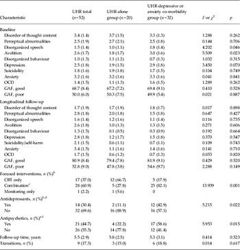
The clinical characteristics of the UHR subjects (cross-sectionally and longitudinally) are detailed in Table 1. Of the 52 UHR subjects, 32 (62%) had an Axis I co-morbid diagnosis of depressive or anxiety disorder in addition to the at-risk signs and symptoms. Specifically, 26 out of 32 (81%) had a co-morbid diagnosis of depression alone or in association with anxiety disorders. Anxiety disorders alone were less frequent, found in six participants out of 32 (19%). UHR participants with and without co-morbidity did not differ on interval between baseline and follow-up assessments. The UHR groups differed at baseline on disorganized speech (F 1,47 = 4.202, p = 0.046), with higher levels in the UHR-co-morbidity group. The UHR-co-morbidity group also had higher levels of avolition (F 1,44 = 5.509, p = 0.023) and depression (F 1,44 = 3.450, p = 0.070, trend level) (Table 1; see online Supplementary Table S1 for the full baseline psychopathological assessment).
Baseline and longitudinal clinical (CAARMS) a , functional (GAF) and outcome profiles of the UHR sample

CAARMS, Comprehensive Assessment of the At-Risk Mental State; GAF, Global Assessment of Functioning; UHR, ultra high risk of psychosis; OCD, obsessive–compulsive disorder; CBT, cognitive–behavioural therapy; OASIS, Outreach and Support in South London service.
Data are given as mean (standard deviation) unless otherwise indicated.
a For the whole CAARMS subscales, see online Supplementary Tables S2 and S3.
b Reliable information on focused interventions and antidepressant medication was missing from six UHR participants (two UHR alone + four UHR co-morbidity).
c Combination of CBT and medication (antidepressants or antipsychotics).
d Antidepressants: exposure to any antidepressant at any point from OASIS intake until discharge from the service.
e Reliable information on antipsychotic medication was missing from five UHR participants (two UHR alone + three UHR co-morbidity).
f Antipsychotics: exposure to any antipsychotic at any point from OASIS intake until discharge from the service.
At follow-up, nine UHR individuals (17%) had developed psychosis, while the remaining 43 (83%) had not. Average follow-up time was 5.5 (sd = 2.9) years. The UHR-alone and UHR-co-morbidity groups did not differ in the number of transitions, or the proportion of good and poor functional outcomes (GAF median split = 67.55). At follow-up, there were no significant symptom differences between the UHR-alone and UHR-co-morbidity groups (Table 1; see online Supplementary Table S1 for the full follow-up psychopathological assessment). However, there was a time × group interaction, with an association between UHR co-morbidity and larger improvements in depression levels (F 1,33 = 4.699, p = 0.037) over time compared with UHR alone. Of note, there were significant differences in the treatments offered to the two groups during their clinical management at the OASIS clinic (i.e. from baseline to follow-up). The UHR-co-morbidity individuals were more likely to be treated with antipsychotics (χ 2 1 = 5.953, p = 0.015) and antidepressants (χ 2 1 = 5.215, p = 0.022), and less likely to be treated with cognitive psychological therapies without adjunctive medications (χ 2 2 = 13.939, p = 0.001) than the UHR-alone group (Table 1).
Impact of depressive and anxiety co-morbid disorder on UHR neuroanatomy
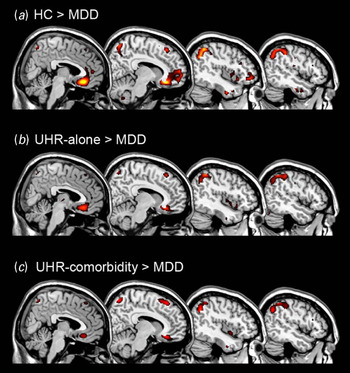
Patients with MDD showed GMV reductions, compared with HC participants with a whole-brain analysis, in a large medial frontal cluster including the orbital gyrus, ACC and medial frontal gyrus, as well as in the middle temporal gyrus and the inferior parietal lobe (online Supplementary Table S2 and Fig. 1 a). MDD patients also showed reductions in medial frontal and inferior parietal clusters compared with UHR-co-morbidity individuals (Fig. 1 b), and similarly with UHR-alone subjects (Fig. 1 c).
Group differences in grey matter volume. Differences between healthy control (HC) participants and major depressive disorder (MDD) patients (a), between patients at ultra high risk (UHR) of psychosis alone and MDD patients (b), and between UHR patients with depressive or anxiety co-morbidity and MDD patients (c) (p < 0.05 after family-wise error correction).

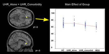
There were GMV reductions in UHR-co-morbidity compared with UHR-alone subjects as revealed by a whole-brain analysis in the medial frontal gyrus and the fusiform gyrus (online Supplementary Table S2). The ROI analysis based on an anatomical ACC mask showed a significant reduction in GMV in the UHR-co-morbidity group relative to the UHR-alone group in a cluster comprising the subgenual prefrontal cortex and the pregenual ACC (online Supplementary Table S2 and Fig. 2). Further analysis confirmed a trend across the four groups, with MDD patients showing the most pronounced decreases relative to HCs, followed by the UHR-co-morbidity and UHR-alone groups (main effect of group: F 3,120 = 15.422, p < 0.001; Bonferroni post-hoc comparisons: MDD < HC, p < 0.001; MDD < UHR co-morbidity, p < 0.001; MDD < UHR alone, p < 0.001; UHR co-morbidity < HC, p = 0.027; UHR co-morbidity < UHR alone, p = 0.046; and HC < UHR alone, p = 0.827). We found no effect of age (F 1,120 = 1.355, p = 0.247) or gender (F1,120 < 1, n.s.) in the analyses.
Effect of co-morbid depression and anxiety on anterior cingulate volume. Differences between ultra-high-risk (UHR) individuals with (UHR co-morbidity) and without (UHR alone) depressive or anxiety co-morbidity, with a region-of-interest approach on the anterior cingulate gyrus (p < 0.05 after family-wise error correction). UHR-co-morbidity had less grey matter volume in the anterior cingulate cortex [MNI (Montreal Neurological Institute) coordinates x, y and z: −8, 27 and −9, respectively]. For visualization purposes, effects are displayed at p < 0.05 uncorrected. The plot shows mean grey matter volumes for the four groups derived from the main effect of group results; values on the y-axis refer to mm3 per voxel. HC, Healthy control; MDD, major depressive disorder.

The results of the comparisons between UHR (combined) versus HC, UHR alone versus HC and UHR co-morbidity versus HC are reported in the online Supplementary Results, and displayed in online Supplementary Fig. S1 a–c.
Baseline and longitudinal correlations of ACC volume with clinical and functional measures
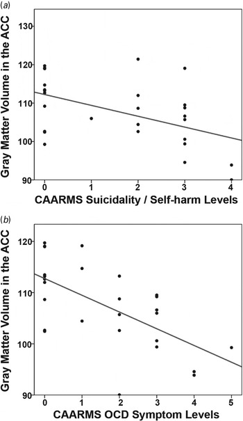
In UHR-co-morbidity participants, and using a priori CAARMS subscales on the basis of previous meta-analytical results (Fusar-Poli et al. Reference Fusar-Poli, Nelson, Valmaggia, Yung and McGuire2014b ), ACC volume was negatively correlated with baseline suicidality/self-harm (r = –0.454, p = 0.013) and OCD symptoms (r = –0.386, p = 0.039). Cook's D test was applied to rule out the influence of potential outliers. This correction strengthened the significance of the correlations in both cases (suicidality/self-harm: r = –0.494, p = 0.009; OCD symptoms: r = –0.628, p < 0.001) (Fig. 3). There were no correlations at baseline or follow-up for either the UHR-alone group or the UHR groups combined. ACC volume did not predict clinical transition or functional outcomes in either UHR subgroup (see online Supplementary Table S3). However, as noted above, this group was receiving more intensive treatments than the UHR-alone group over the duration of the study.
Correlations between anterior cingulate cortex (ACC) volume and baseline clinical profiles determined using the Comprehensive Assessment of the At-Risk Mental State (CAARMS) in patients at high risk of psychosis with co-morbidity diagnoses. The plots show a significant negative correlation between the grey matter volumes in the ACC and baseline levels of CAARMS suicidality/self-harm symptoms (a) and obsessive–compulsive disorder (OCD) symptoms (b) (higher CAARMS values indicate higher severity of symptoms).

For completeness, we tested in a collateral analysis whether the above findings survived after removing UHR subjects with anxiety alone from the UHR-co-morbidity group. Full results are presented in the online Supplementary Results.
Discussion
To our knowledge, this is the first study to examine the impact of depressive and anxiety co-morbidity on the neuroanatomy of subjects at UHR of psychosis.
Based on previous MRI studies of UHR samples (Wood et al. Reference Wood, Pantelis, Velakoulis, Yucel, Fornito and McGorry2008; Fusar-Poli et al. Reference Fusar-Poli, Borgwardt, Crescini, Deste, Kempton, Lawrie, McGuire and Sacchetti2011; Mechelli et al. Reference Mechelli, Riecher-Rossler, Meisenzahl, Tognin, Wood, Borgwardt, Koutsouleris, Yung, Stone, Phillips, McGorry, Valli, Velakoulis, Woolley, Pantelis and McGuire2011), our first hypothesis was that the UHR group as a whole would show GMV decreases relative to HCs, but that stratifying the sample on the basis of co-morbidity would reveal different patterns of GMV change. In line with a recent meta-analysis (Fusar-Poli et al. Reference Fusar-Poli, Borgwardt, Crescini, Deste, Kempton, Lawrie, McGuire and Sacchetti2011), UHR individuals as a whole showed decreased GMV in frontal regions, including the rolandic operculum, inferior frontal gyrus, medial frontal gyrus and orbitofrontal gyrus. However, when they were clinically stratified according to the presence or absence of depressive and anxiety co-morbidity, the co-morbid group showed further reductions in a temporal region. Furthermore, the direct comparison between UHR-co-morbidity and UHR-alone participants at the whole-brain level showed a significant reduction in UHR-co-morbidity participants in the medial frontal gyrus and the fusiform gyrus. In addition, the ROI analysis on this comparison showed a significant reduction in UHR individuals with co-morbid depressive and anxiety disorders in the hypothesized ACC region. As hypothesized, UHR-co-morbidity individuals showed an intermediate effect between patients with MDD alone (who showed the most pronounced decreases in this region) and UHR alone, suggesting that GMV decreases in the ACC are associated with an increased severity of depressive symptomatology. Future studies incorporating measures of severity of depressive symptoms in an MDD group may help expand this finding.
The ACC region implicated in our study comprised the subgenual prefrontal cortex and pregenual ACC. Prior evidence on the neuroanatomical basis of depressive symptomatology has typically highlighted ACC abnormalities as one of the most robust characteristics of the pathophysiology of MDD (Drevets, Reference Drevets2001; Bora et al. Reference Bora, Fornito, Pantelis and Yucel2012; Sacher et al. Reference Sacher, Neumann, Funfstuck, Soliman, Villringer and Schroeter2012). Grey matter abnormalities in the ACC region have also been reported in UHR samples regardless of their co-morbidity status (Borgwardt et al. Reference Borgwardt, McGuire, Fusar-Poli, Radue and Riecher-Rossler2008; Fornito et al. Reference Fornito, Yung, Wood, Phillips, Nelson, Cotton, Velakoulis, McGorry, Pantelis and Yucel2008; Fusar-Poli et al. Reference Fusar-Poli, Borgwardt, Crescini, Deste, Kempton, Lawrie, McGuire and Sacchetti2011; Radua et al. Reference Radua, Borgwardt, Crescini, Mataix-Cols, Meyer-Lindenberg, McGuire and Fusar-Poli2012). The detection of abnormalities in this region, which were more severe in the UHR-co-morbidity group, suggests a dysfunction within the limbic portions of the ACC that are known to modulate subcortical limbic and brainstem structures that mediate emotional and social behaviour (Damasio et al. Reference Damasio, Tranel and Damasio1990). This dysfunction may be key to the pathogenesis of depressive and anxiety co-morbidity in UHR subjects. Histopathological studies in MDD have revealed specific morphometric changes in the frontal cortex including the ACC (Rajkowska et al. Reference Rajkowska, Miguel-Hidalgo, Wei, Dilley, Pittman, Meltzer, Overholser, Roth and Stockmeier1999) that are hypothesized to be due to glial pathology and suggest that such neuroplastic changes could cause a potential dysregulation in the fronto-limbic circuit involved in the pathology of depression (Schroeter et al. Reference Schroeter, Abdul-Khaliq, Krebs, Diefenbacher and Blasig2008, Reference Schroeter, Abdul-Khaliq, Sacher, Steiner, Blasig and Mueller2010). Although largely speculative due to the absence of histopathological evidence from individuals at UHR of psychosis, one possibility for the observed neuroanatomical changes in UHR-co-morbidity subjects would be that they originate as suggested by the histopathologically generated hypothesis of glial pathology in depression, and accompany the morphometric changes typically associated with the UHR. Evidence from a range of methodologies including neuroimaging, lesion analysis and electrophysiological studies of humans and experimental animals all show that reduction in ACC volume and histopathological changes in this area can modulate emotional behaviour and stress responses (Drevets, Reference Drevets2000, Reference Drevets2001). Human lesion studies of the ventral ACC have documented abnormal autonomic responses to emotionally provocative stimuli, decreased emotional experience, and an impact on social behaviour by causing inability to use information regarding the probability of aversive social consequences versus reward in guiding social behaviour (Damasio et al. Reference Damasio, Tranel and Damasio1990). Our results tentatively suggest a neural model of depressive and anxiety co-morbidity in UHR subjects in which neuroanatomical changes in the ACC increase the severity of affective symptomatology. Correlation analysis between the positive subscales and the depression and anxiety subscales of the CAARMS yielded no significant associations in the UHR group as a whole, or in the UHR subgroups, providing further support for the idea that neuroanatomical changes in the UHR-co-morbidity group were not simply due to a higher severity of overall psychopathology. Nevertheless, as the first neuroanatomical evidence of this kind, this finding needs to be replicated in larger samples.
To test our second hypothesis, all UHR participants were followed clinically subsequent to scanning to explore the impact of GMV alterations at baseline on symptoms and functioning. Within the UHR-co-morbidity group, ACC volume was negatively correlated with the baseline suicidality/self-harm and OCD subscales of the CAARMS (showing less volume with increased suicidality and OCD symptoms). The neurobiological association of ACC with suicidal behaviour has been widely supported by neuropathological studies (Hercher et al. Reference Hercher, Canetti, Turecki and Mechawar2010; Torres-Platas et al. Reference Torres-Platas, Hercher, Davoli, Maussion, Labonte, Turecki and Mechawar2011), studies in subjects at high risk for suicide (Wagner et al. Reference Wagner, Schultz, Koch, Schachtzabel, Sauer and Schlosser2012) and studies in patients with previous suicide attempts (Wagner et al. Reference Wagner, Koch, Schachtzabel, Schultz, Sauer and Schlosser2011; Pan et al. Reference Pan, Hassel, Segreti, Nau, Brent and Phillips2013). These findings were detected in the context of an overall clinical improvement of the UHR-co-morbidity subgroup, as indicated by the longitudinal increase in functional levels and by the lack of significant between-group differences in psychopathology at follow-up. These outcomes could be due to spontaneous remission of the co-morbid symptoms, or alternatively to the more intensive treatments offered to these subjects during their clinical management. In fact, the presence of UHR co-morbidity significantly made an impact on the clinical management of UHR subjects during the duration of the study. To the best of our knowledge this is the first report showing that UHR patients with co-morbid depressive or anxiety diagnoses tend to be treated with more medications (antipsychotics and antidepressants) and fewer psychological interventions as stand-alone as compared with the UHR-alone group. Given that antipsychotics and antidepressants in addition to psychological interventions can prevent the onset of psychosis (Fusar-Poli et al. Reference Fusar-Poli, Valmaggia and McGuire2007), the longitudinal results of our cohort do not represent the natural history of the UHR state. Furthermore, treatments for the UHR state are commonly non-specific and may target depression and anxiety and not only attenuated symptoms (O'Connor et al. Reference O'Connor, Sota, Cortesi and Fusar-Poli2007). This model is further supported by recent meta-analytical evidence indicating that ACC structure is particularly sensitive to antipsychotic treatment during the early phases of psychosis (Radua et al. Reference Radua, Borgwardt, Crescini, Mataix-Cols, Meyer-Lindenberg, McGuire and Fusar-Poli2012). Additional functional imaging evidence shows that a few weeks of antipsychotic treatment can modulate the ACC response (Lahti et al. Reference Lahti, Holcomb, Weiler, Medoff, Frey, Hardin and Tamminga2004; Snitz et al. Reference Snitz, MacDonald, Cohen, Cho, Becker and Carter2005). Overall, our results support the notion that the UHR state should be considered as a heterogeneous condition in which individuals present with a combination of psychotic and affective (depression, anxiety) symptoms, some of whom, but not all, have a true vulnerability to schizophrenia (Fusar-Poli et al. Reference Fusar-Poli, Yung, McGorry and van Os2014c ). This is discussed in more detail in two recent and comprehensive reviews (Fusar-Poli et al. Reference Fusar-Poli, Borgwardt, Bechdolf, Addington, Riecher-Rossler, Schultze-Lutter, Keshavan, Wood, Ruhrmann, Seidman, Valmaggia, Cannon, Velthorst, De Haan, Cornblatt, Bonoldi, Birchwood, McGlashan, Carpenter, McGorry, Klosterkotter, McGuire and Yung2013a , Reference Fusar-Poli, Carpenter, Woods and McGlashan2014a ). The present study provides additional evidence for this idea and demonstrates at a neurobiological level the existence of a depressive-anxiety UHR subtype that is characterized by unique neuroanatomical characteristics along with distinctive psychopathological, therapeutic and prognostic features.
There are some limitations to the present study. First, we lacked an additional group of patients affected with anxiety disorders only. This would have allowed us to better delineate the neuroanatomical circuits associated with depressive versus anxiety disorders. Second, because our UHR-co-morbidity sample was receiving distinctive preventative treatments as compared with the UHR-alone group, the natural history of this subgroup could not be fully assessed. Furthermore, we lacked a follow-up scan to clarify the dynamic longitudinal course of the neuroanatomical alterations observed in our UHR-co-morbidity group. Third, the MDD group was older and the proportion of females was higher compared with the UHR and HC groups. Although we modelled these factors as covariates of no interest in GMV analysis, a potential confounding effect cannot be entirely ruled out. Future studies with larger samples are needed to replicate our findings. Information regarding the treatment of UHR subjects during their clinical management at the OASIS clinic (i.e. from baseline to follow-up) was obtained from OASIS patient records.
In summary, our results suggest that depressive and anxiety co-morbidity contributes to specific GMV reductions in UHR subjects, primarily focused in the pregenual ACC. This highlights the neurobiological heterogeneity of UHR, which makes an impact on clinical profiles of UHR individuals both cross-sectionally on their clinical management and longitudinal outcomes. Since co-morbid depressive and anxiety diagnoses confer unique psychopathological, neuroanatomical, treatment and prognostic features to UHR individuals, they should be carefully considered in future imaging studies.
Supplementary material
For supplementary material accompanying this paper visit http://dx.doi.org/10.1017/S0033291714000865
Acknowledgements
This work was supported by the European network of national schizophrenia networks studying Gene–Environment Interactions (project EU-GEI; no. HEALTH-F2-2010-241909). We also acknowledge the St Thomas and Guy's Hospital charitable contributions to the OASIS service, which enabled part of the scanning of this sample.
Declaration of Interest
None.