Due to the continuing progress in the surgical treatment of congenital cardiac disease, many symptomatic patients with cardiac malformations can now benefit from surgery or interventional catheterization during infancy and childhood. The result of such progress has led to improved survival rates of patients with congenitally malformed hearts.Reference Webb1 Still, late complications such as arrhythmias and cardiac failure are common, thus patients require further medical treatment, reoperations, or catheter interventions for residual lesions.Reference Somerville2 The benefit of these measures, especially long term treatment of cardiac failure by beta blockade, have to be proven by clinical trials, as done previously in adults with cardiac failure due to coronary arterial diseaseReference Abrams3–Reference Gottlieb, McCarter and Vogel5 or dilated cardiomyopathies.Reference Brodine, Tung and Lee6, Reference Hara, Inoue and Ogimoto7 But, due to the low mortality rate in adults and grown ups with congenitally malformed hearts, the study end points have to be adopted to the special conditions of this group of patients. Thus, significant surrogate prognostic parameters must be preferred, rather than using death as is typically the case in trials of cardiac failure in adults with coronary arterial disease. In this study, therefore, we used three different parameters to assessment the effects of 6 months of beta-blockade on the cardio-circulatory state of patients who had undergone surgical correction of tetralogy of Fallot.
In this respect, NT-pro brain natriuretic peptide, subsequently to be described simply as brain natriuretic peptide, is regarded as a predictor of morbidity and mortality in patients with cardiac failure because it is independent of other clinical, haemodynamic and neurohumoral risk factors.Reference Hunt, Richards, Nicholls, Yandle, Doughty and Espiner8, Reference Tsutamoto, Wada and Maeda9 Another valid surrogate parameter with an impact on prognosis in patients with cardiac failure is exercise capacity measured by spiroergometry.Reference Mancini, Eisen, Kussmaul, Mull, Edmunds and Wilson10–Reference Stevenson, Steimle and Fonarow12 We also used the effect on right and left ventricular function as quantified by magnetic resonance imaging.Reference Bellenger, Rajappan and Rahman13, Reference Anand, McMurray and Cohn14 Using these surrogates, we report here, as far as we are aware, the first prospective randomized trial of a medical therapy in patients who have undergone surgical corrections for tetralogy of Fallot, seeking to assess the effects of Bisoprolol, a well-tried drug in left ventricular failure,15 on our chosen parameters.
Methods
Patients studied
We performed a prospective, randomized, double-blind, placebo controlled trial for beta blockade in adults and grown ups with congenital cardiac disease. In a pilot study, from May 1, 2003 to May 31, 2004, we evaluated the cardiocirculatory state of 345 patients with congenitally malformed hearts, 141 being females and 204 males. This group included adolescent and adult patients ranging in ages from 14 to 50 years. Of these patients, 94 had undergone surgical correction of tetralogy of Fallot. All patients had their surgical or interventional treatment at the University Hospital of Göttingen. Patients were consecutively recruited from our out-patient clinic for congenital cardiac disease. We performed a prospectively designed protocol for exercise testing, echocardiography, and the assessment of clinical and neurohormonal variables. The group was divided according to their cardiac failure as previously described.Reference Norozi, Wessel and Alpers16 Cardiac failure was assumed to be present if the level of brain natriuretic peptide in the plasma was significantly elevated greater than 100 pg/ml,Reference Hess, Runkel, Zdunek and Hitzler17–Reference Scharhag, Urhausen and Herrmann19 and the peak uptake of oxygen was reduced to less than 25 ml/kg/min.Reference Kruger, Graf, Kunz, Stickel, Hanrath and Janssens20
Of the overall group of 345 patients, 89 (26%) fulfilled the criterions for cardiac failure as defined above. Patients with significant bradycardia, average heart rate in Holter electrocardiogram less than 50 beats per minute, and those who were already on beta-blockade, were excluded from the study. We present the data of the 33 patients who had undergone surgical corrections for tetralogy of Fallot, and who took part in the study. After randomization, these patients had their first examination, including Holter electrocardiography, echocardiography, neurohormonal assessment, cardiac magnetic resonance imaging, and spiroergometry. Depending on the CIBIS II protocol,15 medication was titrated from 1.25 mg/d, 2.5 mg/d, 3.75 mg/d, 5 mg/d, 7.5 mg/d, 10 mg/d Bisoprolol or placebo every two weeks up to the highest tolerated dose. Prior to every augmentation of the dose, the patients were investigated in our outpatient clinic or by the family doctor. Six months after entry, all patients underwent their second examination in our clinic, again including Holter electrocardiography, echocardiography, neurohormonal assessment, cardiac magnetic resonance imaging, and spiro-ergometry.
Informed consent
After adequate explanation of the purpose of the study, informed consent was obtained from all patients. The protocol was approved by the local ethic committee.
Clinical investigation
All patients underwent physical examination and measurements of blood pressure (Dinamap 1846, Critikon Corporation, Tampa, Florida, United States of America) body weight, and height. A standard 12-lead electrocardiogram was recorded to analyse the spontaneous rhythm and the duration of the QRS complex.
Exercise testing
Exercise testing was performed in all patients on an upright bicycle ergometer, beginning with a workload of 0.5 Watts per kilogram body weight. The load was increased by 0.5 Watts per kilogram every 2 minutes. Uptake of oxygen was measured using a breath-by-breath analysis (Oxycon pro™, Jaeger, Hoechberg, Germany) throughout the procedure. All patients were exercised to their maximal capability. Peak uptake of oxygen was determined as the highest value in the terminal phase of exercise. The 6-lead electrocardiogram and heart rate were recorded continuously during the test. Blood pressure was recorded every 2 minutes using a cuff sphygmomanometer.
Neurohormonal assessment
Peripheral samples of venous blood were obtained from all participants, after a rest for at least 15 minutes, prior to exercise testing. The samples were immediately placed on ice, and subsequently centrifuged at 5000 revolutions per minute for 10 minutes. Aliquots of plasma and serum were stored at −80 degrees Celsius until required for further analysis. Atrial natriuretic peptide and endothelin-1 were determined by enzyme immunoassays. Brain natriuretic peptide was determined using immunoassay (Elecsys 2010™, Roche, Mannheim, Germany).
Echocardiography
We performed cross-sectional and M-mode echocardiography via a transthoracic approach, using a Hewlett Packard Sonos 5500™ echocardiograph interfaced with a multifrequency transducer. End-systolic and end-diastolic diameters of the left ventricle were measured from a short axis view by M-mode, and fractional shortening was calculated as a parameter for systolic left ventricular function. Fractional change in area, also used as a parameter for systolic left ventricular function, was calculated from the cross-sectional measurement of the left ventricle. In addition, we measured the right ventricular end-diastolic diameter from a short axis view by M-mode. The velocities of tricuspid valvar regurgitation were measured by continuous-wave Doppler, and served to estimate the right ventricular systolic pressure. We also measured the velocities of flow across the pulmonary valve, using continuous-wave Doppler to estimate the degree of the stenosis.
Cardiac magnetic resonance imaging
All magnetic resonance imaging examinations were performed on a 1.5 Tesla whole body scanner (Magnetom Sonata; Siemens Medical Systems, Erlangen, Germany) equipped with high-performance gradients (maximum amplitude 30 mT/m, slew rate, 300 mT/m/msec). Patients were placed in supine position and a body phase-array coil was placed over the chest. Standard horizontal and vertical long-axis cine magnetic resonance images were obtained with a segmented steady-state free precession sequence (TrueFISP, Siemens). All measurements were prospectively gated by using the R wave as a trigger, and were performed using the breath-hold technique. The investigators were blinded with respect to the medications being received by the patients at the time of their evaluation. Cine magnetic resonance imaging in the cardiac short-axis plane was acquired to assess the volumes of the left and right ventricles, and the myocardial mass. End-systolic, end-diastolic, and myocardial volumes were measured by semi-automatic detection of the epi- and endocardial surfaces. First automatic boarder detection was performed in all slices. Then all slices were revised by two independent observers, with manual correction of the traced surface area performed where appropriate. Volumes of the ventricular chambers were computed as the sum of left ventricular and right ventricular volumes of all slices containing the ventricular chambers.Reference Kuehne, Saeed and Gleason21 The three patients in the cohort who had required insertion of pacemakers were excluded from examination using magnetic resonance imaging because of the potential complications.
Statistical analysis
Data are reported as mean plus or minus standard deviation where appropriate. Because most clinical, neurohormonal and echocardiography variables were not normally distributed, we used the nonparametric Mann-Whitney-Test. The data of comparable groups of patients assigned to receive either bisoprolol or placebo at entry, and after 6 months of medication, was analyzed by the paired t-test. The T-test was also used to compare the data obtained from magnetic resonance imaging for the groups of patients, and also the healthy volunteers. For all parameters, a value for p of less than 0.05 was considered statistically significant. The data analyses were performed using Excel 2000™ and SPSS 12.0™ (Chicago, Illinois).
Results
Characteristics of the patients
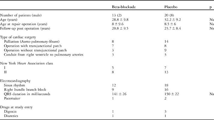
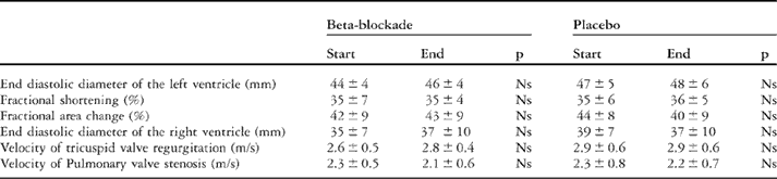
The characteristics, the types of cardiac surgery, and the medication received prior to commencing the study, are shown in Table 1. These medications were continued during the study. In 10 patients, the final dosage was 10 mg, for 1 patient it was 7.5 mg, and 2 Patients ended receiving 5 mg of bisoprolol. In those assigned to the placebo group, 18 patients were taking 10 mg, and 2 patients 7.5 mg of a drug as their final dose. The gradings within the clinical system of the New York Heart Association, and the echocardiographic parameters (Table 2) did not change in either those assigned to receive bisoprolol, or those receiving placebo, before and after intake of the medications.
Characteristics of patients at the beginning of the study.

Values are given as means ± standard deviation; Ns: Not significant.
Echocardiography at entry to, and at the end of, the study.

Values are given as mean ± standard deviation; Ns: Not significant.
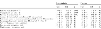
As is shown in Table 3, minimal, average and maximal heart rates as recorded by Holter electrocardiography were significantly reduced in those assigned to beta-blockade. We could find no significant effect, however, of beta-blockade on the variability of heart rate, expressed as slightly increased root-mean-square of successive normal sinus RR interval difference and percentage of successive normal sinus RR intervals greater than 50 milliseconds when taking bisoprolol.
Holter electrocardiography and variability in heart rate at entry to, and at the end of, the study.

Values are given as mean ± standard deviation; Ns: Not significant; Significant correlations are given in bold letters.
Neurohormonal parameters
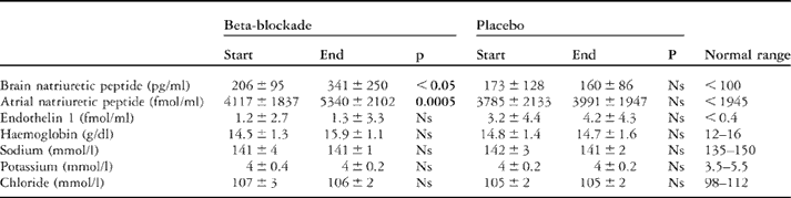
This data is illustrated in Table 4. The natriuretic peptides, and Endothelin-1, were elevated at baseline in both groups of patients. During beta-blockade, levels of both brain and atrial natriuretic peptide were significantly increased, but these measurements remained unchanged in those assigned to receive the placebo. Levels of Endothelin-1 level were unchanged in both groups. Values for haemoglobin and electrolytes were in the normal range on average in both groups, and did not change over the course of the study.
Neurohormonal data and electrolyte metabolism at entry to, and at the end of, the study.

Values are given as mean ± standard deviation; Ns: Not significant; Significant correlations are given in bold letters.
Assessment of cardio-pulmonary exercise capacity
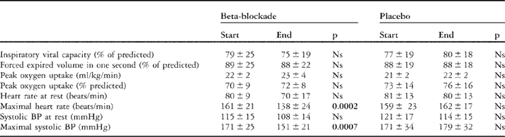
Reduced peak uptake of oxygen remained unchanged both in those assigned to receive the placebo, at 21 plus or minus 2 versus 22 plus or minus 2 ml/kg/min, and in those receiving bisoprolol, these values being 22 plus or minus 2 as opposed to 23 plus or minus 4 ml/kg/min. As is shown in Table 5, vital capacity and forced expired volume in one second were also reduced at baseline in both groups. We found no effect of beta-blockade on these values during spiro-ergometry. Medication with bisoprolol, however, significantly reduced the heart rate at rest, as well as the heart rate and systolic blood pressure during maximal exercise.
The data of spirometry and ergospirometry at entry to,and at the end of, the study.

Values are given as mean±standard deviation; Ns: Not significant; Significant correlations are given in bold letters.
Functional parameters derived from magnetic resonance imaging
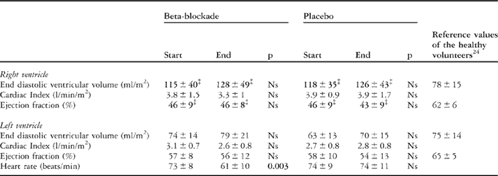
All the data is illustrated in Table 6. At the initial study, those assigned to receive placebo, as well as those to receive bisoprolol, had significantly increased end diastolic right ventricular volumes and decreased right ventricular ejection fraction when compared to reference values, as has been reported previously.Reference Grothues, Moon, Bellenger, Smith, Klein and Pennell22–Reference Rominger, Bachmann, Pabst and Rau24 These parameters remained unchanged during beta-blockade. The end diastolic left ventricular volumes, and ejection fractions, of both groups remained unchanged at roughly normal levels during medication.
Magnet resonance imaging of the left and right ventricle at entry to, and at the end of, the study.

Values are given as mean ± standard deviation; Ns: Not significant; Significant correlations are given in bold letters;
‡: p <0.01 versus healthy volunteers.
Discussion
To our knowledge, this is the first prospective, randomized, double-blind, placebo controlled study for medical treatment of cardiac failure in patients with surgically corrected congenital cardiac defects. The study should help to settle two essential questions relating to the treatment of cardiac failure in this group of patients:
• Does beta-blockade have a favourable effect on relevant prognostic surrogate parameters of cardiac failure, such as values of brain natriuretic peptide in the plasma, right ventricular or left ventricular ejection fractions, and peak uptake of oxygen?
• Is it justified to transfer the positive results of beta-blockade of adults with cardiac failure due to left ventricular dysfunction to this special group of patients, who predominantly suffer from failure of the right heart?
Adverse effects on pulmonary and/or cardiac function could be excluded, because the special indexes measured by spirometry, echocardiography, and magnetic resonance imaging remained unaffected during treatment. An improvement in clinical state as assessed using the grading system of the New York Heart Association, and physical efficiency measured by spiro-ergometry after 6 months of beta blockade, were not noted. Increased right ventricular volume due to the high incidence of pulmonary regurgitation,Reference Vliegen, van Straten and de Roos25, Reference Doughan, McConnell, Lyle and Book26 and reduced right ventricular ejection fraction, also remained unchanged after beta-blockade. These findings could be validated by magnetic resonance imaging and echocardiography.
Surprisingly, we found that beta-blockade with bisoprolol increased significantly the levels of both brain and atrial natriuretic peptides in the plasma of normotensive patients who had undergone surgical correction of tetralogy of Fallot. These results are in contrast to the results of the trials of cardiac failure in adults with left ventricular dysfunction.Reference Frantz, Olson and Grill27–Reference Fung, Yu and Yip29 One possible explanation is that most of these previous studies used carvedilol, which has also alpha-1-blocker activity, in patients with cardiac failure and severely reduced left ventricular ejection fraction below 40 percent, and documented reduced levels of peptides in the plasma of their patients. In our patients, the left ventricular ejection fraction was only slightly reduced, if at all. Thus, we could not document any effect of beta-blockade on left ventricular pump function. Furthermore we used a beta-1 selective blocker, which is reported to enhance the levels of natriuretic peptide in the plasma of hypertensive patients.Reference Deary, Schumann, Murfet, Haydock, Foo and Brown30, Reference van den Meiracker, Lameris, van de Ven and Boomsma31 The significance of the values of the brain peptide after administration of different types of beta-blockade remains controversial. Our results endorse, however, the decision of many paediatric cardiologists not to transfer uncritically the results of the trials of treatment of cardiac failure in adults with left ventricular dysfunction to young people with cardiac failure in the setting of congenitally malformed hearts.
From our results, we conclude that beta-blockade with bisoprolol seems to have no beneficial effect on asymptomatic or mildly symptomatic patients with right ventricular dysfunction secondary to repair of tetralogy of Fallot with residual pulmonary regurgitation and/or stenosis. The possible explanation is that beta blockers initially affect the left ventricle, and do not have an effect on right ventricular function. This could be shown recently in patients with arrhythmogenic right ventricular cardiomyopathy.Reference Hiroi, Fujiu and Komatsu32
Our results underline the necessity of performing prospective randomized studies in adults and grown ups with congenital heart disease with other beta blockers, or with inhibitors of angiotensin converting enzyme, to evaluate their effects on right ventricular function.
There are some limitations to our work. Our study suffers from small number of patients and a follow up of 6 month is probably not long enough to give the final statement about the effect of bisoprolol in patients who have undergone surgical correction of tetralogy of Fallot.
Acknowledgments
We thank Barbara Formanek, who worked as study nurse and Renate Lucyga, Ursula Baumgarten and Sabine Laurenzano for expert technical assistance during this study. This study was supported in part by grants from German Research Foundation (Bonn, Germany) to Armin Wessel (Nr. WE 2670/1-1) and Siegfried Geyer (Nr. GE 1167/3-1) and in part by grants from Merck (Darmstadt, Germany) and Herzkind eV (Braunschweig, Germany) to Reiner Buchhorn.