Introduction
Ligula intestinalis is a widespread pseudophyllidean tapeworm with a three-host life cycle involving copepods (procercoid stage), fish (plerocercoid stage) and piscivorous birds (mature stage). According to the parasite longevity and host involvement, the plerocercoid in fish is the dominant phase of its life cycle (for a review, see Arme, Reference Arme2002). In Europe, the parasite commonly infests cyprinids (Hoole et al., Reference Hoole, Carter and Dufour2010), e.g. roach (Rutilus rutilus), rudd (Scardinius erythrophtalmus), dace (Leuciscus leuciscus), gudgeon (Gobio gobio), bream (Abramis brama), bleak (Alburnus alburnus), minnow (Phoxinus phoxinus), chub (Leuciscus cephalus), tench (Tinca tinca) and silver bream (Blicca bjoerkna). Among these, roach represents a major sentinel species for freshwater pollution monitoring because of its abundance, widespread distribution and contaminant sensitivity (for a review, see Tyler et al., Reference Tyler, Lange, Paull, Katsu and Iguchi2007). The parasite is known to be responsible for various effects on the fish, e.g. physical damage, immune response disturbance, endocrine disruption (Sures, Reference Sures2008) and behaviour alteration (Loot et al., Reference Loot, Brosse, Lek and Guegan2001). The physiological function that is most affected by these effects is reproduction. Indeed, most infected fish present impaired gonadal development (Arme, Reference Arme2002; Geraudie et al., Reference Geraudie, Boulange-Lecomte, Gerbron, Hinfray, Brion and Minier2010a), sexual hormone deregulation along the pituitary–gonadal axis, i.e. gonadotropins (Carter et al., Reference Carter, Pierce, Dufour, Arme and Hoole2005; Trubiroha et al., Reference Trubiroha, Wuertz, Frank, Sures and Kloas2009) and steroid hormones (Hecker & Karbe, Reference Hecker and Karbe2005; Geraudie et al., Reference Geraudie, Boulange-Lecomte, Gerbron, Hinfray, Brion and Minier2010a) and abnormal plasma levels of peripheral-hormone-regulated factors such as vitellogenin (VTG; Hecker & Karbe, Reference Hecker and Karbe2005; Geraudie et al., Reference Geraudie, Boulange-Lecomte, Gerbron, Hinfray, Brion and Minier2010a). All of these disturbances lead to a decrease in fertility of infected individuals, making L. intestinalis a major determinant of the population dynamics of the roach (Kennedy et al., Reference Kennedy, Shears and Shears2001). Over the years, L. intestinalis has proved an invaluable fish–metazoan parasite model for ecological, immunological, ecotoxicological and endocrinological studies (Hoole et al., Reference Hoole, Carter and Dufour2010).
Extensive studies have reported that the range of damage described in ligulosed fish is similar to some of that induced in organisms after exposure to environmental pollutants, especially to endocrine-disrupting compounds (EDC). Endocrine perturbations highlighted in ecotoxicological studies could thus be falsely attributed to EDC exposure in parasitized fish (Sures, Reference Sures2008). Among EDC biomarkers, one of the most relevant is the evaluation of P450 aromatase status, since a wide range of chemicals are known to modulate aromatase expression and/or activity both in vitro and in vivo (Cheshenko et al., Reference Cheshenko, Pakdel, Segner, Kah and Eggen2008). P450 aromatase is a key enzyme involved in androgen–oestrogen conversion. In roach, aromatase is encoded by Cyp19a (also referred to as Cyp19a1a or Cyp19a1 or P450aromA) and Cyp19b (also referred to as Cyp19a1b or Cyp19a2 or P450aromB), expressed in the gonads and the brain, respectively. Over the past few years, development of real-time quantitative polymerase chain reaction (PCR) has made aromatase expression measurement a promising tool for evaluating EDC exposure in fish (Cheshenko et al., Reference Cheshenko, Pakdel, Segner, Kah and Eggen2008). This approach is highly sensitive, time-saving after technical adjustment and does not require radioactivity.
In this context, the aim of the present study was to determine if L. intestinalis infection could affect aromatase expression in roach, in order to: (1) better understand the effects of the parasite on its host; and (2) find out if parasitization can interfere with ecotoxicological data. In order to develop a clearer understanding of how parasitism acts to affect the endocrine system, analyses were conducted in fish caught from a reference site in Normandy (France), which is isolated from any anthropogenic activity.
Materials and methods
Collection and examination of roach
Roach collection (366 individuals) from Venables was performed monthly from January 2006 to April 2007. The Venables site (01°17′47″/49°12′00″, ED50) is a former sand quarry far away from any agricultural or industrial activity, where running water had led to the formation of an isolated arm of the River Seine. A previous study revealed that the sampling site was free from xenoestrogens according to the low plasma vitellogenin levels recorded in male roach (below 30 ng ml− 1). Furthermore, the mutagenicity and the oestrogenicity of the sediment extracts measured by the SOS chromotest and the YES assay (Yeast Estrogen Screening), respectively, were under the detection limit (Geraudie et al., Reference Geraudie, Gerbron, Hill and Minier2010b). Roach (>14 cm long) were caught with a net, anaesthetized with ethyl-3-aminobenzoate methansulphonate (MS222; Sigma-Aldrich, Saint Quentin Fallavier, France) according to the French Ethical Committee recommendations. Fish were considered as ligulosed when a worm was detectable macroscopically in their abdominal cavity. Dissected tissues were immediately frozen in liquid nitrogen and stored at − 80°C until further investigation. The sex of individuals was identified histologically after tissue fixation with 4% formaldehyde, dehydration in ethanol (50–99.9%), xylene clearing, paraffin embedding and section staining (5 μm) in haematoxylin, eosin and safran (Sigma-Aldrich). After macroscopic and microscopic analyses, some roach were randomly chosen to be investigated in the present study. The remaining fish had been included in a previous report (Geraudie et al., Reference Geraudie, Boulange-Lecomte, Gerbron, Hinfray, Brion and Minier2010a). Finally, the transcript dosage in brain tissue was determined in 23 uninfected (14 males, 9 females) and 5 infected (2 males, 3 females) roach, whereas gonadal analysis was performed on 16 uninfected (8 males, 8 females) and 6 infected (3 males, 3 females) fish. All investigated fish had been collected between January and April in both 2006 and 2007.
Molecular analysis
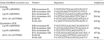
Total RNA was extracted from frozen tissues on ice using RNeasy mini kit according to the manufacturer's recommendations (Qiagen, Courtaboeuf, France). RNA integrity was systematically checked after migration on 1% agarose gels stained with Sybr®Safe (Invitrogen, Cergy-Pontoise, France) and the RNA concentration was measured at 260 nm with an ND-1000 spectrophotometer (NanoDrop Technologies, ThermoScientific, Wilmington, USA). Samples characterized by altered 260/280 or 260/240 ratios were excluded. Finally, 5 samples from parasitized fish and 23 from uninfected fish were retained for further brain expression study whereas 6 samples from parasitized fish and 16 from uninfected fish were retained for further gonad expression analysis. Primers were designed from publicly available sequences (GenBank) using the online Primer3 software (http://fokker.wi.mit.edu/primer3/input.htm) (table 1). From a partial Cyp19b genomic sequence kindly provided by Stephen Pearce (University of Sussex, unpublished sequence), R-B-Aromatase forward (S1, S2) and R-B-Aromatase reverse (AS1, AS2) primers were designed in different exons to avoid amplification from genomic DNA traces. For plasmid cloning (standard curves), RNA (1 μg) was treated with DNase (TURBO DNA-freeTM kit; Ambion Applied Biosystems, Courtaboeuf, France) and was reverse-transcribed by SuperScript III using oligodT(20) and random primers (Invitrogen). Target cDNA was amplified by semi-quantitative PCR in a Mastercycler® epGradient (Eppendorf, Le Pecq, France) with DyNAzyme II master mix (Finnzymes, Espoo, Finland) and specific primers (R-B-Aromatase-S1/R-B-Aromatase-AS1; R-G-Aromatase-S1/R-G-Aromatase-AS1; R-18s-S1/R-18s-AS1) (table 1). After an initial denaturation at 94°C for 2 min, 30 cycles were performed including a denaturation step at 94°C for 30 s, annealing at 55°C for 30 s and extension at 72°C for 30 s. The final extension step was continued for 5 min. DNA was omitted in the negative control. The lack of DNA traces in RNA samples was checked by performing amplification from RNA without previous reverse-transcription. PCR products were analysed on a 1% agarose gel and visualized by Sybr®Safe. Amplicons were purified (QIAquick PCR purification kit, Qiagen), cloned in pGEMt (Promega, Charbonnières, France) and sequenced (MilleGen, Labège, France).
PCR primers designed from GenBank using the online Primer3 software.

For real-time quantitative PCR, RNA (1 μg) was reverse-transcribed using the RT module of the Verso QRT-PCR SYBRGreen 2-Step Fluorescein kit (ABgene, Courtaboeuf, France) according to the manufacturer's instructions. Quantitative PCR was then conducted with the Q-PCR module of the Verso QRT-PCR SYBRGreen 2-Step Fluorescein kit in an i-Cycler IQ Real-Time System (BioRad, Marnes La Coquette, France) from cDNA (100 ng RNA equivalent) using specific primers (R-B-Aromatase-S2/R-B-Aromatase-AS2; R-G-Aromatase-S2/R-G-Aromatase-AS2; R-18s-S2/R-18s-AS2) (table 1) and optimal dNTP concentration (70–280 nm). After an initial denaturation at 95°C for 15 min, 40 cycles were performed including a denaturation step at 95°C for 15 s, annealing at 55°C for 30 s and extension at 72°C for 30 s. A dissociation protocol was added after thermocycling, determining dissociation of the PCR products from 60°C to 100°C (80 cycles of 10 s). The assays systematically included a non-template control and standard curves of three serial dilution points of each plasmid DNA (in steps of 100-fold or 1000-fold, from 5 × 10− 1 ng/μl to 5 × 10− 8 ng/μl, depending on the gene). All samples were analysed in triplicate. The amplicon specificity and the absence of primer dimers were checked for all amplifications using dissociation curves. Data processing was conducted using the Q-Base 1.3.5 software (Hellemans et al., Reference Hellemans, Mortier, De Paepe, Speleman and Vandesompele2007). This program provides a valuable tool for calculation of relative quantities after automatic normalization with one or more reference genes, integrating raw data quality control, inter-run calibration and gene-specific amplification efficiencies (ranged between 1.62 and 1.80 with R 2>0.996). Statistical comparisons were performed using the Student's t-test. Differences were considered as significant at P < 0.05.
Results
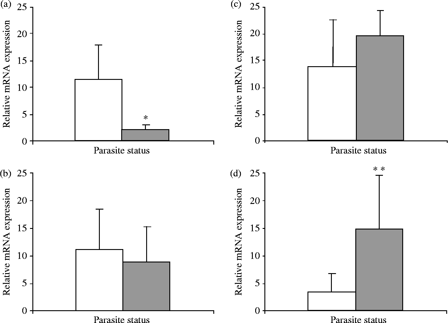
The prevalence of ligulosed fish between January 2006 and April 2007 in the sampled population was 20/366, i.e. 5.5%. A disparate temporal distribution was noted, with a maximal rate of parasitization between January and April for both years, representing 19/209 (9%), whereas only 1 out of 157 fish (0.64%) was parasitized during the rest of the sampling period. Further analyses were thus conducted from infected and uninfected fish caught between January and April in 2006 and 2007. The histological analysis of gonads revealed that sexual maturation may be compromised in infected roach since their gonads displayed more early stages (measured as spermatogonia or primary oocytes) than their uninfected counterparts, i.e. 72.7% versus 33%. Quantitative PCR analysis revealed a general decrease in Cyp19b expression in the brains of infected fish (fig. 1). However, variations were only significant in males, with a fivefold decrease of aromatase expression compared to their uninfected counterparts. No statistical difference was shown between uninfected males and females during the study period. On the contrary, Cyp19a expression in gonads appeared to be increased in ligulosed fish (fig. 1). This enhanced aromatase expression was significant in females, where aromatase expression in infected fish reached five times the level of non-infected ones. No statistical difference was observed between uninfected males and females.
Relative Cyp19b expression in the brain (a, b) and relative Cyp19a expression in the gonads (c, d) of uninfected (white bars) or L. intestinalis infected (grey bars) male (a, c) or female (b, d) roach. Each bar represents the mean normalized expression ± standard deviation of independent biological samples. Student's t-test was used for pairwise comparisons of Cyp19b and Cyp19a expression in uninfected or infected populations. Significant differences are labelled with asterisks (*, P = 0.034; **, P = 0.009).
Discussion
As described above, the general parasitism occurrence was 5.5% for the considered period with a maximum rate in winter/spring (9%) and a minimum rate in summer/autumn (0.64%). These results were in accordance with those reported by Hecker & Karbe (Reference Hecker and Karbe2005) in bream (Abramis brama), as the general prevalence in their reference site was 2.5% with maximum and minimum rates of 5% and 0%, respectively. It was thought possible that the low infestation prevalence may have been linked to the limited impact of pollution in the selected area (Jobling & Tyler, Reference Jobling and Tyler2003). However, this hypothesis can be excluded since Minier and colleagues (Reference Minier, Caltot, Leboulenger and Hill2000) reported similar rates of parasitization in polluted sites in the River Seine close to our reference site, suggesting no correlation between pollution and L. intestinalis occurrence in roach. Moreover, the presence of large plerocercoids in fish between January and April for both years may underlie a seasonal development of L. intestinalis. In accordance with our data, Hecker & Karbe (Reference Hecker and Karbe2005) reported the highest prevalence in the springs of 1999 and 2000. Moreover, Arme & Owen (Reference Arme and Owen1968) showed that L. intestinalis grows markedly in the host body cavity during autumn, supporting the hypothesis of a seasonal cycle of the parasite and justifying the presence of the largest specimens in spring.
In many teleosts, two different genes are known to encode two structurally and functionally different aromatase isoforms: Cyp19a and Cyp19b, preferentially expressed in the gonads and the brain, respectively. On the one hand, in accordance with the recent publication by Trubiroha and colleagues (Trubiroha et al., Reference Trubiroha, Kroupova, Wuertz, Frank, Sures and Kloas2010), we highlighted a decrease of Cyp19b expression in parasitized roach brains in comparison with uninfected ones. The fish population of the present work was the subject of a larger cohort study (Geraudie et al., Reference Geraudie, Boulange-Lecomte, Gerbron, Hinfray, Brion and Minier2010a). Various supplementary biological and biometric data are therefore available for the investigated sampling. In particular, we previously showed a decrease in brain aromatase activity in ligulosed individuals (Geraudie et al., Reference Geraudie, Boulange-Lecomte, Gerbron, Hinfray, Brion and Minier2010a). A reduction of both aromatase activity and expression was thus demonstrated for the first time in the same fish population infected by L. intestinalis, giving a functional relevance to transcript aromatase dosage in the brain. In teleosts, Cyp19b expression is under the strict control of steroids, e.g. direct upregulation by oestrogens due to the presence of the oestrogen response element (ERE) in the promoter of the gene and indirect upregulation by androgens via aromatization (for a review, see Diotel et al., Reference Diotel, Page, Mouriec, Tong, Pellegrini, Vaillant, Anglade, Brion, Pakdel, Chung and Kah2010). We previously detected a reduction of plasma steroid levels (17β-oestradiol, 11-ketotestosterone, progesterone) in ligulosed individuals (Geraudie et al., Reference Geraudie, Boulange-Lecomte, Gerbron, Hinfray, Brion and Minier2010a). This could be responsible for the observed reduction of the cerebral aromatase. If the role of brain aromatase in the regulation of reproductive processes has to be further investigated (Diotel et al., Reference Diotel, Page, Mouriec, Tong, Pellegrini, Vaillant, Anglade, Brion, Pakdel, Chung and Kah2010), its function in sexual and aggressive behaviour, in particular in males, is well established (Cheshenko et al., Reference Cheshenko, Pakdel, Segner, Kah and Eggen2008; Munakata & Kobayashi, Reference Munakata and Kobayashi2010). Brain aromatase reduction may thus contribute to alter mating tactics or hierarchy placement of parasitized individuals within the fish population. Interestingly, ligulosed fish show behaviour alteration such as either shifting away from the bank in unsafe areas (Loot et al., Reference Loot, Brosse, Lek and Guegan2001) or not joining spawning shoals (Orr, Reference Orr1966). This unusual behaviour is responsible for selective predation by birds, facilitating completion of the parasite's life cycle (Loot et al., Reference Loot, Brosse, Lek and Guegan2001).
On the other hand, we demonstrated an enhanced Cyp19a expression in female gonads. To date, no literature data are available as regards gonadal aromatase activity/expression in ligulosed fish. Indeed, L. intestinalis is responsible for impaired gonadal growth, causing aromatase activity to be below the detection limit using current methods (Geraudie et al., Reference Geraudie, Boulange-Lecomte, Gerbron, Hinfray, Brion and Minier2010a). In this context, transcript dosage appears to be an attractive alternative for studying the aromatase status in poorly developed tissues. Moreover, even if the biological relevance of expression variations has to be demonstrated in gonads, the concordance between aromatase activity and Cyp19b expression in the brain is encouraging. Several authors reported an increase of Cyp19a expression during vitellogenesis in many teleosts (for a review, see Cheshenko et al., Reference Cheshenko, Pakdel, Segner, Kah and Eggen2008). One might thus expect to observe a higher expression of Cyp19a in the mature gonads of healthy fish than in immature ligulosed ones. Paradoxically, we demonstrated an enhancement of aromatase expression in parasitized fish. This could be at least partially due to the lack of negative regulation of the Cyp19a gene expression by androgens, since weak systemic 11-ketotestosterone levels were observed in infected roach (Geraudie et al., Reference Geraudie, Boulange-Lecomte, Gerbron, Hinfray, Brion and Minier2010a). Indeed, unlike the Cyp19b promoter, the Cyp19a regulating region only contains ERE half-sites, which prevents an auto-regulatory positive feedback brought about by oestrogens (Guiguen et al., 2010). In contrast, androgens are known to induce inhibition of Cyp19a expression via the androgen response element (ARE; Guiguen et al., 2010). Nevertheless, high levels of Cyp19a transcripts should promote gonad maturation. This process is actually due to the enhancement of oestrogen levels induced by aromatase. Maturation did not succeed in parasitized roach since we demonstrated that oestrogen levels remained weak, probably due to the low levels of circulating aromatizable androgens (Geraudie et al., Reference Geraudie, Boulange-Lecomte, Gerbron, Hinfray, Brion and Minier2010a). Moreover, as commonly depicted in ligulosed fish, we previously reported not only a marked effect of parasitization on gonadal growth – as demonstrated by a 50% decrease in the gonadosomatic index for both sexes – but also the presence of immature but differentiated tissues exhibiting primary oocytes and spermatogonia in most parasitized roach (Geraudie et al., Reference Geraudie, Boulange-Lecomte, Gerbron, Hinfray, Brion and Minier2010a). Given that Cyp19a up-regulation is needed for triggering and maintaining ovarian differentiation, whereas Cyp19a down-regulation is the only necessary step for inducing a testicular differentiation pathway (Guiguen et al., 2010), the Cyp19a expression pattern in sampled fish justifies the observation of well-differentiated sexual tissues in the ligulosed population.
Fecundity and growth reduction in fish after parasitic infection is usually considered to be the result of the drain of energy resources from the hosts by both parasite and immune defence against infection (Hall et al., Reference Hall, Becker and Cáceres2007). In our study, L. intestinalis infection only induced a slight growth alteration as shown by the weak variation in the condition factor between parasitized and unparasitized roach (Fulton index; Geraudie et al., Reference Geraudie, Boulange-Lecomte, Gerbron, Hinfray, Brion and Minier2010a). Thus, the energy diversion seems to be reflected only in the reproductive function, which can be considered as incomplete sterilization since (1) the gonads are still present, even if they are poorly developed and immature; and (2) maintaining Cyp19a expression is associated with sexual differentiation. This ‘parasite strategy’ permits L. intestinalis to maintain a balance between reaping the benefits of its own growth/reproduction and suffering the costs of harming its host (Hall et al., Reference Hall, Becker and Cáceres2007).
In conclusion, Cyp19b and Cyp19a expression in the brain and gonads was affected by L. intestinalis parasitization in roach. In agreement with previous data showing a decrease of aromatase activity (Hecker et al., Reference Hecker, Sanderson and Karbe2007; Geraudie et al., Reference Geraudie, Boulange-Lecomte, Gerbron, Hinfray, Brion and Minier2010a), Cyp19b expression was reduced in the brain of ligulosed individuals, probably contributing to their characteristic behavioural alteration. The concordance between aromatase activity and expression emphasizes the relevance of the PCR approach to evaluate aromatase status in parasitized roach. This technique is highly sensitive and is thus well-suited for aromatase dosage in poorly developed organs, in particular in the gonads of ligulosed individuals. The sustained rate of Cyp19a transcripts in gonads was associated with immature but differentiated sexual tissues in parasitized fish. Differences in aromatase expression in the brain and gonads could result from the drastically different structure of Cyp19b and Cyp19a promoters, as demonstrated in several teleost genes (Callard et al., Reference Callard, Tchoudakova, Kishida and Wood2001). Conducting the study in fish from a site with low contamination enabled us to attribute biological alterations to parasitism, excluding any effect of pollutants. Our results highlight that Cyp19b and Cyp19a expression is markedly impacted by parasitization, even though they are commonly used as EDC exposure biomarkers (Cheshenko et al., Reference Cheshenko, Pakdel, Segner, Kah and Eggen2008). Ligula intestinalis infection must be considered as a major confounding factor for the biomonitoring of endocrine disruption in fish. Consequently, ligulosed fish should be systematically excluded from any ecotoxicological studies, bearing in mind that, at present, only large plerocercoids can be easily identified. Conversely, water quality must be consistently considered by parasitologists in the evaluation of host–Ligula relationships.
Acknowledgements
We gratefully thank Mr and Mrs Hoydrie (Pisciculture de Venables) for their valuable help in fishing. Further, we acknowledge Dr Stephen Pearce (University of Sussex) for having provided the partial Cyp19b genomic sequence. This study is part of the RAED (Risk Analysis associated with Endocrine Disruption in the manche regions; project no. 008) and DIESE (Determination of pertinent Indicators for Environmental monitoring: a Strategy for Europe; project no. 040) research projects supported by grants awarded by the European Interreg IIIA and IVA France (Channel) England programmes.