INTRODUCTION
Human lymphatic filariasis (LF) is a major physical and social burden, and after leprosy, the second leading known cause of disability. It is caused by the tissue dwelling filarial nematodes, namely Wuchereria bancrofti, Brugia malayi and Brugia timori. Over 120 million people are currently infected while approximately 40 million are disfigured and incapacitated by the disease worldwide (World Health Organization, 2014). WHO has targeted the year 2020 for global elimination of LF (World Health Organization, 2010), but the success of this programme will depend upon effectiveness of tools for diagnosis and treatment.
Conventionally, the detection of filarial infections relied exclusively on the detection and identification of microfilariae (MF) in night blood specimens (Nicolas, Reference Nicolas1997). Lately, with the use of circulating filarial antigen (CFA) that detects the presence of viable adult worms (AWs) rather than MF, the sensitivity of diagnosis of bancroftian filariasis has increased as it differentiates the infection free individuals (CFA negatives) from the MF negative individuals which might carry infection (CFA positives) in endemic areas. In bancroftian filariasis the antigen detection tools are based upon monoclonal antibodies incorporating Gib-13, AD-12 and Og4C3 (Forsyth et al. Reference Forsyth, Spark, Kazura, Brown, Peters, Heywood, Dissanayake and Mitchell1985; Faris et al. Reference Faris, Ramzy, Dad, Weil and Buck1993; Chanteau et al. Reference Chanteau, Moulia-Pelat, Glaziou, Nguyen, Luquiaud, Plichart, Martin and Cartel1994; Shah and Mulla, Reference Shah and Mulla2007). Two such CFA detection assays are Og4C3 enzyme linked immune-sorbent assay (ELISA) and the immuno-chromatographic (ICT) card test (Chanteau et al. Reference Chanteau, Moulia-Pelat, Glaziou, Nguyen, Luquiaud, Plichart, Martin and Cartel1994; Weil et al. Reference Weil, Lammie and Weiss1997). ICT has been recommended by international authorities as the diagnostic method of choice for mapping the distribution of bancroftian filariasis for the programmes for elimination of lymphatic filariasis (PELFs) (Gyapong et al. Reference Gyapong, Kyelem, Kleinschmidt, Agbo, Ahouandogbo, Gaba, Owusu-Banahene, Sanou, Sodahlon, Biswas, Kale, Molyneux, Roungou, Thomson and Remme2002). But these tests are unable to detect B. malayi infections and unfortunately no such antigen detection tests are available for brugian filariasis (Weil and Ramzy, Reference Weil and Ramzy2007). In the absence of a sensitive antigen detection test, the less sensitive MF detection in night blood samples remains the only option for identifying the B. malayi infections in endemic areas.
The detection of parasite DNA in mosquito vectors and human blood samples are not practically feasible for PELFs, and antibody detection assays are not very useful in endemic areas as they are unable to distinguish between active and the past infections. IgG4 antibody, however, has been shown to be a better marker of active filarial infections (Jiraamonnimit et al. Reference Jiraamonnimit, Wongkamchai, Boitano, Nochot, Loymek, Chujun and Yodmek2009), so Immunoglobulin G 4 (IgG4) antibody detection assays based upon B. malayi recombinant antigen 1 (BmR1) have been developed and made commercially available (Rahmah et al. Reference Rahmah, Taniawati, Shenoy, Lim, Kumaraswami, Anuar, Hakim, Hayati, Chan, Suharni and Ramachandran2001; Lammie et al. Reference Lammie, Weil, Noordin, Kaliraj, Steel, Goodman, Lakshmikanthan and Ottesen2004; Noordin et al. Reference Noordin, Itoh, Kimura, Ravindran, Mahmud, Supali and Weerasooriya2007). These tests have been reported to detect B. malayi as well as B. timori infections (Fischer et al. Reference Fischer, Bonow, Supali, Ruckert and Rahmah2005). In spite of all advancements, B. malayi filariasis is still lagging behind in antigen detection methods.
The studies regarding antigenaemia status of the three life cycle stages i.e. L3, AW and MF of B. malayi in different clinical presentations and endemic normals are lacking. The present study evaluated stage-specific antigen response in brugian filariasis infected subjects with different clinical spectra to throw light on the immune responses in such patients in B. malayi endemic areas in India. In addition, the treatment group (MF positive but asymptomatic) subjects were also assessed for specific responses before, during and after treatment with diethylcarbamazine (DEC) to assess any change in these markers. The study was approved by the Institutional Ethics Committee, and all the animal experiments were performed after getting approval from the Institutional Animal Ethics Committee of Postgraduate Institute of Medical Education and Research (PGIMER), Chandigarh.
MATERIALS AND METHODS
Study groups
A total of 101 subjects (68 males, 33 females) aged 18 years and above were recruited for the study. The subjects were distributed into the following groups based on the clinical spectra: group I (n = 21), microfilaraemic but asymptomatic individuals without any signs or symptoms of filariasis or any other chronic disease (microfilaraemic, MF); group II (n = 20), acute lymphangitis with or without filarial oedema (adenolympangitis, ADL); group III (n = 20), chronic filarial oedema grade I–IV) without any history of acute lymphangitis during the last 6 months (chronic pathology, CP); group IV (n = 20), apparently healthy persons fromAlleppey (an area endemic for brugian filariasis), Kerela,without apparent filarial infection or any other chronic infection (endemic normal controls, EN); group V (n = 20), apparently healthy subjects from Chandigarh (a non-endemic area), North India, who did not travel to any endemic area for the past 5 years (non-endemic normal controls, NEN).
Samples
Venous blood samples were collected from the above groups after taking written informed consent. For groups I–IV, MF count was done by peripheral blood smear examination on the night blood samples in all the subjects. For group IV, night blood sample were tested for MF on three consecutive days to confirm the negative MF status. Samples of group V were included as controls. Group I subjects were given DEC treatment (6 mg kg−1 body weight, single dose) after collecting blood samples (day 0), and pre- and post-treatment (PT) MF counts were recorded, following which sequential blood samples were collected (days 1, 30, 60, 90, 180 and 365). The ADL and CP patients were not given any anti-filarial drug treatment and blood samples were collected on days 0, 90, 180 and 365. In EN group, after day 0, the blood samples were also collected on day 365.The serum samples collected at Alleppey were stored at −20 °C and were transported to Chandigarh under refrigerated conditions, where the sera were stored at −80 °C till further use.
Maintenance of B. malayi strain
The Black-eyed susceptible strain of Aedes aegypti mosquito originally developed by Prof. W.W. Macdonald of Liverpool School of Tropical Medicine, UK, and the ‘GRA’ Giessen strain of Mastomys natalensis with bright yellow body colour and red eyes (both strains procured from Central Drug Research Institute, Lucknow, U.P., India), were used to maintain the B. malayi strain cyclically. The infective larvae (L3) were separated from mosquitoes while the AW and MF stages were obtained from the Mastomys rats (Murthy et al. Reference Murthy, Tyagi, Roy Chowdhury and Sen1983; Chandrashekar et al. Reference Chandrashekar, Rao, Rajasekariah and Subrahmanyam1984).
Antigen preparation
Extracts/soluble antigens from L3, AW and MF stages of the parasite were prepared by disruption in phosphate buffered saline (PBS) pH 7·2 containing following protease inhibitors:1 mm ethylenediaminetetraacetic acid, 1 m Methyleneglycolbistetraacetic acid, 0·2 mm tosyllysinechloromethylketone, 0·1 mm tosylphenylalanine chloromethylketone and 1 mm phenylmethyl sulphonyl fluoride (Sigma-Aldrich, Bangalore, India).
Infective stage larva (L3) antigen
Briefly, 103 L3 larvae in 1 mL of PBS were disrupted in a ground glass homogenizer. The suspension was sonicated on ice with an ultrasonic homogenizer (Soniprep 150, MSE, UK), six cycles of 30 s each at 20 kHz with 30 s intermittent cooling. The sonicate was centrifuged at 10 000 g at 4 °C for 30 min and the supernatant passed through 0·22 µ m filter. The filtrate was aliquoted and protein estimation done by Lowry's method. The antigen was stored at −80 °C till used (Helmy et al. Reference Helmy, Weil, Faris, Gad, Chandrashekar, Ashour and Ramzy2000).
AW antigen
A total of 100 mixed AWs (both males and females) were disrupted in 1 mL of PBS in a ground glass homogenizer and prepared as mentioned above for L3 antigen (Kurniawan-Atmadia et al. Reference Kurniawan-Atmadia, Sartono, Oartono, Yazdabbakhsh and Maizels1998).
MF antigen
Briefly, 106 MF mL−1 of buffer pH7·2 were directly sonicated without disrupting in ground glass homogenizer. Rest of the procedure was similar as mentioned above.
Hyper-immune sera
The hyper-immune sera against the three stages of the parasite were raised in rabbits. In brief, 6 healthy, male, white New Zealand rabbits of approximately 2 months of age were used for preparation of anti-sera. Two rabbits were used for immunization with each antigen (L3, AW and MF antigens). One mL of each filarial antigen (protein concentration of 325 µg mL−1) mixed with an equal amount of Freund's complete adjuvant was injected subcutaneously on both thighs (0·5 mL on each thigh) once weekly over a period of 4 weeks, followed by two boosters. The rabbits were first bled from ear vein and the sera were checked for the presence of anti-filarial antibodies by gel diffusion test (Parulekar, Reference Parulekar, Kelkar and Khare1984) and then bled by cardiac puncture without sacrificing the animal. The sera were separated and stored at −80 °C till used.
ELISA
The double antibody sandwich ELISA for detection of antigen was carried out as described previously with minor modifications wherever required (Voller et al. Reference Voller, Bartlett and Bidwell1976). The optimum dilutions of the antigen (serum samples), antibody (hyper-immune sera or anti-sera) and second antibody i.e. horseradish peroxidase (HRP) conjugated anti-human IgG (whole molecule) were determined by checkerboard titration method. The microtitre plate wells were coated with optimally diluted hyper-immune sera (100 µL well−1) and incubated at 4 °C overnight. The plate was washed thrice with washing buffer (PBS-Tween 20). The unbound sites of the plate wells were saturated with 1% bovine serum albumin (BSA; Hi-Media, India) and the plate was incubated at 37 °C for 1 h. After washing 100 µL of the test and control sera diluted 1 in 20 were added to duplicate wells and incubated at 37 °C for 1 h. The wells were washed three times and 100 µL of the optimally diluted HRP enzyme labeled anti-human IgG conjugate (Dakopatts, Denmark) was added to each well, followed by incubation at 37 °C for 1 h. After another washing step, 100 µL of substrate (H2O2-OPD mixture) was added to each well and the plate was incubated at room temperature for 15–30 min. The reaction was stopped with 6% H2SO4 and read at 490 nm in an ELISA reader (Emax™, California). The mean optical density (OD) value of the two readings was taken as the reading for that sample.
Interpretation of results
All samples were tested in duplicate and the mean of two OD values was taken as absorbance value for that sample. ELISA-ratio (or E-ratio) system was adopted to minimize batch to batch or plate to plate variations. The individual OD values were divided by the mean + 2 standard deviation (s.d.) of normal controls run in the same plate, the ratios thus obtained known as E-ratios. These E-ratios, unlike OD values, remain almost unaffected by the variations due to plates, temperature, incubation periods, reagents or other common factors. Before statistical analyses, all OD values were converted to E-ratios and using the above methodology the cut-off E-ratios were calculated (Baveja and Chattopadhyay, Reference Baveja and Chattopadhyay2002).
To check antigen specificity, 100 serum samples from various protozoan and helminthic infections: amoebiasis (n = 20), toxoplasmosis (n = 20), malaria (n = 20), cysticercosis (n = 20) and echinococcosis (n = 20) were tested against the antigens.
Statistical analysis
The student's t-test and McNemar test were carried out to calculate the P-values and find the significance of difference in microfilaraemia and also in L3, AW and MF pre-treatment and post-treatment (PT) antigenaemia. A value P ⩽ 0·05 was taken to be significant.
RESULTS
All the 101 subjects (68 males and 33 females) were adults (18 years and above). There was no statistically significant difference in antigen detection in both the sexes (P = 0·27).
Microfilarial antigenaemia
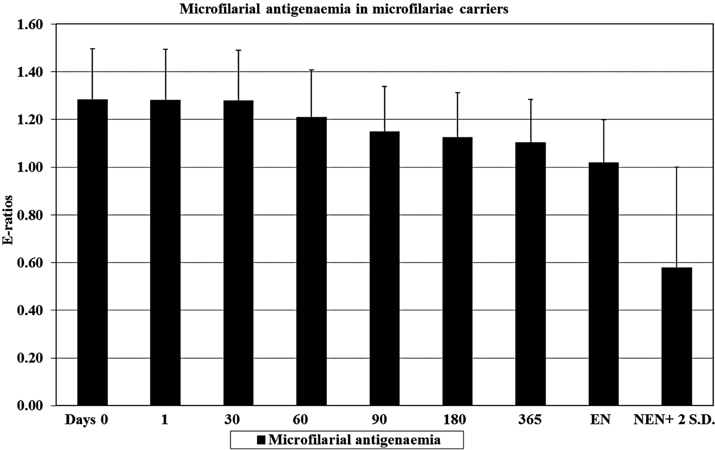
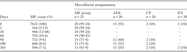
In MF group patients a gradual decrease in antigenaemia (mean E-ratios) was observed following treatment. The microfilarial antigenaemia did not show significant difference on day 0, when compared with samples collected on day 1 and 30 PT (P ⩾ 0·16), but significant differences were noted when day 0 compared with day 60 PT onwards up to day 365 PT (P < 0·0001). Twenty (95·24%) out of 21 sera from MF group subjects were found to be antigen positive on day 0 (pre-treatment) and thirteen (61·9%) serum samples remained antigen positive on day 365 PT (Fig. 1, Table 1).
Microfilarial antigenaemia in MF group on days 0 (pre-treatment), 1, 30, 60, 90, 180 and 365. MF group: D 0 vs 1, d 30 (P ⩾ 0·16), d 0 vs 60 upto 365 (P < 0·0001), MF group d 0 vs EN and NEN (P < 0·0001), MF group d 365 vs EN (P = 0·21), MF group d 365 vs NEN (P = 0·02). Abbreviations: EN, endemic normal controls; MF, microfilaraemic group; NEN, non endemic normal controls.

Comparative analysis of the pre- and post-treatment microfilaraemia and MF stage-specific antigenaemia in the treatment group (MF); CFA detected in patients [number (%)] with different clinical spectra of brugian filariasis

ADL, adenolympangitis; CFA, circulating filarial antigen; CP, chronic pathology; EN, endemic normal controls; MF, microfilaria; n, number.
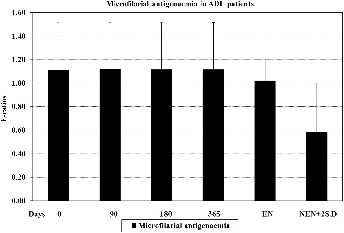
In ADL patients no significant differences in antigenaemia response were observed between samples collected on day 0 and days 90, 180 and 365 values (P ⩾ 0·10). Eleven (55%) out of 20 sera were having positive antigenaemia response on day 0, 180 and 365, while on day 90, 12 (60%) were microfilarial antigen positive (Fig. 2, Table 1).
Microfilarial antigenaemia in ADL patients on days 0, 90, 180 and 365. MF group: Day 0 vs 90 upto 365 (P ⩾ 0·10), ADL d 0 vs EN (P = 0·27), ADL d 0 vs NEN (P = 0·23). Abbreviations: ADL, adenolympangitis; EN, endemic normal controls; MF, microfilaraemic group; NEN, non endemic normal controls.

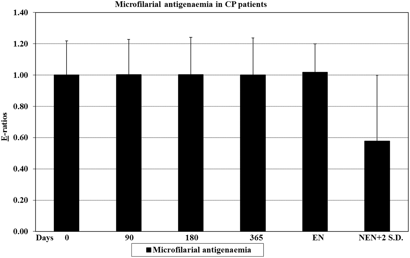
The CP patients did not show any significant difference in antigenaemia response of serum samples collected on day 0 to days 90, 180 and 365 (P ⩾ 0·72). Antigen was detected in 2 (10%) out of 20 serum samples (Fig. 3, Table 1).
Microfilarial antigenaemia in CP patients on days 0, 90, 180 and 365. MF group: Day 0 vs 90 upto 365 (P ⩾ 0·72), ADL d 0 vs EN (P = 0·98), ADL d 0 vs NEN (P = 0·97). Abbreviations: ADL, adenolympangitis; CP, chronic pathology; EN, endemic normal controls; MF, microfilaraemic group; NEN, non endemic normal controls.

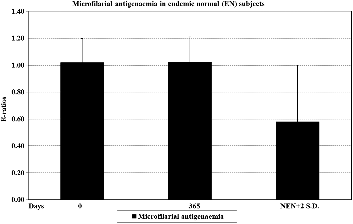
In EN controls no significant difference in mean E-ratios was noted. The difference in antigenaemia response was not significant between the serum samples collected on two time points i.e. on day 0 and 365 (P = 0·51). The differences in antigenaemia response on day 0 or 365 were also non-significant when compared with NEN controls (P ⩾ 0·70). Two (10%) out of 20 sera from EN control subjects were found to be antigen positive (Fig. 4, Table 1).
Microfilarial antigenaemia in EN controls on days 0 and 365. MF group: day 0 vs 365 (P = 0·51), EN d 0 and 365 vs NEN (P ⩾ 0·70). Abbreviations: EN, endemic normal controls; MF, microfilaraemic group; NEN, non endemic normal controls.

The results of other hyper-immune sera i.e. L3 and adult antigenaemia showed similar results (data not shown).
Comparative analysis
Highly significant differences were observed in microfilarial antigenaemia response in MF carriers on day 0 when compared with EN (P < 0·0001) and NEN (P < 0·0001) controls. On day 365 PT, the difference to endemic controls became non-significant (P = 0·21) but it remained significant with NEN controls (P = 0·02). Microfilarial antigenaemia in ADL and CP patients did not show any significant difference on day 0 when compared with EN (P = 0·27 and 0·98, respectively) and NEN subjects (P = 0·23 and 0·97, respectively). Almost similar were the results of L3 and adult antigenaemia (data not shown).
Kinetics of microfilaraemia and antigenaemia in asymptomatic MF carriers
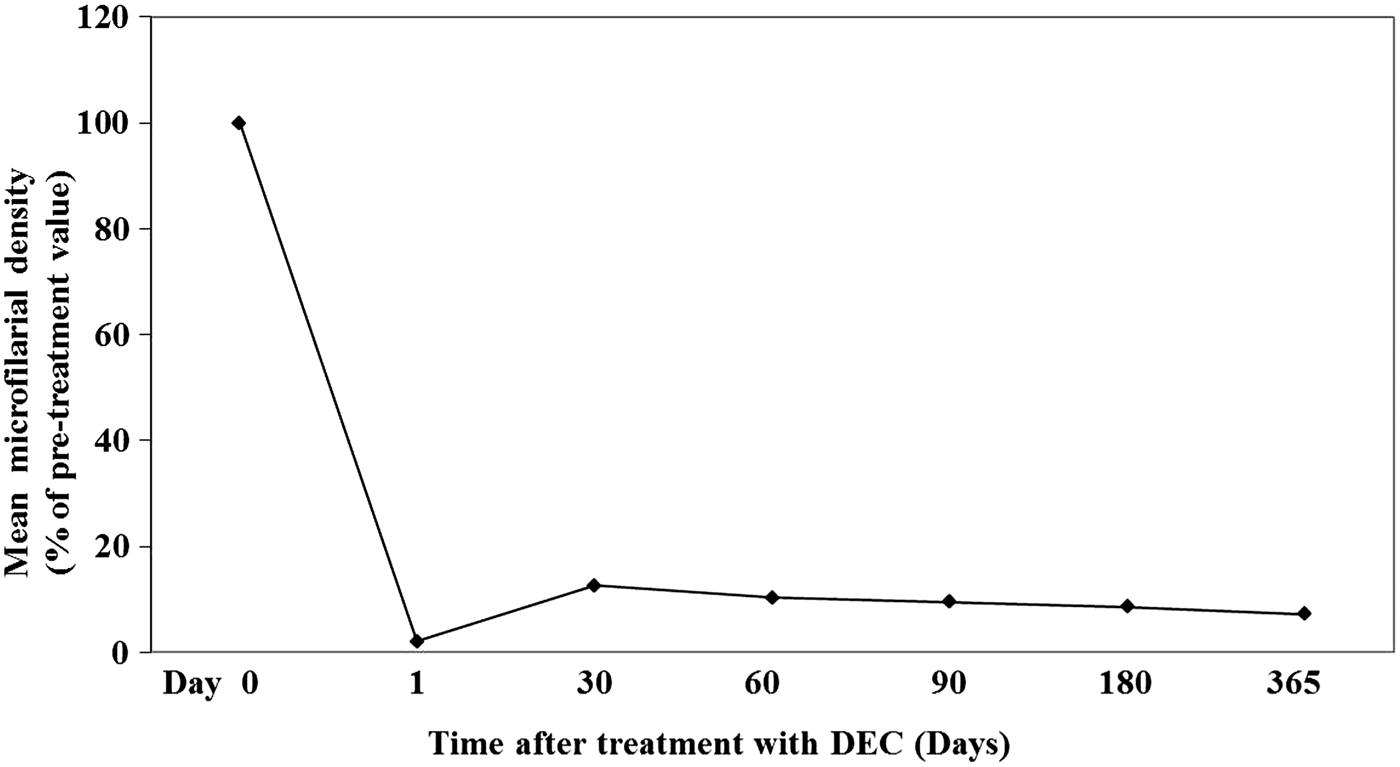
The pattern of clearance of microfilaraemia following treatment in microfilaraemic individuals is illustrated in Fig. 5 and Table 1. The kinetics of clearance indicated drastic decrease from 100 to 2·2% after 24 h (day 1) PT. Although, on day 365 PT, mean MF density was 7·3% of pre-treatment value, yet L3, AW and microfilarial antigenaemia could be detected in 66·7, 90·5 and 61·9% MF carriers.
Effect of single dose of DEC (6 mg kg−1 body weight) on clearance of microfilaraemia in B. malayi MF group subjects. The mean MF count of 21 individuals on day 0 (pre-treatment) was considered as 100% and the subsequent PT MF counts were calculated in respect to that. MF count on day 0 vs days 1, 30, 60, 90, 180 and 365 (P < 0·0001). Abbreviations: DEC, diethylcarbamazine; MF, microfilaria; PT, post-treatment.

Specificity
No cross-reactivity was observed for amoebiasis, toxoplasmosis, malaria, cysticercosis and echinococcosis, demonstrating 100% specificity for other non-filarial parasites.
DISCUSSION
Antigen detection methods have been reported of immense value in the detection of active bancroftian filariasis. For detection of W. bancrofti, monoclonal antibody (mo-Ab) based assay recognizing a 200 kDa AW antigen has been used, while another assay utilizing mo-Ab Og4C3 is thought to recognize an antigen shared both by AWs and MF (Turner et al. Reference Turner, Copeman, Gerisi and Speare1993; Rocha et al. Reference Rocha, Addiss, Ribiero, Norões, Baliza, Medeiros and Dreyer1996). However, stage-specific antigenaemia response in brugian filariasis following specific treatment in MF carriers has not been studied. In the present study, it was observed that in MF carriers 90·5% (19/21) individuals had positive pre-treatment L3 antigenaemia, which showed significant decrease after DEC treatment up to day 365, although, in 14 (66·7%) individuals antigenaemia persisted on day 365, in agreement with previous reports of bancroftian filariasis (Lalitha et al. Reference Lalitha, Ravichandran, Suba, Kaliraj, Narayanan and Jayaraman1998; Schuetz et al. Reference Schuetz, Addiss, Eberhard and Lammie2000). AW antigenaemia was detected in 95·2% (20/21) MF carriers, and no significant decrease in antigenaemia response was found between day 0 (pre-treatment) and day 30 PT. Although differences were significant day 60 onwards, yet on day 365, antigenaemia decreased to 90·47% (19 samples positive). The microfilarial antigenaemia status on day 0 was also similar to that of AW antigenaemia (95·2% positivity) but after treatment there was a rapid decrease, and on day 365, 61·9% samples remained microfilarial antigen positive compared with L3 (66·7%) and AW antigenaemia (94·5%). This suggests that detection of microfilarial antigenaemia response may serve a better prognostic marker as compared with L3 and AW antigenaemia. A direct relationship between the microfilaraemia and antigenaemia (more the MF count, higher the antigen reactivity) was observed. The only serum sample from MF group which was CFA negative (AW and microfilarial antigenaemia), was from the patient with least MF count.
DEC has been the drug of choice against W. bancrofti, B. malayi and B. timori. A single dose of 6 mg kg−1 is as effective as the previously recommended 12 days course, both against the MF as well as adults (Gyapong et al. Reference Gyapong, Kumaraswami, Biswas and Ottesen2005). It was observed that in MF carriers the decrease in L3 and microfilarial antigenaemia on day 365 PT was significant (P < 0·05), while this decrease in AW antigenaemia was not statistically significant (P > 0·1), indicating that the ineffective macrofilaricidal response of the single dose of DEC.
L3 antigenaemia was detected in 50% (10/20) ADL patients on day 0 which persisted up to day 365. AW antigenaemia was detected in 14 (70%), and on day 90 one more sample showed antigen reactivity which remained positive up to day 365. Microfilarial antigenaemia response was detected in 11 (55%) subjects on day 0, 12 (60%) on day 90 and 11 (55%) on days 180 and 365. Thus the antigen positivity at 4 time points i.e. on days 0, 90, 180 and 365 was 55, 60, 55 and 55%, respectively. In acute and sub-acute bancroftian filariasis patients in Wardha, India, 30–40% antigen positivity has been reported (Cheirmaraj et al. Reference Cheirmaraj, Reddy and Harinath1990).
ADL is an acute manifestation of LF in filariasis endemic areas after years of repeated and intense exposure to the infected vectors. So this group of patients was expected to exhibit CFA pattern similar to that of MF carriers, but a much lesser antigenaemia was observed compared with MF group individuals. In bancroftian filariasis a diminished antibody response during filarial fever has been reported due to immune-complex formation (Kar et al. Reference Kar, Mania and Kar1993), which could be the reason of suppressed antigen detection in the present report, however it needs further investigation. The CP patients are usually amicrofilaraemic and the AW status is very difficult to detect (Ravindran et al. Reference Ravindran, Satapathy, Sahoo and Mohanty2003). In the present study, out of 20 patients, L3, AW and microfilarial antigenaemia was detected in 2 (10%), 5 (25%) and 2 (10%), respectively. The L3 and microfilarial antigenaemia was detected in a lesser percentage of patients as compared with AW, and these were also AW antigenaemic. It can be thus postulated that this was due to some common antigenic epitopes among the three life-stages of the parasite. These results are consistent with earlier study in CP patients in bancroftian filariasis whereby approximately 20% antigen positivity has been reported (Cheirmaraj et al. Reference Cheirmaraj, Reddy and Harinath1990). All the antigen positive sera in our study were from CP grade I and II patients, while none from CP grade III or IV showed positive antigen response, which suggests that the young AWs in comparatively new infections have strong reproduction rate and so exhibit high microfilaraemia.
It was observed that L3, AW and microfilarial antigenaemia was detected in 3 (15%), 3 (15%) and 2 (10%) EN controls, respectively. This suggests that these subjects who are thought to be putatively immune are not truly immune as 10–15% of these individuals have CFA indicating that few of them harbour filarial AWs, which may be of single sex or the cryptic filarial infections. Although all the EN subjects were MF negative by night blood examination but 10–15% individuals were antigenaemic suggesting that CFA detection can identify very low worm burden and even the pre-patent phase of infection.
In conclusion, the present study indicates that a single annual dose of 6 mg kg−1 DEC may not be effective in complete clearance of brugian infection and that antigen detection rather than MF could be used as marker of infection as well as to monitor response following specific treatment. The study also highlights that EN subjects, the ‘putatively immune’ individuals, are not truly immune as also shown by others (Lalitha et al. Reference Lalitha, Eswaran, Gnanasekar, Rao, Narayanan, Scott, Nutman and Kaliraj2002). Also, among the three stage-specific antigenaemic responses, the detection of microfilarial antigenaemia appears to be highly sensitive and may serve as a better prognostic marker as compared with L3 and AW antigenaemia.
ACKNOWLEDGEMENT
We thank Central Drug Research Institute, Lucknow, India, for providing the Brugia malayi susceptible strains of the tiger mosquito, Aedes aegypti originally developed by Prof. W.W. Macdonald of Liverpool School of Tropical Medicine, U.K., and the ‘GRA’ Giessen strain of Mastomys natalensis.
FINANCIAL SUPPORT
This research received no specific grant from any funding agency, commercial or not-for-profit sectors.