INTRODUCTION
It is generally known that depression is associated with dementia or reduced cognitive function, although the specifics and underlying mechanisms of this relationship are unknown (Byers & Yaffe, Reference Byers and Yaffe2011; Jorm, Reference Jorm2000). In recent years, several studies have proposed the hypothesis that depression and dementia are linked through a common pathology (Panza et al., Reference Panza, Frisardi, Capurso, D’Introno, Colacicco, Imbimbo, Santamato, Vendemiale, Seripa, Pilotto, Capurso and Solfrizzi2010; Singh-Manoux et al., Reference Singh-Manoux, Dugravot, Fournier, Abell, Ebmeier, Kivimäki and Sabia2017; Steffens, Reference Steffens2017). Among potential common pathologies, cerebrovascular disease has the most convincing support as the link between depression and dementia (Byers & Yaffe, Reference Byers and Yaffe2011; Gorelick, Reference Gorelick2004; Reppermund, Reference Reppermund2016).
White matter hyperintensities (WMHs) are one of the most important indicators of cerebrovascular disease associated with a development of depression and cognitive impairment (Taylor, Aizenstein, & Alexopoulos, Reference Taylor, Aizenstein and Alexopoulos2013). Numerous studies have reported that WMHs are significantly associated with the severity of depressive symptoms (De Groot et al., Reference De Groot, De Leeuw, Oudkerk, Van Gijn, Hofman, Jolles and Breteler2000; Taylor et al., Reference Taylor, MacFall, Payne, McQuoid, Steffens, Provenzale and Krishnan2005; Tupler et al., Reference Tupler, Krishnan, McDonald, Dombeck, D’Souza and Steffens2002) and that more severe WMHs at baseline are predictive of a greater risk of developing depression during follow-up (Godin et al., Reference Godin, Dufouil, Maillard, Delcroix, Mazoyer, Crivello, Alpérovitch and Tzourio2008; Teodorczuk et al., Reference Teodorczuk, Firbank, Pantoni, Poggesi, Erkinjuntti, Wallin, Wahlund, Scheltens, Waldemar, Schrotter, Ferro, Chabriat, Bazner, Visser, Inzitari and O'Brien2010). In addition to depression, studies have found that WMHs also significantly predict subsequent cognitive impairment (Brickman et al., Reference Brickman, Honig, Scarmeas, Tatarina, Sanders, Albert, Brandt, Blacker and Stern2008; Kim et al., Reference Kim, Noh, Cho, Yoon, Ye, Kim, Jeon, Lee, Kim, Choe, Lee, Kim, Lee, Na and Seo2013; Prins et al., Reference Prins, van Dijk, den Heijer, Vermeer, Jolles, Koudstaal, Hofman and Breteler2005). Several studies have reported that impaired cognitive function is associated with depression in patients with mild cognitive impairment (MCI, Brunet et al., Reference Brunet, Hudon, Macoir, Belleville, Rousseau, Bouchard, Verret, Chertkow, Chayer, Kergoat and Joubert2011; Dong et al., Reference Dong, Han, Jeon, Yoon, Jeong, Huh, Pae, Patkar and Steffens2016) and Alzheimer’s disease (AD) with and without depression exhibited significant differences in the rate of cognitive impairment during the follow-up period (Rapp et al., Reference Rapp, Schnaider-Beeri, Wysocki, Guerrero-Berroa, Grossman, Heinz and Haroutunian2011; Spalletta et al., Reference Spalletta, Caltagirone, Girardi, Gianni, Casini and Palmer2012). Taken together, these findings suggest that there may be developmental pathways that encompass the causal relationship between WMHs, depressive symptoms, and cognitive function.
WMHs are classified as periventricular WMHs (PWMHs) that are attached to the ventricle and deep WMHs (DWMHs) that are in the subcortical white matter far from the ventricle (Kim, MacFall, & Payne, Reference Kim, MacFall and Payne2008). Categorical distinction between PWM and DWMH can be arbitrary (DeCarli, Fletcher, Ramey, Harvey, & Jagust, Reference DeCarli, Fletcher, Ramey, Harvey and Jagust2005) and PWMHs/DWMHs dichotomy itself is still controversial (Kim et al., Reference Kim, MacFall and Payne2008). However, it is also argued that PWMHs and DWMHs are different entities and their distinction can provide valuable information on the pathological aging process (Griffanti et al., Reference Griffanti, Jenkinson, Suri, Zsoldos, Mahmood, Filippini, Sexton, Topiwala, Allan, Kivimäki, Singh-Manoux, Ebmeier, Mackay and Zamboni2018). Specifically, previous studies suggested that cognitive function is more strongly related to PWMHs than deep DWMHs (De Groot et al., Reference De Groot, De Leeuw, Oudkerk, Van Gijn, Hofman, Jolles and Breteler2002; Yoon et al., Reference Yoon, Shim, Cheong, Hong, Lee, Park, Ahn, Kim, Kim, Choi and Yang2014). However, a stronger correlation of depression with DWMHs than with PWMHs has been reported (De Groot et al., Reference De Groot, De Leeuw, Oudkerk, Van Gijn, Hofman, Jolles and Breteler2000; Krishnan et al., Reference Krishnan, O’Brien, Firbank, Pantoni, Carlucci, Erkinjuntti, Wallin, Wahlund, Scheltens, Elisabeth and Inzitari2006). Since the two types of WMHs appeared to have different properties in previous studies, it is necessary to treat the two types of WMHs as separate variables. A number of studies have been published on the strong correlation between depression and dementia. However, the pathophysiological mechanism through which the two variables are associated remains unknown. The results of previous studies have suggested that the relationship between WMHs and cognitive function may be mediated by depressive symptoms (De Groot et al., Reference De Groot, De Leeuw, Oudkerk, Van Gijn, Hofman, Jolles and Breteler2000, Reference De Groot, De Leeuw, Oudkerk, Van Gijn, Hofman, Jolles and Breteler2002; Kim et al., Reference Kim, MacFall and Payne2008; Krishnan et al., Reference Krishnan, O’Brien, Firbank, Pantoni, Carlucci, Erkinjuntti, Wallin, Wahlund, Scheltens, Elisabeth and Inzitari2006: Yoon et al., Reference Yoon, Shim, Cheong, Hong, Lee, Park, Ahn, Kim, Kim, Choi and Yang2014). However, to the best of our knowledge, no study has yet been conducted on the mediating effects of depressive symptoms by simultaneously considering all three variables (WMHs, depressive symptoms, and cognitive function) in an integrated manner. This is because previous studies have focused on verifying the relationship between any of these two variables instead of using a comprehensive approach, which has led to limitations in identifying the pathological development of cognitive impairment mediated by depressive symptoms.
In this study, we attempted to confirm the pathological development path of cognitive impairment by adopting an integrated approach to overcome the limitations of previous studies and to examine the relationship among the three variables at the same time. Specifically, this study focused on WMHs and tried to test the fit of the models in which WMHs cause cognitive dysfunction through depressive symptoms. In addition, we attempted to test whether the relationship between WMHs and cognitive function mediated depressive symptoms in such a model. Considering the different properties of the two types of WMHs, PWMHs and DWMHs were treated as separate variables, which was expected to help build more sophisticated models.
METHOD
Participants
The Clinical Research for Dementia of South Korea (CREDOS) is a comprehensive epidemiologic study of dementia in Korea. The CREDOS is a multicenter cohort study from 30 nationwide hospitals in South Korea and has been funded by the Ministry of Health, Welfare, and Family Affairs. From September 2005 to June 2010, outpatients visiting the clinics for memory disorders at different CREDOS participating centers were consecutively included. Among the participants within the CREDOS study, we included 1158 patients who were diagnosed with mild-to-moderate dementia. Patients with severe dementia or MCI were not included in the study. The severity of dementia was determined according to the scores on the Clinical Dementia Rating scale (CDR) (Choi et al., Reference Choi, Na, Lee, Hahm, Jeong, Yoon, Yoo, Ha and Han2001), which was assessed by neuropsychologists after consent to participation in the study. Patients with a CDR score <3 (0.5, 1, 2) were included but patients with a CDR score ≧3 (3, 4, 5) were excluded because in such patients, the impairment of cognitive function is so severe that it is difficult to adequately report depressive symptoms. Individuals with CDR scores of .5 were individually reviewed and only those with mild dementia were included in the present study. The study was approved by the institutional review board of the participating centers, and informed consent was obtained from all participants after providing a full description of the study. The participants underwent extensive neuropsychological tests, brain magnetic resonance imaging (MRI), and laboratory tests that included the thyroid function test (TFT), a test measuring serum levels of vitamin B12 and folate, and the venereal disease research laboratory test. Each patient had a reliable caregiver who was able to provide the investigators with the requested information. The possibility of exclusion for each patient was assessed by structured interviews with neurologists or neuropsychologists based on results of several tests. We excluded patients with idiopathic Parkinson’s disease, diffuse Lewy body disease, corticobasal degeneration, and progressive supranuclear palsy. Those with clinical evidence of stroke, and territory infarctions or strategic infarctions, and those exhibiting high MRI signal abnormalities related to radiation injury, multiple sclerosis, vasculitis, or leukodystrophy were also excluded. Other exclusion criteria were as follows: 1) not within the age range of 60–85; 2) presence of mental retardation or psychosis; 3) presence of disorders that could have confounded participants’ cognitive state, such as untreated thyroid illnesses, syphilis, or metabolic encephalopathy; 4) current or past neurological or psychiatric illnesses such as schizophrenia, epilepsy, brain tumors, encephalitis, or severe head trauma; 5) physical illnesses or disorders that could interfere with the clinical study, such as hearing or vision loss, aphasia, uncontrolled diabetes, hypertension, malignancy, or renal disorders; 6) history of possible mild or moderate traumatic brain injury; head trauma with loss of consciousness longer than 1 hr, traumatic intracranial hemorrhage or subarachnoid hemorrhage; and 7) presence of an abnormal result on the TFT such as a deficiency in vitamin B12 or folate. We included patients with AD or subcortical ischemic vascular dementia (SIVD). Patients with AD met the probable AD criteria proposed by the National Institute for Neurological and Communicative Disorders and Stroke–Alzheimer’s Disease and Related Disorder Association (NINCDS-ADRDA) (McKhann et al., Reference McKhann, Drachman, Folstein, Katzman, Price and Stadlan1984); patients with vascular dementia (VD) met the Diagnostic and Statistical Manual of Mental Disorders-IV (DSM-IV) criteria (Frances, Pincus, & First, Reference Frances, Pincus and First1994). Diagnosis of AD and VD was performed using a diagnostic interview conducted by neurologists and psychiatrists. AD and VD share vascular risk factors (Diniz, Butters, Albert, Dew, & Reynolds, Reference Diniz, Butters, Albert, Dew and Reynolds2013), and dichotomy between AD and VD has been much criticized because of simplistic division (Park et al., Reference Park, Lee, Na, Kim, Cheong, Moon, Shim, Park, Ku, Choi, Joo, Lee, Go, Kim, Kim, Cha, Lee and Seo2011). Thus, instead of dichotomizing the patients into AD or SIVD, the CREDOS study applied its own classification system. Specifically, we reclassified all patients using two criteria: the degrees of cognitive impairment and subcortical vascular ischemic lesion observed on MRI. Cognitive impairment was defined by CDR score; subcortical ischemic lesion severity was categorized as minimal, moderate, or severe according to the subcortical ischemic lesion rating criteria using combination score of DWMHs and PWMHs. According to these procedures, patients with a CDR score of .5, 1, or 2 were classified into three categories according to the severity of small vessel disease (SVD) seen on MRI as follows: AD, AD with SVD, and SIVD.
Measures
MRI was performed based on the MRI acquisition for CREDOS registration; T1 axial, T2 axial, fluid attenuated inversion recovery (FLAIR) axial, gradient echo (GRE) axial, and thin-slice (.5–1 mm) T1 coronal series. We acquired MRI scans with a 1.5-T MRI scanner (GE Signa, Milwaukee, WI, USA). FLAIR (repetition time = 750–1000 ms; echo time = 25–40 ms; excitations = 2,4; matrix of 256 × 128) or T2-weighted GRE pulse sequences (repetition time = 450 ms; echo time = 15 ms; excitations = 1; matrix of 256 × 224) were performed. We evaluated the severity of WMHs on the T2 axial or FLAIR images. WMHs were rated using the Fazekas scale (Fazekas, Chawluk, Alavi, Hurtig, & Zimmerman, Reference Fazekas, Chawluk, Alavi, Hurtig and Zimmerman1987). Ratings on a three-point scale were assessed such that PWMHs were rated as 1 (cap or band < 5 mm), 2 (5 mm ≤ cap or band < 10 mm), or 3 (10 mm ≤ cap or band); and DWMHs were rated as 1 (maximum diameter < 10 mm), 2 (≥10 mm, <25 mm), or 3 (≥25 mm). Three trained neurologists rated all the scans from all participating centers. The inter-rater reliability of the CREDOS WMH visual rating scale was found to be very high [intraclass correlation coefficient (ICC) = .726 – .905]. Reflecting the view that ordinal independent variables can be analyzed within the structural equation model as continuous variables (Heckman, Reference Heckman1977; Winship & Mare, Reference Winship and Mare1983), we input these scores of the two types of WMHs as measurement variables into the model.
The cognitive function was constructed as a latent variable using five measurement variables in the model from structural equation modeling (SEM), in order to adequately fit the theoretical conceptualization that cognitive function is a constitutional concept composed of individual subcognitive functions. Each measurement variable was calculated using the domain score of a standardized neuropsychological battery, the Seoul Neuropsychological Screening Battery (SNSB; Kang, Na, & Hahn, Reference Kang, Na and Hahn2003), which is composed of five domains (attention, language, visuo-spatial, memory, and executive function). We used the sum of the scores derived from the tests belonging to each domain as the domain score. The attention domain score was derived from the score for a forward and backward digit span task (ranges, 0–9 and 0–9) (Kang, Jin, & Na, Reference Kang, Jin and Na2002). The visuo-spatial domain score was evaluated using the score for a copy task of the Rey-Complex Figure Test (RCFT) (range, 0–36) (Meyers & Meyers, Reference Meyers and Meyers1995). The language domain score was drawn from the score for a repetition task (range, 0–15) and the Korean version of the Boston Naming Test (K-BNT) (range, 0–60) (Kang, Kim, & Na, Reference Kang, Kim and Na1999). The memory function score resulted from the sum of an immediate-recall task, a delayed-recall task, and a recognition task from the Seoul Verbal Leaning Test (SVLT) (ranges, 0–36, 0–12, and 0–24) (Kang & Na, Reference Kang and Na2003a), and the RCFT (ranges, 0–36, 0–36, and 0–24) (Meyers & Meyers, Reference Meyers and Meyers1995). The executive function score was created by summing the go-no-go (range, 0–20) (Kang & Na, Reference Kang and Na2003b) contrasting programs (range, 0–20) , (Kang & Na, Reference Kang and Na2003c), phonemic and semantic Controlled Oral Word Association Test (COWAT) (ranges, 0–60 and 0–90) (Kang et al., Reference Kang, Jin, Na, Lee and Park2000), and the Stroop letter/color reading test scores (ranges, 0–112 and 0–112) (Kim et al., Reference Kim, Kim, Son, Lee, Yu, Lee, Hong and Kim2004). A high score in the SNSB tests indicates a high level of cognitive function. Since we used composition scores for each cognitive domain, we calculated the ICCs for each domain. The ICC was .677 for attention, .541 for language, .712 for memory, and .739 for executive function (Figure 2). The visuo-spatial domain score was calculated using only the score for a copy task of the RCFT, and ICC was not presented.
The severity of depressive symptoms was assessed using the Korean form of the Geriatric Depression Scale (KGDS). The KGDS was specifically designed to assess geriatric depression and is known to be reliable and valid for such uses, with an area under the curve (AUC) of .902, and a Cronbach’s alpha of .921 (Cho et al., Reference Cho, Bae, Suh, Hahm, Kim, Lee and Kang1999). In a validation study, 88 outpatients or inpatients who were 55 years old or older were assessed with the KGDS (Cho et al., Reference Cho, Bae, Suh, Hahm, Kim, Lee and Kang1999). Depression was diagnosed through a diagnostic interview by two psychiatrists, and the optimal cutoff was determined to be 17 points based on the diagnosis. KGDS has also been found to have internal consistency and acceptable AUC in subsequent studies (Kim et al., Reference Kim, Prince, Shin and Yoon2001). The internal consistency (Cronbach’s alpha) calculated from the data included in this study was .917. The variable KGDS used in this study was used as a continuous variable, so the range of scores was from 0 to 30 points.
Statistical analyses
To explore possible models, we performed SEM using AMOS (Analysis of Moment Structure) version 21.0. We set age, sex, and years of education as control variables for both the mediating variable and dependent variable, in order to rigorously verify the model fit and the effects in the model. The standard chi-square test, Tucker–Lewis Index (TLI), Comparative Fit Index (CFI) and Root-Mean-Square Error of Approximation (RMSEA), as well as the standardized root-mean-square residual (SRMR), were used to evaluate the model fit. If the TLI and CFI are approximately .90 or higher, the fit of the model is considered good. In the case of the RMSEA and the SRMR, below .05 indicates good fit, from .05 to .10 indicates mediocre fit, and greater than .10 indicates poor fit (Hooper, Coughlan, & Mullen, Reference Hooper, Coughlan and Mullen2008).
We set up a research model and slightly modified competition models based on the relationships that consistently showed causal associations in the results of previous studies. To compare these models, we performed the chi-square difference test. We used the bootstrap method to verify the mediating effects based on an empirical distribution in which the number of bootstrap samples was 10,000.
RESULTS
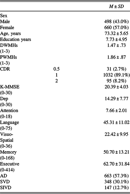
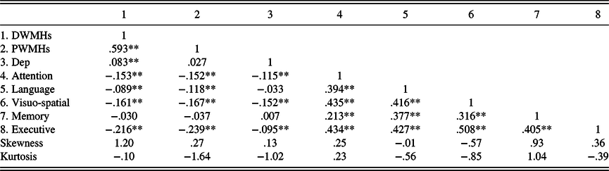
The demographic and clinical characteristics of all participants are displayed in Table 1. When the absolute values of the skewness and kurtosis do not exceed 2 and 7, respectively, the assumption of normality is satisfied (Finch, West, & MacKinnon, Reference Finch, West and MacKinnon1997). As shown in Table 2, the variables used in this study met the assumption of normality. Because the rating of WMHs uses an ordinal scale, the Spearman correlation coefficient was calculated (Table 2).
Demographic and clinical characteristics of participants (N = 1158)

AD = Alzheimer’s Disease; SVD = small vessel disease; SIVD = subcortical ischemic vascular disease; DWMHs = deep white matter hyperintensities; PWMHs = periventricular white matter hyperintensities; CDR = Clinical Dementia Rating; K-MMSE = the Korean version of the Mini-Mental Status Examination; Dep = the severity of depressive symptoms. The range of the minimum and maximum values is given at the bottom of the test.
Spearman correlation between measurement variables (N = 1158)

DWMHs = deep white matter hyperintensities; PWMHs = periventricular white matter hyperintensities; Dep = the severity of depressive symptoms.
*p < .05, **p < .01, ***p < .001.
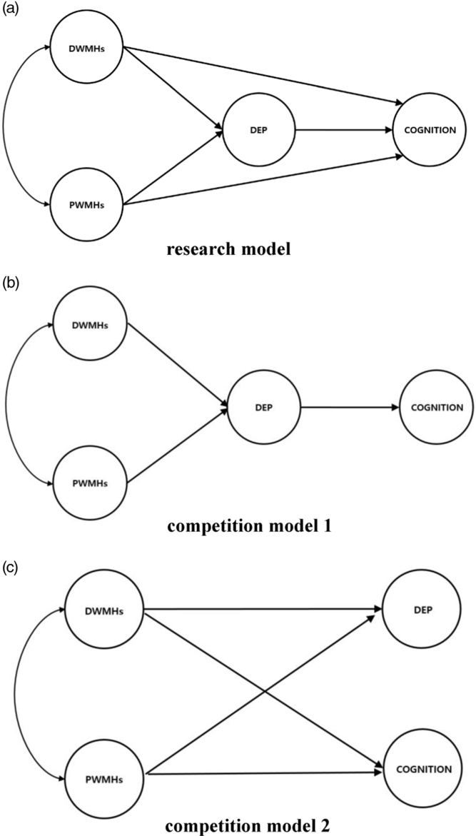
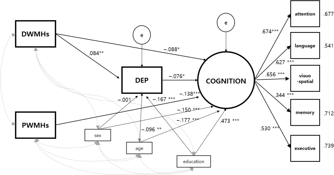
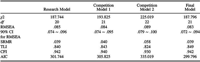
Data for 1158 participants were analyzed by Structural Equation Analysis. The fit of the research model and the competition models were evaluated through SEM, controlling for age, sex, and education years. First, we set up the research model with both direct and indirect paths between the two types of WMHs and cognitive function (Figure 1). Next, we set up a competition model 1 that excludes direct paths from the research model. Finally, we set up a competition model 2 that excludes the path between depression and cognitive function. The fitness of each model is shown in Table 3. The research model included both direct effects of WMHs on potential common pathology, depression symptoms and cognitive impairment, and the indirect effect of WMHs via depressive symptoms. Competition model 1 assumed that WMHs will only indirectly affect cognitive impairment and was far from a common pathology model. Competition model 2 focused on shared vulnerabilities, excluding the indirect effect of WMHs. The three models were in a nested relationship with each other, so we conducted a chi-square difference test. As a result, the competition model 1 is the better fit than competition model 2[Δχ2 (1) = 31.194, p < .001]. When comparing competition model 1 to the research model, the research model showed a better fit [Δχ2 (1) = 6.081, p = .014]. In spite of that, the direct path from PWMHs to depressive symptoms was not significant. After removing this path, then we obtained the final model as the best fitting model (see Figure 2). The final model showed an acceptable fit to the data [χ2 (21) = 187.796; RMSEA = .083 (90% confidence interval: .072–.094); SRMR = .039; TLI = .849; CFI = .942] (Figure 2).
A research model and the competition models on the relationship of WMHs, depressive symptoms and cognitive function.

The Mediation model of depressive symptoms on the relationship between WMHs and cognitive function.

Statistics of fit in each model

RMSEA = Root-Mean-Square Error of Approximation; 90% CI for RMSEA = confidence interval for the RMSEA; SRMR = Standardized Root Mean square Residual; CFI = Comparative Fit Index; TLI = Tucker–Lewis Index; AIC = Akaike Information Criterion.
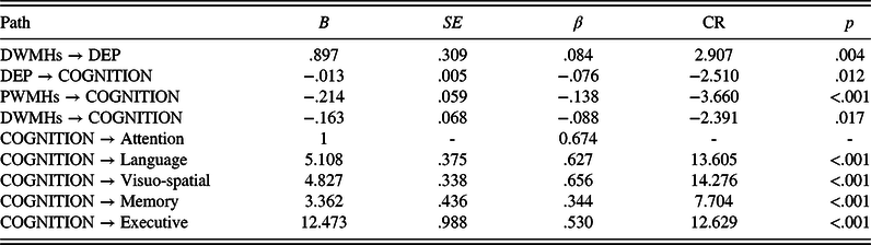
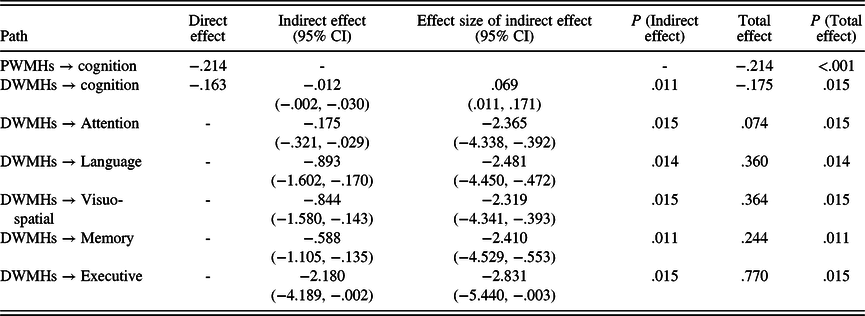
Table 4 shows the non-standardized and standardized coefficient estimates and the standard error for each direct path in the final model. The latent factor, cognition, was well represented by each indicator (subcognition in each domain) and the factor loadings all being significant (p < .001). This model includes a significant direct effect from DWMHs to depressive symptoms (b = .897, p = .004), from depressive symptoms to cognition (b = −.013, p = .012), from PWMHs to cognition (b = −.214, p < .001), and from DWMHs to cognition (b = −.163, p = .017). Table 5 shows the overall effects of PWMHs and DWMHs. The indirect effect of DWMHs to cognition, which affects cognitive function via depressive symptoms, was significant (b = −.012, p = .011, 95% confidence interval: −.002 to −.030). An effect size of the indirect effect, which represents the ratio of indirect effect to total effect, was .069 (95% confidence interval: .011–.171). The total effect of DWMHs was significant (b = −.175, p = .015) and that of PWMHs was also significant (b = −.214, p < .001). The indirect effect of DWMHs on cognitive function in the attention domain, language domain, visuo-spatial domain, memory domain, and executive domain was all significant (respectively, b = −.214, p < .001; b = −.214, p < .001; b = −.214, p < .001; b = −.214, p < .001; b = −.214, p < .001). Finally, the squared multiple correlation (SMC) of cognition was .444, indicating that this model accounts for 44.4% of the variance in cognition. Moreover, effect size was very large at an f squared value of .798 as estimated from the SMC.
Path coefficients of the direct path (N = 1158)

DWMHs = deep white matter hyperintensities; PWMHs = periventricular white matter hyperintensities; Dep = the severity of depressive symptoms; b = non-standardized coefficient estimates; SE = standard error; β = standardized coefficient estimates; CR = critical ratio.
Overall effects of PWMHs and DWMHs for cognition (N = 1,158)

All coefficients are non-standardized coefficient estimates. The PWMHs are not shown in the table because they do not have an indirect effect on the cognitive function in each domain. DWMHs = deep white matter hyperintensities; PWMHs = Periventricular white matter hyperintensities; CI = confidence interval. The effect size of indirect effect represents the ratio of indirect effect to total effect.
DISCUSSION
The purpose of this study was to model the relationship among the two types of WMHs, depressive symptoms, and cognitive impairment in terms of mediating models and to identify mediating effects of depression. Using SEM, we arrived at a model that included only direct pathways between PWMHs and cognitive function and both direct and indirect pathways (via depressive symptoms) between DWMHs and cognitive function, which most closely reflected the relationship among the variables.
This study was not limited to only examining the relationship between any two variables (e.g., WMHs-depression, or WMHs-cognition) as was the case in previous studies. Instead, our study is the first exploratory attempt, to the best of our knowledge, to construct a model that most closely reflects the relationships among all three variables, taking into account their interactions and the role of depression as a mediating variable. The modeling of the relationship between variables was performed by exploring several models derived from the theoretical basis. Specifically, the research model and the two competitive models were investigated and a final model, with a slight modification of the research model, was obtained (Figure 1). Two competitive models emphasized either direct or indirect effect on cognitive impairment of WMHs (or direct effect), whereas the final model that best reflected the relationships among the variables was the model in which DWMHs affected cognitive function directly and indirectly through the depressive symptoms. On the other hand, PWMHs only directly affected cognitive function. The most important feature of the supported model in the present study is that the depressive symptoms mediated the developmental pathway from DWMHs to cognitive impairment, which suggests that people with pathology of deep white matter lesions are more likely to experience depressive symptoms at some time point in the process leading to cognitive impairment. In addition, previous studies have proposed that depressive symptoms should be regarded as a prodromal symptom to the onset of dementia (Almeida, Hankey, Yeap, Golledge, & Flicker, Reference Almeida, Hankey, Yeap, Golledge and Flicker2016; Snowden et al., Reference Snowden, Atkins, Steinman, Bell, Bryant, Copeland and Fitzpatrick2015). However, it is still unknown why depression is a prodrome to dementia. On the other hand, in this study, DWMHs, the causative pathogenesis of cognitive impairment, was found to be mediated by depressive symptoms, providing a rational basis for understanding why depressive symptoms appear before the onset of dementia. In patients with depression, the hippocampus may be vulnerable due to hypothalamic-pituitary-adrenal (HPA) axis hyperactivity (Checkley, Reference Checkley1996) and this suggests that depression may be a factor that precedes cognitive impairment, especially memory impairment (Brunet et al., Reference Brunet, Hudon, Macoir, Belleville, Rousseau, Bouchard, Verret, Chertkow, Chayer, Kergoat and Joubert2011; Johnson et al., Reference Johnson, Mauer, Jahn, Song, Wyshywaniuk, Hall, Balldin and O’Bryant2013). On the other hand, the results of this study differ from those of previous studies that emphasize the direct effect of depression on cognitive function (Jorm, Reference Jorm2001; Ownby, Crocco, Acevedo, John, & Loewenstein, Reference Ownby, Crocco, Acevedo, John and Loewenstein2006), assuming a pathway from WMHs to cognitive function and that the pathway is mediated by depressive symptoms. Thus, this study contributes to expanding the understanding of developmental pathology in dementia beyond the results of previous studies. In addition, this study can be applicable to dementia patients with vascular factors because it was performed on a relatively large population of patients, such as those with AD and SVD rather than patients with a narrow range of conditions, such as only AD.
While previous studies have shown that the magnitude of the effects of the two types of WMHs on cognitive impairment is different, no studies have so far investigated whether this difference is associated with the mediation of cognitive impairment by depressive symptoms. However, this study first found that only DWMHs affected cognitive function through mediation of depressive symptoms. Meanwhile, the current study found that DWMHs significantly contributed to cognitive impairment, which stands in contrast to the results of previous studies that reported that DWMHs are not related to cognitive impairment (De Groot et al., Reference De Groot, De Leeuw, Oudkerk, Van Gijn, Hofman, Jolles and Breteler2002; Park et al., Reference Park, Lee, Na, Kim, Cheong, Moon, Shim, Park, Ku, Choi, Joo, Lee, Go, Kim, Kim, Cha, Lee and Seo2011). While previous studies have examined only the direct effects between DWMHs and cognitive function, the current study examined an indirect effect mediated by depressive symptoms in addition to the direct effect. The previous studies (De Groot et al., Reference De Groot, De Leeuw, Oudkerk, Van Gijn, Hofman, Jolles and Breteler2002; Park et al., Reference Park, Lee, Na, Kim, Cheong, Moon, Shim, Park, Ku, Choi, Joo, Lee, Go, Kim, Kim, Cha, Lee and Seo2011) measured only global cognitive function with the Mini Mental State Examination and used the measurement variable without the process of correcting for the measurement error. On the other hand, in this study, not only the cognitive function scores were measured for each domain but also the latent variables were used for the analysis after correcting the measurement error using such measurement variables. Therefore, it is possible that the differences in the results may have been caused by differences in the measurement tools and statistical analysis methods. Although both types of WMHs are included in vascular diseases, they have different patterns in relation to depression and cognitive function. Periventricular white matter lesions are predominantly affected by chronic hemodynamic insufficiency including hypotension, hypoperfusion, and atrophy (Wen & Sachdev, Reference Wen and Sachdev2004; Wen, Sachdev, Li, Chen, & Anstey, Reference Wen, Sachdev, Li, Chen and Anstey2009), while deep white matter lesion volumes are predominantly affected by ischemic SVD (Kim et al., Reference Kim, MacFall and Payne2008), which is a risk factor for depression (Brookes et al., Reference Brookes, Herbert, Lawrence, Morris and Markus2014). Considering these points, the differential relevance of DWMHs to depressive symptoms could be attributable to the relationship with SVD. On the other hand, as cholinergic fiber tracts, which have a significant effect on cognitive function, pass near the ventricles, PWMHs may be correlated with damage to cholinergic fiber pathways (Selden, Gitelman, Salamon-Murayama, Parrish, & Mesulam, Reference Selden, Gitelman, Salamon-Murayama, Parrish and Mesulam1998). It is difficult to consider that cognitive function is controlled by a specific region of the brain but the strong association between cognitive function and PWMHs presented in this study is plausible considering the role of the cholinergic system in maintaining cognitive function in patients. These could explain the differential consequences of the two types of lesions. However, further research that focused on specific pathways and regions is needed in order to delineate the effects of specific WMHs. These results suggest that clinicians should consider the possibility of depression leading to cognitive dysfunction rather than limiting it to an issue that is merely an emotional distress for patients with depressive symptoms and DWMHs. We also investigated the relationship between specific cognitive domains and DWMHs, where the total effect of DWMHs and the indirect effects via depressive symptoms were the largest for executive function. Considering that DWMHs is a representative of vascular disease, the results of this study are consistent with findings related to vascular depression (Krishnan et al., Reference Krishnan, O’Brien, Firbank, Pantoni, Carlucci, Erkinjuntti, Wallin, Wahlund, Scheltens, Elisabeth and Inzitari2006). On the other hand, our modeling results showed that, unlike DWMHs, PWMHs do not have an indirect effect on the global cognitive latent variable via depressive symptoms, so the cognitive variable of specific domains is also deviated from the influence of this indirect effect. The results of this study suggest that DWMHs may have a stronger association with vascular depression than PWMHs.
This study has several limitations. First, although the model was built by established empirical studies supporting each pathway, the interpretation of the results may be somewhat limited due to the cross-sectional design. Although the model is used in terms of model design, it is composed of cross-sectional samples. The interpretation of the results of this study as implying causality must be cautious, and a longitudinal study is needed to confirm causality. Second, the model proposed in this study did not include impairments in activities of daily living (ADL), another core feature of dementia. Research that verifies the fit of the model including cognition and ADL impairments would likely produce a more accurate model of the process from WMHs to dementia development through depressive symptoms. Third, it is necessary to consider that the large sample size contributed to the significance of the present results. Large sample sizes are likely to overestimate the effect rather than the actual effect. In order to compensate for this, we presented both the significance and the effect size. Additionally, it is also necessary to consider that large sample sizes have the advantage of improving statistical power and providing reliable and repeatable results. Fourth, another limitation of this study is that an investigation on the history of depression was not performed. Current research suggests that depressive symptoms are derived from WMHs and cause cognitive impairment. Considering that late-onset depression is more likely to be associated with dementia than early-onset depression (Hashem, A Gomaa, & Khalaf, Reference Hashem, A Gomaa and Khalaf2017; Koenig, Bhalla, & Butters, Reference Koenig, Bhalla and Butters2014), the depressive symptoms included in our model may have a stronger association with late-onset depression than early-onset depression. Therefore, if information on early onset and late onset can be obtained using the data on the patient’s history of depression, it is expected that the validity of the same mediating model supported by this study will be enhanced. A final limitation is that this study assessed the severity of depression on a continuous basis and did not perform MDD diagnosis. Further research is therefore needed to determine whether this conclusion is applicable to patients with clinical depression.
CONCLUSION
This study presented a mediation model that included important information on the pathological pathways among two types of WMHs, depressive symptoms, and cognitive impairment beyond the framework of previous research. In addition, our model supports the possibility that both types of WMHs are involved in different pathways that affect cognitive impairment.
ACKNOWLEDGEMENTS
This research was supported by the Hallym University Research Fund 2017(HURF-2017-37) and by a National Research Foundation of Korea grant funded by the Korea government (MSIP) (No. 2017R1A2B4008920).
CONFLICT OF INTEREST
The authors report no conflict of interest.
SOURCE OF FUNDING
This research was supported by the Hallym University Research Fund 2017(HURF-2017-37) and by a National Research Foundation of Korea grant funded by the Korea government (MSIP) (No. 2017R1A2B4008920).