Introduction
For optimal efficiency in vitro embryo production (IVEP), it is essential to understand the factors that interfere with its stages, which include oocyte maturation, oocyte fertilization, and in vitro culture (Egashira et al., Reference Egashira, Ihara, Khatun, Wada, Konno, Tatemoto and Yamanaka2019). Factors influencing the in vitro maturation (IVM) stage include high oxygen tension, light interference, temperature variations, and the absence of antioxidants naturally present in follicular fluid (El-Sanea et al., Reference El-Sanea, Abdoon, Kandil, El-Toukhy, El-Maaty and Ahmed2021). Consequently, the production of reactive oxygen species (ROS) and subsequent oxidative stress are associated with the low efficiency of oocyte maturation and embryonic development in vitro. Controlling ROS production is, therefore, critical (Khazaei and Aghaz, Reference Khazaei and Aghaz2017).
Despite the established IVEP technique, the quality of embryos produced in vitro remains inferior to those produced in vivo, with production rates from standard protocols not exceeding 40%. This necessitates the exploration of additional tools to enhance results and improve the survival potential of in vitro-produced embryos to levels comparable to in vivo-produced ones (Viana et al., Reference Viana, Siqueira, Palhao and Camargo2012).
Resveratrol is associated with a reduction in ROS production, either directly or by modulating signalling pathways that produce these free radicals (Kim et al., Reference Kim, Park and Woo2019). However, there is still limited knowledge about the influence of resveratrol on signalling pathways related to free radical production. A study in human medicine indicated that this antioxidant downregulated the increase in hypoxia-inducible factor 1α (HIF-1α) levels and the activated phosphorylation of p38 mitogen-activated protein kinase (MAPK) and c-Jun N-terminal kinase (JNK) in interleukin 1 beta (IL-1β)-stimulated rat synovial (RSC-364) cells, thereby reducing free radical production (Yang et al., Reference Yang, Chang, Yang, Yuan, Xu, Ho and Li2018). Additionally, literature reports link resveratrol with enzymes such as sirtuin 1 (SIRT1) and 5 (SIRT5), peroxisome proliferator-activated receptor-gamma coactivator 1 alpha (PGC-1α), nuclear respiratory factor 1 (NRF1) and 2 (NRF2), enhancing the levels of genes associated with antioxidants and ATP production. Resveratrol is also considered a chemical activator for mitochondrial biogenesis. Through these functions, resveratrol improves oocyte and embryonic quality during IVEP, related to decreased ROS production, and increased glutathione (GSH) and SIRT1 (Iwata, Reference Iwata2021).
Nanoparticles have diverse applications in health and other fields, including bioseparation, adsorption, catalysis, controlled drug release, chemical markers, hormones, peptides, dyes, and cosmetics (Shen et al., Reference Shen, Yang, Li, Zhou, Zhang, Li, Chen, Wang, Zhang and Zhao2014). Silica nanoparticles have gained prominence over the last decade as drug carriers (Vazquez et al., Reference Vazquez, Gonzalez, Ferrari and Castro2017).
The objective of this study was to evaluate the effects of different concentrations of resveratrol (0.5 μM and 1 μM) and its conjugation with silica nanoparticles (0.5 μM and 1 μM) on bovine oocytes maturation and embryo production. Parameters assessed included cumulus oophorus and oocyte size, ROS production, oocyte nuclear maturation, cleavage rate, blastocyst formation, and embryonic cell apoptosis.
Material and methods
Places and animals
Ovaries were collected from cows post-slaughter at Frigorífico Luciana, located in Uberlândia, Minas Gerais, Brazil. The transport of the ovaries was conducted in a thermos container maintained at a temperature between 34 °C and 38 °C, ensuring delivery within a maximum period of two hours. Upon collection, the ovaries were heated with a 0.9% saline solution to maintain the appropriate temperature. Subsequently, they were transported to the Animal Reproduction Laboratory at the Federal University of Uberlândia for further processing.
Silica nanoparticles and resveratrol loading
The silica nanoparticles were synthesized according to the methodology described by Arantes et al. (Reference Arantes, Pinto, Leite, Longo and Camargo2012). The synthesis process utilized tetraethyl orthosilicate (TEOS, Sigma-Aldrich, 131903) in an alcoholic medium, with particle size controlled by varying the initial molar amounts of TEOS, ammonia, and water via a 23 factorial design. The synthesis involved dissolving TEOS in ethanol, adding an ammonia solution, and optionally adding water. This mixture was then stirred for 24 h, followed by centrifugation and drying. The resulting nanoparticles had an average size of approximately 20 nm and a spherical shape, as confirmed by scanning electron microscopy.
Resveratrol (Sigma-Aldrich, R5010) was loaded onto the silica nanoparticles following the method outlined by Summerlin et al. (Reference Summerlin, Qu, Pujara, Sheng, Jambhrunkar, McGuckin and Popat2016). This process involved adding resveratrol to a TEOS ethanol solution, dispersing it ultrasonically, and stirring the mixture. Hydrofluoric acid was then added to induce gelation, followed by a period of aging and solvent replacement with deionized water. The mixture was subsequently freeze-dried, effectively trapping the resveratrol molecules within the aerogel network during the gelation process.
Collection and selection of oocytes
Upon arrival at the laboratory, the temperature of the ovaries was maintained between 36 °C and 37 °C. The ovaries were washed three times in sterile 0.9% NaCl solution. Ovarian follicles were then aspirated using a disposable 10 or 20 mL syringe with a 40 × 1.2 mm (21G) needle. The aspirated follicular fluid was stored in 15 ml Corning tubes in a water bath at 37 °C. After allowing 15 min for sedimentation, oocyte retrieval commenced. The pellet was transferred to a Petri dish (100 × 20 mm), and oocytes were selected under stereomicroscopes at 15× magnification according to the criteria of Leibfried and First (Reference Leibfried and First1979). Only oocytes classified as grades I and II were utilized in the experiment.
In vitro maturation
Grade I and Grade II oocytes were transferred to another 35 mm Petri dish, where they were washed once in TCM-199 HEPES medium base wash and then washed twice with maturation medium. The maturation medium consisted of TCM-199 medium supplemented with 1 mg/ml of BSA, 1 μM of pyruvate, 0.5 μg/ml of follicle-stimulating hormone (FSH, Folltropin-V, Bioniche, Ontario, Canada), 5 μg/ml luteinizing hormone (LH), 1 μg/ml 17β-oestradiol, and 100 μM cysteamine. After washing, the oocytes were transferred to 100 μl drops of maturation medium under mineral oil and incubated at 38.5 °C in a humid atmosphere containing 5% CO2 for 22 to 24 h.
Grade I and II oocytes were randomly distributed into one of five treatments: control (0 μM resveratrol; n = 159), resveratrol 0.5 (0.5 μM resveratrol; n = 158), resveratrol 1.0 (1 μM resveratrol; n = 155), nanoparticles 0.5 (nanoparticles conjugated with 0.5 μM resveratrol; n = 159), and nanoparticles 1.0 (nanoparticles conjugated with 1 μM resveratrol; n = 158). Seventeen replicates were performed during the experiment, with a total of 789 oocytes.
Evaluation of reactive oxygen species and oocyte maturation
After maturation, the oocytes were mechanically denuded to remove cumulus cells, followed by three washes in phosphate-buffered saline (PBS) containing 0.4% BSA. Subsequently, 26 oocytes from each treatment group were incubated in the absence of light in a medium containing 10 μM of 2′,7′-dichlorodihydrofluorescein diacetate (DCF) for 30 min at 38.5 °C under 5% CO2 to mark ROS production. After labelling with DCF, the oocytes were washed three times in PBS and fixed in 4% paraformaldehyde for 15 min at 36 °C. Post-fixation, they were washed again in PBS drops and placed on a microscope slide with one drop of FluoroShield mounting medium containing 4′,6-diamidino-2-phenylindole (DAPI, Sigma-Aldrich, F6057).
The measurement of ROS was performed using LSM 510 Meta Confocal microscopy with DCF labelling. DCF is a highly fluorescent compound that, when in contact with ROS, generates a green fluorescence on the oocytes. This fluorescence was quantified by reading the slides with oocytes stained with DCF using confocal microscopy.
Oocyte maturation was evaluated in 19 oocytes from each treatment group using DAPI staining and examined with the epifluorescence microscope EVOs® FL. DAPI is a fluorescent marker that strongly binds to adenine-thymine-rich regions (ATR), labelling the germinal vesicle (presence of a spherical nucleus surrounded by a nuclear membrane and decondensed chromosomes), metaphase plate, and polar body. In this study, oocytes were considered immature if they exhibited the germinal vesicle phase or metaphase I (highly condensed chromosomes and metaphase plate formation). Mature oocytes were those in metaphase II (presence of a metaphase plate and the release of the first polar body).
In vitro fertilization and cultivation
To carry out in vitro fertilization, TALP-SPERM, TALP-FERT, and Percoll® gradient media were prepared. After maturation, the oocytes were washed in two drops of TALP-FERT medium or IVF medium containing amikacin (83 mg/ml), pyruvate solution (0.11 mg/ml), PHE (2 μM penicillamine, 1 μM hypotaurine, 1 μM epinephrine), heparin (10 μg/ml), and bovine serum albumin (BSA) essential fatty acids (6 mg/ml). The oocytes were then transferred to the IVF plate in 100 μl drops of the same TALP-FERT medium, maintaining the same distribution as in the IVM process. The fertilization plate was placed in the incubator until sperm capacitation was completed.
Fertilization was performed using semen from the same bull for all replicates. The semen straws were thawed, and the spermatozoa underwent selection and qualification using the Comcoll® discontinuous centrifugation method, adapted from Parrish et al. (Reference Parrish, Krogenaes and Susko-Parrish1995). The gradient was prepared by placing 500 μl of TALP-SPERM medium and 500 μl of 90% Percoll® solution in a 15 ml tube, forming a 45% Percoll® layer. Below this layer, 1 ml of 90% Percoll® solution was added, creating a total of 2 ml of gradients. The semen was then deposited on the gradient and centrifuged at 2400 rpm for 30 min.
Sperm motility, vigour, and concentration were evaluated using a Neubauer chamber under a light microscope. Sperm counts were performed to determine the volume of TALP-SPERM medium to be added to the IVF tube to adjust the sperm concentration to 100,000 sperm per 100 μl drop. Subsequently, the volume of TALP-FERT medium to be added to the semen was calculated to achieve a concentration of 25 × 106 spermatozoa/ml, with an insemination dose of 4 μl per drop. The day of IVF was considered day 0 (D0) of embryonic development. Fertilization was carried out on the IVF plate in drops containing TALP-FERT medium, and the COCs (Cumulus-Oocyte Complexes) along with the spermatozoa were incubated for 12 to 18 h under the same conditions as maturation.
For the in vitro culture (CIV) step, the potential zygotes were denuded and washed three times in Synthetic Oviduct Fluid (SOF) medium supplemented with pyruvate solution (0.11 mg/ml), amikacin (83 mg/ml), fatty acid-free BSA with low endotoxin (6 mg/ml), and fetal bovine serum (2.5%). They were then transferred to the CIV plate, maintaining the same drop/treatment separation, with 10 to 15 zygotes per 100 μl drop of SOF medium. The zygotes were incubated at 38.5 °C in a humid atmosphere containing 5% CO2 for seven days. Forty-eight hours after fertilization, the number of cleaved structures (cleavage rate) was counted. Seven days later, the number of blastocysts (blastocyst rate) was evaluated.
Assessment of plasma membrane integrity
To assess the integrity of the plasma membrane, 23 embryos from each treatment group at D7 were washed three times in PBS with fetal bovine serum. They were then stained with propidium iodide (PI) for 15 min and subsequently fixed in paraformaldehyde for another 15 min. The embryos were evaluated using LSM 510 Meta Confocal microscopy with PI staining, which generates a red fluorescent label. Propidium iodide is responsible for staining the DNA in cells that are dead or have compromised membranes.
Statistical analysis
Statistical analysis was conducted using SigmaPlot® version 11.0.1 (Systat Software Inc., USA). To measure the oocytes and cumulus cells, the Image J program was utilized. Cumulus oophorus size and oocyte size were considered dependent variables and assessed with ANOVA using a generalized linear mixed model, fitted with the GLIMMIX procedure of SAS. This model employed a Gaussian distribution and an identity link function, with replication as the random effect and treatment (control, resveratrol 0.5 μM, resveratrol 1.0 μM, nanoparticles 0.5 μM, nanoparticles 1.0 μM) as the independent variable.
The effects of treatment on germinal vesicle, metaphase I, and metaphase II were analysed using the chi-square or Fisher’s exact test. Comparison of means for variables such as ROS production, PI, cleavage rate, and blastocyst rate was performed using Fisher’s LSD test. Data were presented as percentages and mean values (± standard error of the mean). All statistical analyses were conducted using SAS Studio (SAS Institute Inc., Cary, NC).
Distributions and normality tests were performed using the univariate procedure, with normality assessed via the Kolmogorov-Smirnov method. Statistical significance was defined as P ≤ 0.05 for the main effects and interactions.
Results
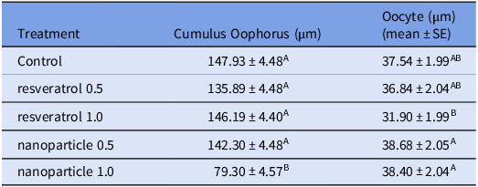
The nanoparticle 1.0 treatment significantly reduced the size of cumulus oophorus (P < 0.05) compared with all other treatments, which did not differ significantly among themselves (P > 0.05). The oocyte diameter for the resveratrol 1.0 treatment was significantly smaller (P < 0.05) compared with the other treatments. The nanoparticle 0.5 and 1.0 treatments resulted in the largest oocyte diameters, which were statistically higher than the resveratrol 1.0 μM treatment (P < 0.05) and comparable to the control (P > 0.05; Table 1).
Number of measurement (mean ± standard error of the mean) of cumulus oophorus and oocyte measurement according to treatment using resveratrol (0.5 and 1.0 μM) and nanoparticle conjugated with resveratrol (0.5 and 1.0 μM)

A,B Means followed by different letters in the columns differ statistically (P < 0.05).
SE = Standard Error.
Control = no treatment.
resveratrol 0.5 = resveratrol 0.5 μM.
resveratrol 1.0 = resveratrol 1 μM.
nanoparticle 0.5 = silica nanoparticles + resveratrol 0.5 μM.
nanoparticle 1.0 = silica nanoparticles + resveratrol 1 μM.
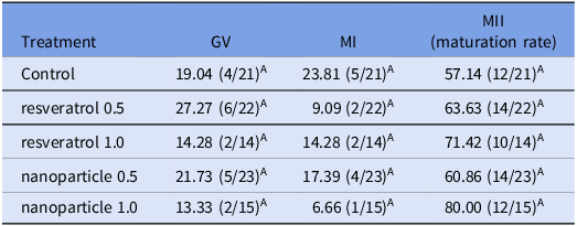
There was no significant difference in the percentage of oocytes in the germinal vesicle (GV) phase among the treatments (P > 0.05). Similarly, no significant differences were observed among the treatments in the metaphase I (MI) phase (P > 0.05). Despite the treatments with higher concentrations of resveratrol (resveratrol 1.0 and nanoparticle 1.0) resulting in a higher maturation rate, this increase was not statistically different from the other treatments (P > 0.05; Table 2).
Percentage (mean ± standard error of the mean) of oocytes in the germinal vesicle (GV), metaphase I (MI), metaphase II (MII) phases according to the treatments after the maturation process

A,B Means followed by different letters in the columns differ statistically (P < 0.05).
Control = no treatment.
resveratrol 0.5 = resveratrol 0.5 μM.
resveratrol 1.0 = resveratrol 1 μM.
nanoparticle 0.5 = silica nanoparticles + resveratrol 0.5 μM.
nanoparticle 1.0 = silica nanoparticles + resveratrol 1 μM.
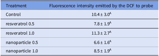
The fluorescence intensity emitted by DCF did not show statistically significant differences (P > 0.05) among the control, resveratrol, and nanoparticle treatments. These results suggest that the oxidative stress levels, as indicated by DCF fluorescence, were similar across all treatments after the oocyte maturation process (Table 3).
Fluorescence intensity emitted by the DCF oocytes to probe (mean ± standard error of the mean) after the maturation process according to the treatments

A,B Means followed by different letters in the columns differ statistically (P < 0.05).
Control = no treatment.
resveratrol 0.5 = resveratrol 0.5 μM.
resveratrol 1.0 = resveratrol 1 μM.
nanoparticle 0.5 = silica nanoparticles + resveratrol 0.5 μM.
nanoparticle 1.0 = silica nanoparticles + resveratrol 1 μM.
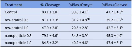
The resveratrol 1.0 treatment showed a significantly reduced cleavage rate compared with the other groups (P < 0.05). The control, resveratrol 0.5, nanoparticle 0.5, and nanoparticle 1.0 treatments had similar cleavage stages (P > 0.05; Table 4).
Proportion (mean ± standard error of the mean) of cleavage and blastocyst production (Blas.) in oocyte maturation treatments using resveratrol (0.5 and 1.0 μM) and nanoparticle conjugated with resveratrol (0.5 and 1.0 μM)

A,B Means followed by different letters in the columns differ statistically (P < 0.05).
Blas. = blastocyst.
Control = no treatment.
resveratrol 0.5 = resveratrol 0.5 μM.
resveratrol 1.0 = resveratrol 1 μM.
nanoparticle 0.5 = silica nanoparticles + resveratrol 0.5 μM.
nanoparticle 1.0 = silica nanoparticles + resveratrol 1 μM.
Blastocyst rates were similar among the control, resveratrol 0.5, nanoparticle 0.5, and nanoparticle 1.0 treatments (P > 0.05). However, there was a significant reduction in the blastocyst rate in the resveratrol 1.0 treatment (P < 0.05). The percentage of blastocyst production relative to the number of cleaved embryos was consistent across treatments (Table 4).
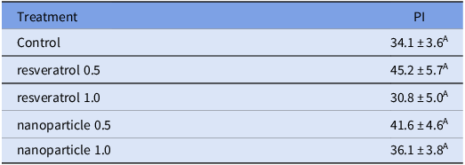
The data indicate that the cell degeneration rates, as measured by PI fluorescence emission, did not differ significantly (P > 0.05) among the control, resveratrol, and nanoparticle treatments. These results suggest that the various maturation treatments did not have a statistically significant impact on the cell degeneration rate of the embryos produced from the treated oocytes (Table 5).
Cell degeneration rate (mean ± standard error of the mean) measured by the fluorescence emission of PI emitted by the embryos produced from the oocytes submitted to the different maturation treatments

A,B Means followed by different letters in the columns differ statistically (P < 0.05).
Control = no treatment.
resveratrol 0.5 = resveratrol 0.5 μM.
resveratrol 1.0 = resveratrol 1 μM.
nanoparticle 0.5 = silica nanoparticles + resveratrol 0.5 μM.
nanoparticle 1.0 = silica nanoparticles + resveratrol 1 μM.
Discussion
The present study shows that the size of the cumulus oophorus is directly linked to the use of resveratrol and, consequently, the cleavage stage rate. High concentrations of resveratrol, such as 1 μM, can lead to a decrease in nuclear maturation rates by damaging cumulus-oocyte complexes (COCs). However, further studies are necessary to understand the mechanisms through which resveratrol affects cumulus cells. It is known that when COCs are co-cultured with oocytes, the nuclear maturation process improves (Broi et al., Reference Broi, Giorgi, Wang, Keefe, Albertini and Navarro2018; Virant-Klun et al., Reference Virant-Klun, Bauer, Ståhlberg, Kubista and Skutella2018). Oocytes release growth factors that influence cumulus cells, enhancing COC function and determining their phenotype, which is crucial for adequate oocyte capacitation (Turathum et al., Reference Turathum, Gao and Chian2021). Providing appropriate substrates for COCs during in vitro maturation (IVM) improves embryonic development potential (Boruszewska et al., Reference Boruszewska, Sinderewicz, Kowalczyk-Zieba, Grycmacher and Woclawek-Potocka2015). Moreover, oocyte cyclic adenosine monophosphate (cAMP) levels during IVM are related to increased communication between oocytes and cumulus cells via gap junctions (Luciano et al., Reference Luciano, Franciosi, Barros, Dieci and Lodde2018).
In this study, the resveratrol 1.0 treatment led to a decrease in COC size but was beneficial for oocyte growth (Table 1). Additional research is needed to explore the relationship between these findings. Other substances added to the oocyte maturation medium also have dose-dependent effects, potentially harming COCs at higher concentrations. For instance, Butafosfan is crucial for catalysing various cellular reactions and reducing metabolic stress but is harmful to cleavage rate and embryonic development at higher doses (Hax et al., Reference Hax, Rincón, Schneider, Pegoraro, Collares, Pereira, Pradieé, Del Pino and Corrêa2019).
The treatments did not affect the rate of nuclear maturation, indicating that the concentrations of mesoporous silica nanoparticles bound to resveratrol need to be adjusted to observe their impact on the maturation process. Piras et al. (Reference Piras, Ariu, Falchi, Zedda, Pau, Schianchi, Paramio and Bogliolo2020) found that resveratrol at different concentrations (1 μM, 2.5 μM, 5 μM) during cat oocyte nuclear maturation did not alter maturation percentages but efficiently reduced ROS levels and increased the rate and number of cells in blastocysts. Wang et al. (Reference Wang, Zhu, Liang, Xu, Liao, Lu and Lu2019) reported that 5 μM resveratrol during pig oocyte maturation increased polar body extrusion rates compared with control and 0.1 μM resveratrol. Similarly, Liu et al. (Reference Liu, Sun, Zhao, Liu, Ma, Li, Huai, Zhao and Liu2018) observed that 1 μM resveratrol increased polar body emission rates and maturation in oocytes. Additionally, resveratrol (2 μM) combined with melatonin during IVM enhances oocyte nuclear maturation (Lee et al., Reference Lee, Jin, Taweechaipaisankul, Kim and Lee2018).
Previous studies with antioxidants and nanoparticles reported reduced ROS production and cell degeneration in in vitro embryo production (Remião et al., Reference Remião, Lucas, Domingues, Silveira, Barther, Komninou, Basso, Jornada, Beck, Pohlmann, Varela Junior, Seixas, Campos, Guterres and Collares2016). Nanoparticles have been shown to cross protective barriers in developing oocytes (Hou and Zhu, Reference Hou and Zhu2017). In this study, ROS production, evaluated by DCF fluorescence intensity, was similar across all treatments after oocyte maturation. High ROS levels during oocyte maturation can compromise viability by altering oxidative phosphorylation and ATP synthesis, leading to cytogenetic changes (Russo et al., Reference Russo, Monaco, Rubessa, El-Bahrawy, El-Sayed, Martino, Beneult, Ciannarella, Dell’Aquila, Lacalandra and Uranio2014). Adequate antioxidant concentrations during IVM can reduce ROS production, improving oocyte and embryonic quality (Zabihi et al., Reference Zabihi, Shabankareh, Hajarian and Foroutanifar2019; Kwak et al., Reference Kwak, Cheong, Jeon, Lee, Choi, Jeung and Hyun2012).
Considering that ROS are naturally produced during cell metabolism and that oocytes lose antioxidant defences when removed from their natural environment, controlling ROS production during in vitro processes is crucial. Factors such as oxygen tension during IVM can also influence oxidative stress, decreasing intracellular Ca2+ and affecting oocyte activation and fertilization (Jiao et al., Reference Jiao, Cao, Cui, Lian, Miao, Wu, Han and Tan2013).
The integrity of the plasma membrane, evaluated by PI staining, showed no differences between treatments, contrasting with Wang et al. (Reference Wang, Zhu, Liang, Xu, Liao, Lu and Lu2019), who found that resveratrol during oocyte maturation decreased programmed cell death. Wu et al. (Reference Wu, Chen, Dong, Han, Zhu, Lui, Yang, Xie and Jin2023) also reported reduced cell degeneration and ROS production with resveratrol.
The resveratrol 1.0 treatment negatively affected cleavage and blastocyst/oocyte rates, but there was no difference in blastocyst percentages relative to cleaved embryos. This suggests that resveratrol’s harmful effects occur during oocyte maturation. Once oocytes mature and cleave, subsequent embryonic development is unaffected by resveratrol. Low concentrations of resveratrol during oocyte maturation are linked to higher cleavage and blastocyst formation rates (Lee et al., Reference Lee, Jin, Taweechaipaisankul, Kim and Lee2018). However, antioxidants like resveratrol have limitations such as low stability, solubility, light sensitivity, and dose-dependent effects (Shaito et al., Reference Shaito, Posadino, Younes, Hasan, Halabi, Alhababi, Al-Mohannadi, Abdel-Rahman, Eid, Nasrallah and Pintus2020).
The gradual release of resveratrol from nanoparticles may explain the absence of harmful effects on oocyte maturation at 1 μM concentration. The cleavage and blastocyst rates were similar between the control and resveratrol nanoparticles treatments, suggesting that resveratrol concentrations used were insufficient. Higher concentrations of resveratrol can have anti-proliferation and pro-apoptotic effects (Saunier, Reference Saunier, Antonio, Regazzetti, Auzeil, Laprévote, Shay, Coumoul, Barouki, Benelli, Huc and Bortoli2017; Jang et al., Reference Jang, Song, Lee, Moon and Moon2021). Resveratrol’s inhibition of various phosphodiesterases at specific concentrations can increase cytosolic cAMP and intracellular Ca2+ levels, delaying oocyte maturation (Park et al., Reference Park, Ahmad, Philp, Baar, Williams, Luo, Ke, Rehmann, Taussig, Brown, Kim, Beaven, Burgin, Manganiello and Chung2012).
Resveratrol supplementation during IVM regulates oocyte bioenergetic/redox status and cytoplasmic organelle distribution, affecting active mitochondria proportion and ATP concentration (Piras et al., Reference Piras, Menéndez-Blanco, Soto-Heras, Catalá, Izquierdo, Bogliolo and Paramio2019). The observed lack of significant effects on ROS emission, nuclear maturation, and embryonic cell apoptosis might be due to the concentrations of resveratrol used, which varied less compared with other studies with more distinct resveratrol concentrations.
Conclusion
In conclusion, while resveratrol shows promise in enhancing oocyte quality and embryonic development at lower concentrations, higher doses may impair these processes. The use of nanoparticles for antioxidant delivery represents a promising avenue, but precise dosing and delivery mechanisms require further investigation to maximize efficacy and safety in bovine oocyte maturation and embryo production.
Acknowledgment
This study was funded in part by the Coordination for the Improvement of Higher Education Personnel – Brazil (CAPES) – Funding Code 001. Author R.M. Santos received a scholarship from the National Council of Scientific and Technological Development (CNPq) (PQ – Research productivity, Process no. 306873/2022-2).
Competing interests
The author(s) declare none