Introduction
Macrolide antibiotics such as erythromycin, clarithromycin and roxithromycin are frequently used in the treatment of upper and lower respiratory tract infections. In recent years, there has been increasing interest in the immunomodulatory actions of macrolides, especially in chronic rhinosinusitis and nasal polyposis. Over the last decade, there have been numerous studies, conducted by Japanese and other investigators, on the efficacy of long-term, low-dose therapy with 14-membered ring macrolides for the treatment of chronic rhinosinusitis and nasal polyposis. These reports assessed different pharmacological actions, such as nasal polyp shrinkage,Reference Ichimura, Shimazaki, Ishibashi and Higo1, Reference Yamada, Fujieda, Mori, Yamamoto and Saito2 dispersion of bacterial biofilms,Reference Cervin and Wallwork3 suppression of mucin secretion,Reference Rhee, Majima, Arima, Jung, Jinn and Min4 and suppression of the production of the cytokines interleukin (IL) 8Reference Yamada, Fujieda, Mori, Yamamoto and Saito2, Reference Nonaka, Pawankar, Saji and Yagi5 and IL-1β.Reference Miyanohara, Ushikai, Matsune, Ueno, Katahira and Kurono6
Nasal polyposis is a chronic inflammatory disease of nasal and paranasal cavity mucosa. The condition is characterised by oedematous masses of hyperplastic epithelium and lamina propria prolapsing into the nose, leading to nasal obstruction, hypersecretion, loss of smell, headache and reduced quality of life.Reference Pawankar7
Histopathological studies of nasal polyps have demonstrated predominantly eosinophilic tissue infiltration. Increased expression and production of a variety of proinflammatory cytokines and chemokines have also been demonstrated in nasal polyp epithelium and lamina propria.Reference Bachert, Gevaert, Holtappels and Van Cauwenberge8 The mechanisms responsible for selective accumulation of eosinophils in polyps are unknown. Nasal polyp fibroblasts could play a role in the recruitment of eosinophils through the release of the ‘regulated on activation, normal T cell expressed and secreted’ chemokine (also known as RANTES and CCL5), and of other chemokines.Reference Nonaka, Pawankar, Saji and Yagi9
The ‘regulated on activation, normal T cell expressed and secreted’ chemokine is a member of the CC chemokine family. This heparin-binding polypeptide has been reported to be a selective chemotactic factor for eosinophils, memory T lymphocytes and monocytes.Reference Meyer, Bartels, Görögh, Sticherling, Rudack and Ross10 Studies of this chemokine have demonstrated that it also induces transendothelial migration of eosinophils and leads to activation of eosinophils, resulting in the release of cytotoxic agents such as superoxide and eosinophilic cationic protein.Reference Meyer, Bartels, Görögh, Sticherling, Rudack and Ross10, Reference Saji, Nonaka and Pawankar11 In addition to these toxic mediators, activated eosinophils are also capable of producing a variety of cytokines and chemokines, especially IL-4, IL-5, IL-6 and the ‘regulated on activation, normal T cell expressed and secreted’ chemokine.Reference Bachert, Gevaert, Holtappels and Van Cauwenberge8 This suggests that eosinophils appear to extend their own lifespan and to increase their tissue infiltration in an autocrine fashion.
Nasal secretions contain small amounts of cytokines and chemokines. As these inflammatory mediators play an important role in the pathophysiology of airway disease, the assessment of their concentrations in nasal fluid may help to elucidate both the mechanisms underlying nasal polyp formation and the immunomodulatory effects of macrolide treatment.
In this prospective study, we examined whether long-term, low-dose clarithromycin treatment affected the levels of the cytokines and chemokines which modulate eosinophil function. Specifically, we measured the concentrations of IL-4, IL-5, IL-6 and the ‘regulated on activation, normal T cell expressed and secreted’ chemokine in the nasal secretions of non-allergic and allergic patients with bilateral nasal polyps; we also measured the levels of eosinophilic cationic protein, a well-described and standardised marker of eosinophil activity.
This study aimed to assess whether there was any relationship between changes in the levels of these mediators and changes in nasal polyp size, following clarithromycin therapy.
Materials and methods
Patients
Forty patients with bilateral nasal polyps (22 non-allergic and 18 with co-morbid allergic rhinitis) were included in this prospective, non-placebo-controlled study.
The study was conducted according to the Declaration of Helsinki, and written, informed consent was obtained from all subjects. The study was approved by the ethics committee of the Military Medical Academy, Belgrade, Serbia.
All patients were examined, treated and followed up by the same rhinologist for the entire duration of the study.
The diagnosis of nasal polyposis was based on the documented medical history and on the results of clinical examination, nasal endoscopy (using a rigid optic 0° and 30° endoscope; Storz, Tuttlingen, Germany) and computed tomography (CT) of the paranasal sinuses, according to the current European Guidelines.Reference Fokkens, Lund and Mullol12
We excluded from the study any patients with lower airway obstruction symptoms, bronchial asthma, aspirin sensitivity, antrochoanal or sphenochoanal polyps, cystic fibrosis, or primary ciliary dyskinesia. None of the patients in this study had an acute respiratory tract infection, and none had received oral or topical corticosteroids, antibiotics or antihistamines in the three weeks preceding study enrolment. No patient had undergone nose or sinus surgery in the six months prior to study commencement.
Allergy determination
Skin-prick tests were performed on all patients to test for sensitivity to commonly inhaled allergens, i.e. birch, timothy, mugwort (Artemisia vulgaris), dog hair, cat hair, horse hair, mites (Dermatophagoides farinae and D pteronyssinus), moulds (Alternaria alternata, Aspergillus fumigatus and Cladosporium herbarum), Olea europaea, Parietaria judaica, Plantago lanceolata and Platanus acerifolia. Skin-prick tests were performed on the volar part of the forearm. A negative control (0.9 per cent natrium chloridum solution) and a positive control (1 mg/ml histamine dihydrochloride solution) were included in each skin-prick test. Reactions were read after 15 minutes. A test was considered positive if the diameter of the resultant weal was 3 mm greater that the diameter of the negative control.
Drug treatment
Patients received a single daily 500 mg oral dose of clarithromycin (a 14-membered ring macrolide antibiotic) for eight weeks. No concomitant medication was used during this time.
The exclusion criteria for long-term, low-dose clarithromycin treatment were: pregnancy, macrolide hypersensitivity, age less than 18 years, and liver or gastrointestinal dysfunction.
Sampling of nasal secretions and inflammatory mediator analysis
Nasal fluid samples were collected from the nasal cavities of all 40 patients (i.e. 22 non-allergic and 18 allergic patients, all with nasal polyposis) before and within seven days after clarithromycin treatment, using the modified absorption technique. This involved placing cotton-wool sticks (10 mm long, 4 mm wide; Institute of Virology, Vaccines and Sera, ‘Torlak’, Belgrade, Serbia) against the nasal cavity wall posterior to the mucocutaneous junction for 60 seconds, as previously described.Reference Bachert, Van Kempen and Van Cauwenberge13, Reference Perić, Vojvodić, Radulović, Vukomanović-Đurđević and Miljanović14 Because nasal secretions can be scanty in healthy subjects, control samples were not collected in this study.
All samples were placed in a 2 ml Eppendorf tube containing 1 ml of transfer medium (phosphate-buffered saline containing gentamycin 50 µg/ml, penicillin G 340 U/ml and fungizone 500 µg/ml) for 30 minutes to allow diffusion of mediators into the medium. Samples were then stored at 4°C for a maximum of 2 hours before processing. Nasal fluid was centrifuged at 1000 g for 10 minutes to separate the cellular components. After centrifugation, supernatants were portioned and stored at −70°C until mediator analysis.
The levels of the T helper 2 cytokines IL-4, IL-5 and IL-6 were measured using a commercial flow cytometric kit (Flow Cytomix; Bender MedSystems, San Diego, California, USA) and a flow cytofluorimeter (Beckman Coulter XL-MCL, Brea, California, USA), according to the manufacturers' instructions. Sensitivities were 20 pg/ml for IL-4 detection, 30 pg/ml for IL-5 detection and 21 pg/ml for IL-6 detection.
A quantitative sandwich immunoassay (R&D Systems, Minneapolis, Minnesota, USA) was performed to measure the concentration of the ‘regulated on activation, normal T cell expressed and secreted’ chemokine. The minimum measurable concentration was 5 pg/ml.
The concentration of the eosinophilic cationic protein was measured using fluorescent enzyme immunoassay (UniCAPTM100, Pharmacia Diagnostics AB, Uppsala, Sweden). The minimum detectable concentration was 2 µg/l.
Subjective symptom score
To investigate the effects of clarithromycin, the patients were asked to assess their nasal polyposis symptoms (i.e. obstruction, anosmia, sneezing, rhinorrhoea and itching) on the day of study enrolment and within seven days of clarithromycin treatment completion. They were instructed to score each of these five symptoms from zero to three, according to the method of Tsicopoulos et al., Reference Tsicopoulos, Shimbara, de Nadai, Aldewachi, Lamblin and Lassalle15 i.e. 0 for no symptoms, 1 for mild symptoms, 2 for moderate symptoms and 3 for severe symptoms. Thus, the maximal possible nasal symptom score was 15.
On the basis of their symptom scores, patients were categorised as clarithromycin treatment ‘responders’ and ‘non-responders’. Patients with a total score improvement of 1 or more after treatment were considered responders.
Nasal polyp size
Changes in polyp size were assessed using nasal endoscopy with a rigid endoscope (0° and 30°; Storz, Tuttlingen, Germany), with the patient in a sitting position. Topical anaesthesia and decongestion were not used.
The endoscopic physical findings were assessed before and within seven days after clarithromycin administration, using the scoring system of Lildholdt et al. Reference Lildholdt, Rundcrantz, Bende and Larsen16 Nasal polyp size was scored in relation to fixed anatomical landmarks, with 0 indicating no polyposis, 1 mild polyposis (i.e. small polyps not reaching the upper edge of the inferior turbinate), 2 moderate polyposis (i.e. medium-sized polyps reaching between the upper and lower edges of the inferior turbinate) and 3 severe polyposis (i.e. large polyps reaching below the lower edge of the inferior turbinate). The maximal endoscopic score was 6, bilaterally. Treatment results were divided into the following two categories: ‘improvement’ and ‘non-improvement’. Improvement was defined as observed shrinkage of nasal polyps giving a minimum score improvement of 2, bilaterally, after treatment.
Statistical analysis
Data are presented as means ± standard deviation. Wilcoxon's signed rank test was used to analyse differences in the levels of the T helper 2 cytokines, the ‘regulated on activation, normal T cell expressed and secreted’ chemokine and the eosinophilic cationic protein, as well as differences in pre- and post-treatment nasal symptom scores and nasal polyp size scores. Between-group comparisons were analysed using the non-parametric chi-square test. A p value of 0.05 or less was considered to be statistically significant. The Statistical Package for the Social Sciences software program was used for statistical analysis.
Results
The study group comprised six female and 16 male non-allergic patients with nasal polyps (mean age, 44.36 ± 14.15 years; age range, 25 to 72 years) together with eight female and 10 male allergic patients with nasal polyps (mean age, 46.22 ± 12.37 years; age range, 19 to 65 years).
In the non-allergic patients, the mean nasal symptom score improved from 12.32 ± 2.34 before clarithromycin treatment to 9.13 ± 4.09 after treatment (p < 0.05). In the allergic patients, the mean nasal symptom score decreased from 12.77 ± 2.76 before clarithromycin treatment to 10.16 ± 3.47 after treatment (p < 0.05). Following clarithromycin treatment, the proportion of responders was 67.83 per cent in the non-allergic group and 55.55 per cent in the allergic group; this difference was not statistically significant (chi-square test).
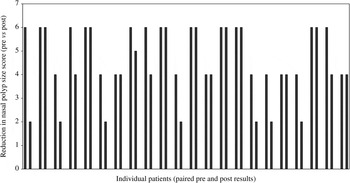
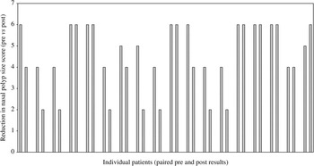
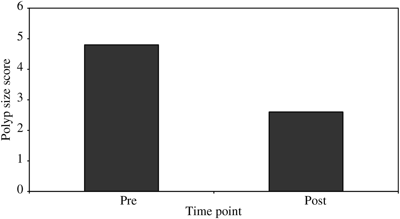
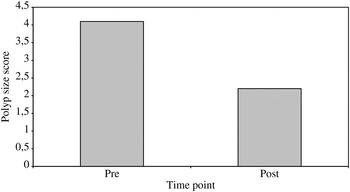
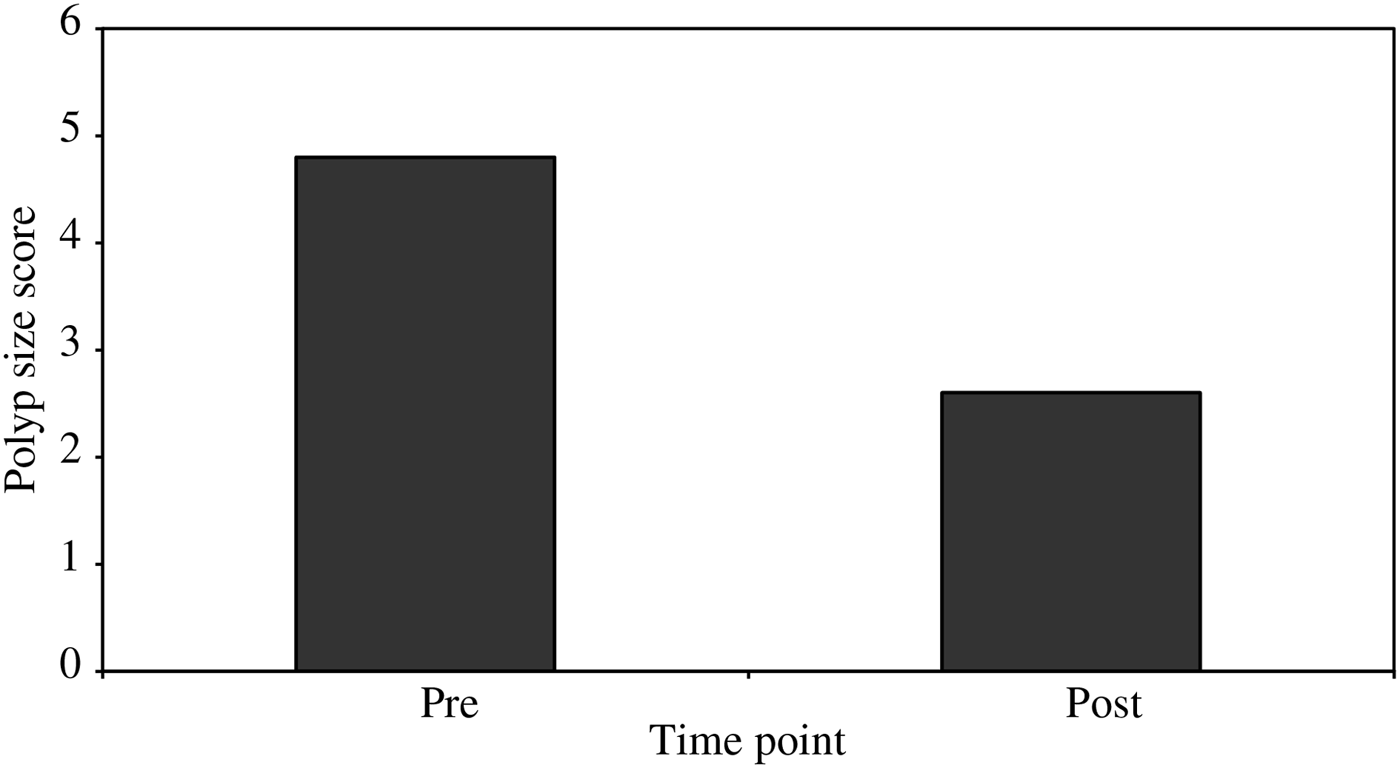
After clarithromycin treatment, nasal polyps of various sizes persisted in all 40 patients. However, the non-allergic patients showed a significant reduction in endoscopically assessed polyp size score after treatment (from 5.09 ± 1.02 to 4.12 ± 1.78; p < 0.05). The allergic patients also showed a significant reduction in polyp size score after treatment (from 5.05 ± 1.01 to 4.00 ± 1.00; p < 0.05). The nasal polyp size decreased in 45.45 per cent (10 of 22) of the non-allergic patients (Figure 1) and in 50 per cent (nine of 18) of the allergic patients (Figure 2), but this difference was not statistically significant (chi-square test). The improvement group showed a statistically significantly greater reduction in nasal polyp size, compared with the non-improvement group. In the non-allergic patients from the improvement group, polyp size score decreased from 4.80 ± 1.01 to 2.60 ± 0.67 (p < 0.01) (Figure 3). In the allergic patients from the improvement group, polyp size score decreased from 4.10 ± 0.82 to 2.20 ± 0.54 (p < 0.01) (Figure 4).
Reduction in nasal polyp size score in non-allergic patients, comparing scores before (pre) and after (post) clarithromycin treatment. Each pair of bars represents the nasal polyp size score for an individual patient before (left bar) and after (right bar) treatment.

Reduction in nasal polyp size score in allergic patients, comparing scores before (pre) and after (post) clarithromycin treatment. Each pair of bars represents the nasal polyp size score for an individual patient before (left bar) and after (right bar) treatment.

Mean polyp size scores in non-allergic patients of the improvement group before (pre) and after (post) clarithromycin treatment, showing a significant reduction (p < 0.01).
Mean polyp size scores in allergic patients of the improvement group before (pre) and after (post) clarithromycin treatment, showing a significant reduction (p < 0.01).

In the non-allergic patients, the polyp size score decreased from 6 to 2 in one patient, from 6 to 4 in three patients, and from 4 to 2 in six patients (Figure 1). In the allergic patients, polyp size score decreased from 5 to 2 in one patient, from 6 to 4 in two patients, and from 4 to 2 in six patients (Figure 2). We found that clarithromycin treatment was more effective in reducing the size of smaller polyps than bigger polyps.
We found no significant differences in the nasal secretion levels of IL-4 and IL-5 before versus after clarithromycin treatment.
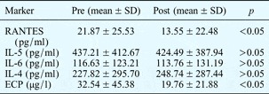
In the non-allergic patients, clarithromycin treatment resulted in significantly lower nasal secretion concentrations of both the ‘regulated on activation, normal T cell expressed and secreted’ chemokine (p < 0.05) and the eosinophilic cationic protein (p < 0.05), compared with pre-treatment levels (Table I).
Nasal fluid markers: non-allergic nasal polyp patients

Pre = pre-treatment; post = post-treatment; SD = standard deviation; RANTES = ‘regulated on activation, normal T cell expressed and secreted’ chemokine; IL = interleukin; ECP = eosinophilic cationic protein
In the allergic patients, clarithromycin treatment resulted in significantly lower concentrations of this same chemokine (p < 0.05) and of IL-6 (p < 0.05), compared with pre-treatment levels (Table II).
Nasal fluid markers: allergic nasal polyp patients

Pre = pre-treatment; post = post-treatment; SD = standard deviation; RANTES = ‘regulated on activation, normal T cell expressed and secreted’ chemokine; IL = interleukin; ECP = eosinophilic cationic protein
In the non-allergic patients in the improvement group, clarithromycin treatment resulted in significantly reduced concentrations of the ‘regulated on activation, normal T cell expressed and secreted’ chemokine (p < 0.01); however, in the non-allergic patients in the non-improvement group, a less statistically significant reduction was seen (p < 0.05).
In the non-allergic patients in the improvement group, clarithromycin treatment resulted in a significantly reduced concentration of both the ‘regulated on activation, normal T cell expressed and secreted’ chemokine (p < 0.01) and the eosinophilic cationic protein (p < 0.01), compared with pre-treatment levels. However, in the non-allergic patients in the non-improvement group, a less statistically significant reduction in both these proteins was seen (p < 0.05) (Table III).
Non-allergic patients: nasal fluid markers vs nasal polyp shrinkage

Results are shown as mean ± standard deviation unless otherwise specified. See text for definition of improvement and non-improvement groups. Pre = pre-treatment; post = post-treatment; RANTES = ‘regulated on activation, normal T cell expressed and secreted’ chemokine; ECP = eosinophilic cationic protein
In the allergic patients in the improvement group, clarithromycin treatment resulted in a significantly reduced concentration of the ‘regulated on activation, normal T cell expressed and secreted’ chemokine (p < 0.05), compared with pre-treatment levels (Table IV). However, in the allergic patients in the non-improvement group, clarithromycin treatment caused no significant difference in the levels of this chemokine (p > 0.05).
Allergic patients: nasal fluid markers vs nasal polyp shrinkage

Results are shown as mean ± standard deviation unless otherwise specified. See text for definition of improvement and non-improvement groups. Pre = pre-treatment; post = post-treatment; RANTES = ‘regulated on activation, normal T cell expressed and secreted’ chemokine; ECP = eosinophilic cationic protein
In the allergic patients, clarithromycin treatment resulted in a significant decrease in IL-6 levels in both the improvement and non-improvement groups (p < 0.05 for both), compared with pre-treatment levels. There was no significant difference in the IL-6 level reduction seen in these two groups (Table IV).
Discussion
Nasal polyposis is an example of extreme immune dysregulation.Reference Pácová, Kučera, Astl and Martínek17 Both clinical and experimental studies indicate that nasal polyp formation and growth are activated and perpetuated by an integrated process involving the mucosal epithelium, lamina propria and inflammatory cells, which in turn may be initiated by both infectious and non-infectious inflammation.Reference Norlander, Brönnegård and Stierna18 Various toxic agents, infectious agents and allergens, encountered at the level of the nasal and paranasal mucosa, may activate innate immune mechanisms and lead to induction of pro-inflammatory cytokines.
Fundová and colleagues' resultsReference Fundová, Filipovský, Funda, Hovorka, Holý and Navara19 suggest that nasal polyp pathogenesis and growth may be promoted by dysregulation of innate immune mechanisms (e.g. signalling via toll-like receptors and induction of nuclear factor κB and therefore cytokine production) and defective homeostasis of epithelial cells (with prolonged cell survival).
Lo et al. Reference Lo, Di Palma, George and McCombe20 suggested that local release of carbon monoxide may play a role in the pathogenesis of nasal polyposis.
Because of the prominent eosinophilic inflammation associated with ethmoidal nasal polyps, and the potential of eosinophils to elicit tissue damage and subsequent remodelling, it is likely that a better understanding of the mechanisms underlying the migration, activation and maintenance of eosinophils in nasal polyp tissue will be central to understanding the aetiology and pathogenesis of nasal polyps. Bonfils et al. Reference Bonfils, Badoual, Bonfils, Gallast and Malinvaud21 found that eosinophilic infiltration influences the outcome of nasal polyposis surgery, and that it could be considered as a risk factor for resistance to medical treatment.
Long-term, low-dose administration of erythromycin, clarithromycin and roxithromycin has recently been reported to be very effective for patients with chronic upper respiratory tract infection. Ichimura et al. Reference Ichimura, Shimazaki, Ishibashi and Higo1 found that roxithromycin administered at 150 mg/day for at least eight weeks shrank the nasal polyp size in 52 per cent of the 20 investigated patients. Yamada et al. Reference Yamada, Fujieda, Mori, Yamamoto and Saito2 showed that clarithromycin administered at 400 mg/day for eight to 12 weeks resulted in marked shrinkage of polyps in 40 per cent of their 20 patients. The bacterial culture results reported by Cervin et al. Reference Cervin, Kalm, Sandkull and Lindberg22 suggest that the risk of selecting resistant bacteria is low. In this study, bacterial cultures were positive in a small number of patients, but this was not always linked to an increase in symptoms. This could be due to the fact that, in addition to their direct bacteriostatic effect, macrolides may in some cases reduce the virulence of bacteria without eradicating them.
Ichimura et al. Reference Ichimura, Shimazaki, Ishibashi and Higo1 reported that the efficacy of macrolide therapy was not related to allergic symptoms. Haruna et al. Reference Haruna, Shimada, Ozawa, Fukami and Moriyama23 performed an investigation which included 68 patients with chronic rhinosinusitis and nasal polyps. Comparison of allergy examination findings and macrolide therapeutic efficacy found no correlation between the presence of nasal allergy signs and macrolide treatment efficacy. We too found that there was no evidence of a relationship between the presence of atopy and the clinical efficacy of macrolide treatment.
The mechanisms of polyp shrinkage during macrolide treatment are not well known. Nonaka et al. Reference Nonaka, Pawankar, Tomiyama and Yagi24 demonstrated that in vivo roxithromycin treatment directly suppressed nasal polyp fibroblast proliferation and that this effect was persistent, indicating that roxithromycin may prevent the progression of nasal polyps by inhibiting the development of fibrosis. Park et al. Reference Park, Park, Cho, Lee and Lee25 showed that macrolides can inhibit nasal polyp fibroblast differentiation and collagen production via their antioxidant effect.
In our nasal polyp patients, the nasal fluid concentrations of the ‘regulated on activation, normal T cell expressed and secreted’ chemokine and the eosinophilic cationic protein were reduced after treatment with clarithromycin; these reductions were generally statistically significant in both allergic and non-allergic patients, apart from a non-significant reduction in eosinophilic cationic protein levels in allergic patients. In non-allergic patients, decreases in both these concentrations were associated with polyp shrinkage. In allergic patients, polyp shrinkage was associated with reduced ‘regulated on activation, normal T cell expressed and secreted’ chemokine levels but not with reduced eosinophilic cationic protein levels. These findings underline the importance of the ‘regulated on activation, normal T cell expressed and secreted’ chemokine for nasal polyp pathogenesis and growth.
Bartels et al. Reference Bartels, Maune, Meyer, Kulke, Schlüter and Röwert26 showed that expression of ‘regulated on activation, normal T cell expressed and secreted’ chemokine messenger RNA was elevated in non-allergic and allergic nasal polyps, compared with healthy nasal mucosa. Meyer et al. Reference Meyer, Bartels, Görögh, Sticherling, Rudack and Ross10 clearly showed that this chemokine is expressed and secreted in nasal polyps; in their study, patients with greater tissue eosinophilia had significantly greater gene expression and production of this chemokine, compared with patients with non-eosinophilic nasal polyps.Reference Meyer, Bartels, Görögh, Sticherling, Rudack and Ross10 Saji et al. Reference Saji, Nonaka and Pawankar11 demonstrated that nasal polyp fibroblasts synthesised this chemokine via stimulation with the proinflammatory cytokines IL-1β and tumour necrosis factor α (TNF-α). Suzaki et al. Reference Suzaki, Asano, Yu and Hisamitsu27 clearly showed that roxithromycin exerted suppressive effects on ‘regulated on activation, normal T cell expressed and secreted’ chemokine production in nasal polyp fibroblasts, via inhibition of messenger RNA expression in response to TNF-α stimulation in vitro.
Thus, we speculate that the observed reduction in nasal secretion levels of the ‘regulated on activation, normal T cell expressed and secreted’ chemokine following clarithromycin therapy could be a result of two associated processes: suppression of nasal polyp fibroblast proliferation, and suppression of the production of this chemokine in nasal polyp fibroblasts.
Our results demonstrate that clarithromycin treatment decreases the level of eosinophilic cationic protein in nasal secretions, which is in accordance with results presented by Cervin et al. Reference Cervin, Wallwork, Mackay-Sim, Coman and Greiff28 However, in allergic patients we found no differences between the levels of eosinophilic cationic protein before and after clarithromycin treatment. This finding suggests that, in allergic nasal polyposis patients, eosinophil function and metabolism are under the influence of other cytokines.
Our results demonstrate that clarithromycin treatment has no significant effect on IL-5 and IL-4 concentrations.
Previous data point to IL-5 being one of the key proteins in the pathogenesis of tissue eosinophilia, enhancing the differentiation, activation, expansion, mobilisation and in situ survival of eosinophils.Reference Bachert, Wagenmann, Rudack, Höpken, Hillebrandt and Wang29 The main sources of IL-5 are eosinophils and mast cells.Reference Bachert, Wagenmann, Rudack, Höpken, Hillebrandt and Wang29 Preliminary findings presented by Fan et al. Reference Fan, Wang and Takenaka30 suggest that T-cell-derived IL-5 and autosecretion of IL-5 from activated eosinophils could account for the extension and persistence of eosinophilic inflammation in nasal polyps. Thus, it seems that, in allergic patients, eosinophilic cationic protein is usually increased predominantly because of IL-5 up-regulation. Treatment with clarithromycin evidently cannot suppress this strong influence.
• Macrolide antibiotics have anti-inflammatory and immunomodulatory effects on nasal polyposis
• This study assessed the effect of long-term clarithromycin on nasal fluid markers in non-allergic and allergic nasal polyposis patients
• ‘Regulated on activation, normal T cell expressed and secreted’ chemokine levels fell in both groups, eosinophilic cationic protein levels fell in non-allergic patients and interleukin-6 levels fell in allergic patients
• Clarithromycin showed a strong anti-inflammatory effect and variable immunomodulatory effect in these patients
Of the other T helper 2 cytokines, IL-4 has been shown to deliver signals that support or directly trigger selective influx of eosinophils.Reference Bachert, Wagenmann, Rudack, Höpken, Hillebrandt and Wang29
Interleukin 6 is an important T helper 2 type cytokine involved in the induction of immunoglobulin E synthesis as well as mast cell proliferation and maturation.Reference Liu, Hong, Shun, Hsiao, Wang and Wang31 Interleukin 6 also stimulates fibroblast proliferation and collagen synthesis.Reference Liu, Hong, Shun, Hsiao, Wang and Wang31 Suzaki et al. Reference Suzaki, Asano, Yu and Hisamitsu27 clearly showed that roxithromycin could suppress IL-6 production from nasal polyp fibroblasts via inhibition of IL-6 messenger RNA expression induced by TNF-α stimulation in vitro. However, in our study clarithromycin treatment reduced the level of IL-6 only in allergic nasal polyposis patients. Our explanation is that clarithromycin has different immunomodulatory effects depending on the atopic status of patients with nasal polyps.
Conclusion
Our findings demonstrate that clarithromycin has a strong anti-inflammatory effect in nasal polyposis patients. Our results suggest that macrolide treatment has different immunomodulatory and similar clinical effects in allergic and non-allergic nasal polyposis patients. On the other hand, these results may be due to the fact that allergic and non-allergic patients with similar clinical findings have different mediator profiles in their nasal secretions, implying clear differences in polyp pathogenesis. Allergies have no influence on the clinical efficacy of macrolide treatment.
Decreased nasal secretion levels of the ‘regulated on activation, normal T cell expressed and secreted’ chemokine were associated with polyp shrinkage in both non-allergic and allergic patients. These results indicate the importance of this chemokine in the pathogenesis of both allergic and non-allergic nasal polyposis, probably related to its chemotactic activity for eosinophils.
Acknowledgements
This study was supported by grants from the Military Medical Academy Research Fund. It was performed as part of a project of the Institute of Medical Research, Military Medical Academy, Belgrade, Serbia (VMA/11-14/B-6).