Milk plays a critical role in supporting the development and health of infant mammals as well as fulfilling the dietary needs of humans by providing a rich source of essential nutrients. According to the reports of the Food and Agriculture Organization (FAO), world milk production reached 928 million tons in 2021, which is 1.5 percent higher than in 2020 (FAO, 2021). However, maintaining sustainable and profitable milk production from dairy cows is only possible with good udder health. Cow mastitis, which is inflammation of the mammary gland, is the most prevalent disease and remains a major problem in the dairy industry worldwide, resulting in losses in milk quality and quantity, reduced milk production, negative animal welfare, costly veterinary treatments and reduced farm income. Although viruses and fungi can cause mastitis, Gram-positive and Gram-negative bacteria are the most frequent agents. Escherichia coli (E. coli, Gram-negative) and Staphylococcus aureus (S. aureus, Gram-positive) are two of the most common bacterial causes of mastitis. In addition, S. aureus affects human health as a result of possible zoonotic transmission. According to scientific research in this field, the zoonotic risks associated with S. aureus mastitis may become a global problem in the future (Rainard et al., Reference Rainard, Foucras, Fitzgerald, Watts, Koop and Middleton2018). In addition, mastitis is a human health issue because widespread antibiotic treatments in cows result in antimicrobial resistance in cattle, and bacterial toxins also have an effect on consumers. S. aureus is the main etiological agent causing contagious mastitis and prolonged antibiotic treatments because it causes a weaker immune reaction in the udder. Consequently, S. aureus mastitis may often become persistent (Günther et al., Reference Günther, Petzl, Bauer, Ponsuksili, Zerbe, Schuberth, Brunner and Seyfert2017; Zaatout et al., Reference Zaatout, Ayachi and Kecha2020).
Medications are often ineffective in treating S. aureus infections due to the occurrence of antibiotic-resistant strains, such as methicillin-resistant S. aureus (Pietrocola et al., Reference Pietrocola, Nobile, Rindi and Speziale2017). The innate immune system is induced by contact between pathogens that enter the mammary gland and somatic cells in the milk, which causes the transcriptional activation of important response genes. The innate immune system is the initial immune defense when mastitis-causing pathogens reach the teat end of the mammary gland. The first predominant innate defense mechanism starts in the epithelial cells, which have pattern recognition receptors (PRRs). The second defense mechanism is endothelial cells, which regulate leukocyte migration and activation (Sordillo, Reference Sordillo2018). If the innate immune response is insufficient, the bacteria multiply and become established in the mammary gland tissue once inside the teat cistern. Innate immunity, despite having a ‘non-specific’ nature, begins when PRRs on the outside or inside of host cells bind particular bacterial molecules called pathogen-associated molecular patterns (PAMPs: Wellnitz and Bruckmaier, Reference Wellnitz and Bruckmaier2012). These PAMPs are recognized by toll-like receptors (TLRs), which are linked to Gram-positive (peptidoglycan linkages) and Gram-negative (lipopolysaccharide linkages) mastitis-causing bacteria. Among the TLR family, TLR2 is particularly significant for mammary defense because it recognizes domains of lipoteichoic acid (LTA) and peptidoglycans (PGN) from Gram-positive bacteria, such as S. aureus (Oviedo-Boyso et al., Reference Oviedo-Boyso, Valdez-Alarcón, Cajero-Juárez, Ochoa-Zarzosa, López-Meza, Bravo-Patiño and Baizabal-Aguirre2007). S. aureus generates a variety of peptides that block particular functions of the host's innate immune system, which serves as the initial immune defense (Pietrocola et al., Reference Pietrocola, Nobile, Rindi and Speziale2017). There is evidence of changes in gene and protein expression levels that are different for different infectious pathogens. However, the pathophysiology and host response to mastitis are not fully understood (Umesh et al., Reference Umesh, Guttula and Gupta2022).
There is a lot of evidence to suggest that miRNAs are crucial to the growth and function of innate and adaptive immune cells. Numerous innate immune system cells, including monocytes, dendritic cells, macrophages, natural killer cells and granulocytes, express miRNAs. Thus, miRNAs regulate immune responses at various phases of the innate immune network, including cytokine/chemokine synthesis and release, adhesion molecule expression, miRNA transport and feedback regulation of immunological homeostasis (Momen-Heravi and Bala, Reference Momen-Heravi and Bala2018). Several genetic studies have investigated miRNA expression profiles to understand the role of miRNAs in the molecular mechanisms underlying innate immunity against S. aureus-associated mastitis (Dysin et al., Reference Dysin, Barkova and Pozovnikova2021). For example, the expression level of miR-21 has been found to be up-regulated in cows with S. aureus-associated mastitis (Chen et al., Reference Chen, Liu, Li, Wang, Liu, He, Yang, Niu, Wang and Guo2014). Also, similar data were obtained by Jin et al. (Reference Jin, Ibeagha-Awemu, Liang, Beaudoin, Zhao and Guan2014), who determined the miR-21-3p up-regulated expression level after 24 h of S. aureus infection in bovine mammary epithelial cells.
Despite considerable research on mammary gland infection, the molecular basis of S. aureus mastitis has not yet been elucidated. Therefore, the identification of miRNAs from bovine milk and their potential impact on S. aureus mastitis resistance in the dairy cow innate immune system remain unclear. Consequently, the purpose of the present study was to investigate the expression pattern of certain important miRNAs associated with S. aureus pathogens in subclinical mastitis infections in dairy cows to elucidate the molecular basis of S. aureus mastitis related to innate immunity. The main research question of this study is: what is the effectiveness of bioinformatic analyses in identifying miRNAs associated with S. aureus pathogen and bovine species-specific innate immune system in subclinical mastitis infections in cows? Additionally, what is the effectiveness of bioinformatics analyses in validating the expression of miRNAs belonging to these hub genes that are identified?
Material and methods
Study design
The research strategy conducted in this study first identifies hub genes related to the innate immune system specific to the S. aureus pathogen in subclinical mastitis infections in cows by in-silico analysis, and the experimental validation of the miRNAs of these determined hub genes follows.
Ethics statements
The study was approved by the Ankara University Animal Research Ethical Committee (Approval and work permit number: 2022-11-107). Milk samples were collected using standard farm techniques without causing discomfort or stress to the animals.
In-silico analysis
A detailed description of all in-silico analyses is provided in the online Supplementary File.
Animal sampling
A total of 30 Holstein cows were used in this study from the experimental farm of Ankara University, Faculty of Veterinary Medicine. All the animals were milked twice a day and fed the same ration; also, the milk yield of each individual was recorded twice per day.
A clinical assessment of the udder was performed, examining for signs of swelling, redness, heat, and pain. Cows showing clinical mastitis symptoms were excluded from the study before group assignment. A California mastitis test (CMT) was performed, and the scores were evaluated as 0, (+), (+ +), and (+ + +) for ‘negative,’ ‘weak positive,’ ‘positive,’ or ‘strong positive,’ respectively (Barnum and Newbould, Reference Barnum and Newbould1961). The 10 cows that had ‘weak positive’ and ‘positive’ test results were assigned as subclinical mastitis cows prior to further analysis. The 20 cows that had ‘negative’ CMT results were assigned as healthy controls. The description of the symptoms and parameters used for the group of subclinical animals is shown in online Supplementary Table S1. Milk samples were collected under aseptic conditions from each cow prior to an evening milking in four separate 50 ml centrifuge tubes for SCC, milk composition, S. aureus identification, and miRNA isolation analyses. Using the Milkotester milk analyzer, the fat, protein, lactose, density and non-fat solids values were determined (Milkotester Master Classic LM2). The Somatos Mini SCC device was used to do a somatic cell count, healthy cows being defined as having SCC <1 × 10⁵ cells/ml and cows with subclinical mastitis as SCC ≥2 × 10⁵ cells/ml were selected. For the bacteriological examination, 0.5 ml of the milk samples were seeded from each sample onto a blood agar plate and incubated aerobically at 37°C for 24 h. The cultivated colony from blood agar was then subcultured onto Baird-Parker agar (Baird-Parker Agar, Code: CM1127, ThermoFisher) for the selective isolation of Gram-positive Staphylococci spp., and incubated at 37°C for the next 24 h. Following that, the coagulase and catalase test were used to identify S. aureus colonies (online Supplementary Fig. S6). The results of these various tests allowed us to define the two experimental groups: six cows with subclinical mastitis infected with S. aureus and ten healthy control cows. A detailed description of these sixteen cows is given in online Supplementary Table S2.
miRNA extraction
The miRNeasy serum/plasma advanced kit (Qiagen, USA, Cat. No. 217204) was utilized for miRNA isolation from milk. The isolation was performed according to the kit's protocol. Thermo NanoDrop 2000 spectrophotometry assessed the quality and amount of miRNAs at A260 / A280 nm wavelengths. RNA was considered pure when the A260/A280 nm absorbance ratio was 1.90 or greater.
miRNA expression analysis
The expression levels of determined miRNAs (bta-miR-15a, bta-miR-16b, bta-miR-23a, bta-miR-27a-3p, bta-miR-103, bta-miR-146b and bta-mir-374b, with bta-miR-148a used for normalization) were measured using miRNA PCR Assays (miRCURY LNATM Qiagen), which included a reverse transcription stem-loop followed by a real-time PCR for quantifying mature miRNA. Each sample's total RNA was reverse transcribed to cDNA using a reverse transcription kit miRCURY LNA RT kit (Qiagen, ABD, Cat. No:339340) during the reverse transcription step. Then, using the miRCURY LNA SYBR green PCR kit (Qiagen, USA Cat. No: 339347) and the CFX 96 real-time PCR device, the cDNAs of the samples were analyzed by qPCR. The reactions and thermal cycle conditions were applied according to the manufacturer's protocols for each sample. Additionally, all experiments were applied in two technical replicates.
Statistical analysis
Lai et al. (Reference Lai, Fujikawa, Maemura, Ando, Kitahara, Endo, Yamato, Koiwa, Kubota and Miura2017) and Schanzenbach et al. (Reference Schanzenbach, Kirchner, Ulbrich and Pfaffl2017) determined that the most abundant bovine milk miRNA was miR-148-3p. Moreover, Tzelos et al. (Reference Tzelos, Ho, Charmana, Lee and Donadeu2022) used miR-148-3p as the endogenous control to determine the geometric mean in the qPCR assay in cattle. Based on the literature, we used miR-148-3p for normalization. The gene expression calculations were performed using the Livak and Schmittgen (Reference Livak and Schmittgen2001) method. Due to the limited sample size, the Wilcoxon rank-sum test was used to analyze the distribution of quantitative measures among groups that did not meet the requirements for a normal distribution. All statistical analyses were performed using SPSS 22 software.
Results
In-silico analysis
The gene sets that play a role in the categories ‘innate immune response’ and ‘defense response of Gram-positive bacteria’ for Bos taurus cattle were identified from the GO databases. A total of 62 genes were determined (online Supplementary Fig. S2 and Table S3). The KOBAS online analysis database further confirmed the functional enrichment analysis; KOBAS was used to perform the KEGG and Reactome pathways and GO analysis on the overlapping genes (online Supplementary Table S5). The PPI network of the 62 genes consisted of 145 edges and 58 nodes. There were three significant modules (MCODE 1-3) determined from the PPI network (online Supplementary Fig. S3). Enrichment analysis revealed that the majority of Module 1 genes (TLR2, MYD88, CASP4, P2RX7, PYCRAD, NOD1, TBK1, RIPK2, IL18, and LTA) were associated with defense responses to Gram-positive bacteria at the GO BP cluster (GO: 0050830) (online Supplementary Fig. S3). After determining the PPI of the 62 genes, hub genes related to the innate immune system specific to the S. aureus pathogen in subclinical mastitis infections in cows were ascertained. According to the results, the top 5 hub genes were identified, which are TLR2, MYD88, CASP4, NOD2, and TBK1, respectively (online Supplementary Fig. S4).
Target microRNAs of identified hub genes (TLR2, MYD88, CASP4, NOD2, and TBK1) were determined by the miRNet database (online Supplementary Table S6). The miRNet database results were evaluated together with the miRNA pathway analysis results (online Supplementary Fig. S1). After the evaluation, for TLR2: bta-miR-146b; for MYD88: bta-miR-15a, bta-miR-16b, bta-miR-23a, bta-miR-27a-3p, bta-miR-103; for CASP4: bta-miR-103 and bta-mir-374b; for NOD2, bta-miR-27a-3p; and for TBK1, bta-miR-16b, bta-miR-103, and bta-mir-374b, were determined for validation by RT-qPCR (online Supplementary Fig. S5).
Validation of determined miRNAs by RT-qPCR
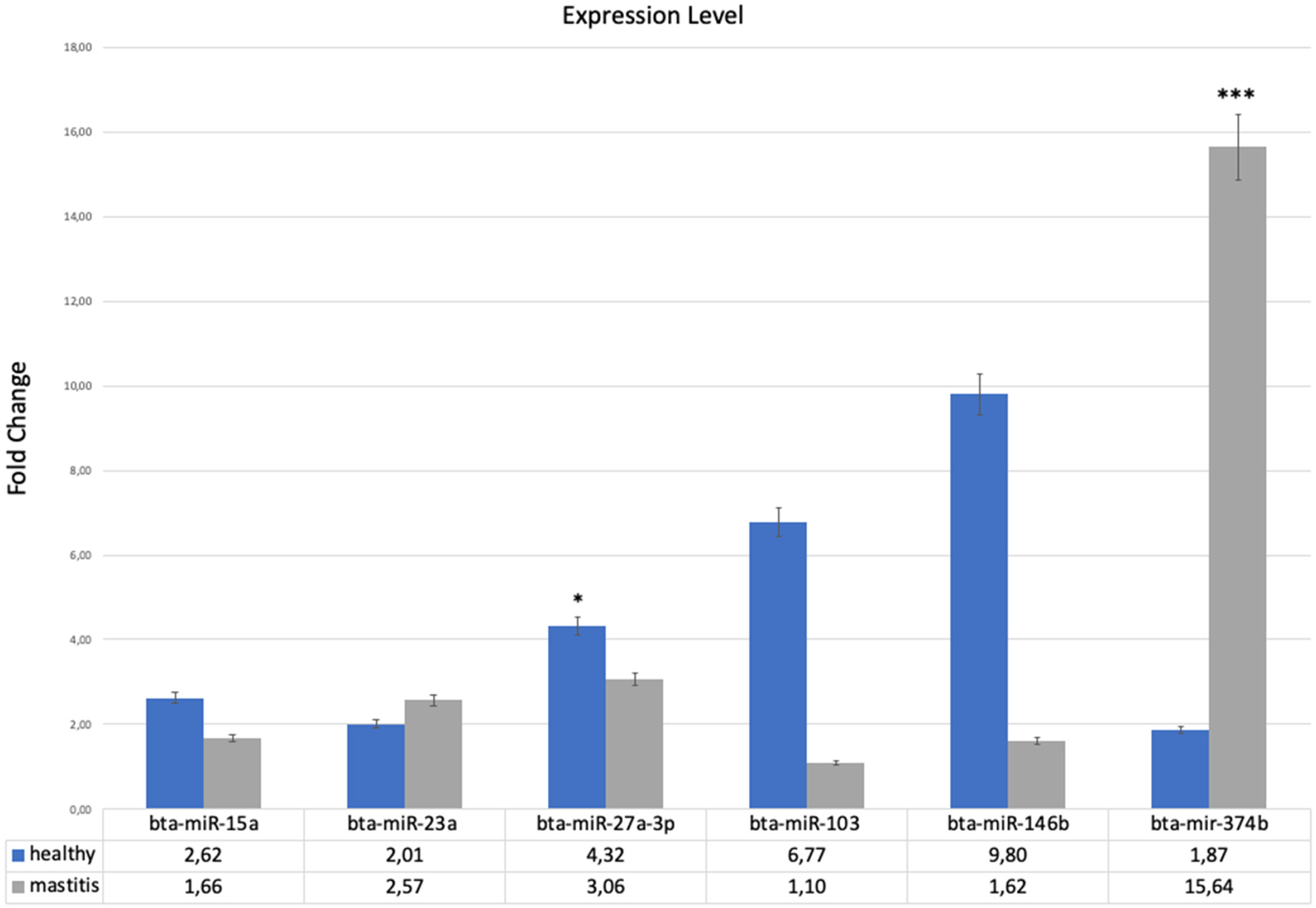
To validate the bioinformatically determined miRNAs, RT-qPCR analyses were applied to the 10 healthy and six S. aureus-infected subclinical mastitis milk samples after miRNA isolation from milk. The results of RT-qPCR for bta-miR-15a, bta-miR-23a, bta-miR-27a-3p, bta-miR-103, bta-miR-146b, and bta-mir-374b in milk samples are represented in Fig. 1. The expression of bta-miR-27a-3p, bta-miR-103, and bta-miR-146b was up-regulated in healthy cows in comparison to subclinical mastitis cows, whereas the reverse was true for the expression of bta-mir-374b. These results suggest that bovine milk infected with S. aureus contains innate immune system-related miRNAs whose expression levels may be affected by mastitis. On the other hand, attempts to analyze bta-miR-16b under the experiment's conditions ended in failure.
Fold changes in the relative expression levels of miRNAs studied in healthy and S. aureus subclinical mastitis groups. *P < 0.05; ***P < 0.001.

In this study, the fold change analysis used (logFC) >2 or <2 as the criterion for gene expression changes. Consequently, it was determined that the fold changes of bta-miR-27a-3p, bta-miR-103, bta-miR-146b, and bta-mir-374b were greater than 2 when comparison was drawn between healthy and S. aureus-infected subclinical mastitis groups. However, when the two groups were compared, no difference was found in the expression levels of bta-miR-15a and bta-miR-23a. Moreover, the expression levels of bta-miR-27a-3p were found to be statistically different when the healthy and subclinical mastitis cows were compared. The KEGG analysis can facilitate our understanding of the target genes and functions as miRNAs, allowing us to examine related miRNAs by targeting their genes in more detail. Based on the results of KEGG enrichment analysis of the target microRNAs of identified hub genes, NOD-like receptor signaling pathways (bta04621; MYD88, TBK1, CASP4, and NOD2) and Toll-like receptor signaling pathways (bta04620; TLR2, MYD88, and TBK1) were identified as the statistically most significant pathways (online Supplementary Fig. S7).
Discussion
This study identified hub genes related to the innate immune system specific to the S. aureus pathogen in subclinical mastitis infections in cows by in-silico analysis and verified the miRNAs in milk between healthy and subclinical mastitis cows, obtaining reproducible results. Our findings revealed that all bioinformatically determined miRNAs, except for bta-miR-16b, were validated by RT-qPCR in healthy and subclinical mastitis cows. Comparing the expression levels of the studied miRNAs showed significant fold changes for bta-miR-27a-3p, bta-miR-103, bta-miR-146b and bta-mir-374b. However, we found that only the fold changes of bta-miR-27a-3p and bta-mir-374b were statistically significant when comparing the expression levels in healthy cows to those with subclinical mastitis infected with S. aureus. Interestingly, the expression of bta-mir-374b was found to be significantly up-regulated in S. aureus infected cows. To the best of our knowledge, this is the first study to bioinformatically determine hub genes specific to the innate immune system and the S. aureus pathogen in subclinical mastitis infections and validate the determined miRNAs in milk between healthy and subclinical mastitis-infected cows.
Enrichment analysis of the target microRNAs of the identified hub genes revealed that the NOD-like receptor signaling pathway (bta04621) and the Toll-like receptor signaling pathway (bta0462) were the most statistically significant pathways. In this study, TLR2 was identified as the target gene of bta-miR-146b, which is functional in the TLR pathway; the expression of bta-miR-146b was up-regulated in healthy cows compared to S. aureus infected. There is a negative correlation between miRNAs and their target genes because the miRNAs interact with the 3′-UTR mRNA and inhibit protein synthesis or gene expression by degrading the target mRNAs. TLRs are expressed on innate immune cells and respond to Gram-positive or Gram-negative bacterial membrane components. Therefore, we speculated that TLR2 could promote the expression of the TLR pathway, which is the specific family of PRRs responsible for detecting microbial pathogens and generating innate immune responses, in cows with subclinical mastitis infected with S. aureus. It has been confirmed that TLR2 is a target gene of miR-146 (Jurkin et al., Reference Jurkin, Schichl, Koeffel, Bauer, Richter, Konradi, Gesslbauer and Strobl2010; Arenas-Padilla et al., Reference Arenas-Padilla, González-Rascón, Hernández-Mendoza, Calderón de la Barca, Hernández and Mata-Haro2022). Thus, an increased bta-miR-146b in healthy cows could probably be associated with decreased TLR2 expression. In this context, we speculated that bta-miR-146b might regulate the TLR pathway via positive feedback by increasing TLR2 protein levels in cows infected with S. aureus.
Contrary to our results, previous studies have demonstrated that bta-miR-46b expression levels were significantly up-regulated in mastitis-infected dairy cattle. Wang et al. (Reference Wang, Luoreng, Zan, Raza, Li, Li and Liu2016) reported that there was a significant upregulation of bta-miR-146a and bta-miR-146b expression levels in bovine mammary tissues infected with subclinical, clinical and experimental mastitis. Consistent with the results of that study but contrary to our results, Lai et al. (Reference Lai, Fujikawa, Maemura, Ando, Kitahara, Endo, Yamato, Koiwa, Kubota and Miura2017) found that the expression level of miR-146a was significantly up-regulated in California mastitis test positive (CMT+) milk. Moreover, Tzelos et al. (Reference Tzelos, Ho, Charmana, Lee and Donadeu2022) reported that the levels of bta-miR-146a in whole milk were significantly correlated with the mRNA levels of known inflammatory markers in milk cells. Additionally, the authors confirmed a significant effect of lactation number for bta-miR-146a (higher during lactation 1 than later lactations). However, in those studies (Wang et al., Reference Wang, Luoreng, Zan, Raza, Li, Li and Liu2016; Lai et al., Reference Lai, Fujikawa, Maemura, Ando, Kitahara, Endo, Yamato, Koiwa, Kubota and Miura2017; Tzelos et al., Reference Tzelos, Ho, Charmana, Lee and Donadeu2022), the specific bacterial pathogen for the cause of mastitis was not determined. This situation is one of the limitations of comparing our results with earlier studies. As mentioned before, there are many factors, such as viruses and fungi, that could cause mastitis, but Gram-positive and Gram-negative bacteria are the most frequent agents. We focused on the S. aureus bacterial infection of cows with subclinical mastitis. Similar to our results, Srikok et al. (Reference Srikok, Patchanee, Boonyayatra and Chuammitri2020) reported that the expression level of miR-146a was reduced in milk from cows with mastitis compared to milk from healthy cows. These findings suggest that bta-miR-146b might play a major role in the innate immune system in cows with subclinical mastitis infected with S. aureus. However, there was no statistically significant difference in bta-miR-146b expression level between groups in our study. Nevertheless, a larger number of samples than those used to evaluate our study should be analyzed in future studies.
Gram-positive pathogens are recognized by ligands (PAMPs) binding to innate immune receptors and triggering diverse innate signaling pathways. When pathogens pass through the physical barrier of the teat canal, the host's innate immune system uses PRRs, notably TLRs, to detect and respond to the microorganisms, which initiate a range of host defense mechanisms. The interaction between the PAMPs and TLRs is essential for starting protection against microorganisms. The extracellular region of TLR2 can recognize and bind LTA and PGN of Gram-positive bacteria, such as S. aureus (Kawai and Akira, Reference Kawai and Akira2010; Bai et al., Reference Bai, Wang, Lin, Dong, Gao, Ji, Zhang, Zhao and Zhang2022). According to the molecular mechanism of TLR2 in the cow mammary glands, TLR2 could promote inflammation and inhibit immunological responses by regulating the expression of some immune factors. Additionally, TLR2 contributes to apoptosis by regulating CASP8 expression through the MYD88 pathway. Also, activated TLR2 induces the activation of MYD88 (Bai et al., Reference Bai, Wang, Lin, Dong, Gao, Ji, Zhang, Zhao and Zhang2022). In this study, MYD88 and TBK1 are identified as other hub genes that are functional in the TLR pathway. MYD88 was identified as the target gene of the bta-miR-15a, bta-miR-16b, bta-miR-23a, bta-miR-27a-3p and bta-miR-103; TBK1 was identified as the target gene of the bta-miR-16b, bta-miR-103, and bta-mir-374b. Among those miRNAs, bta-miR-103 and bta-mir-374b were investigated as immune-related milk microRNAs in cows with subclinical mastitis infected with S. aureus for the first time in this study. We found that there was no significant difference in the expression levels of bta-miR-15a and bta-miR-23a between the groups. We also observed that the expression level of bta-mir-374b was significantly up-regulated in S. aureus infected cows. CASP4 and TBK1 were identified as the target genes of bta-mir-374b. Therefore, it is possible that the upregulation of bta-mir-374b in the S. aureus infected cows was functionally related to the expression change of the target genes (CASP4 and TBK1).
Also, it was interesting to see that only the fold change of bta-miR-27a-3p and bta-mir-374b was statistically significant between the groups. Although not statistically significant, the expression of bta-miR-103 was up-regulated in healthy cows compared to S. aureus infected. Most of the TLR-initiated inflammatory responses except TLR3 depend on a TLR signaling pathway that is mediated by the recruitment of intracellular signaling adaptor proteins, including MYD88. Additionally, the signaling within immune cells is produced according to instructions from the MYD88 gene. Consequently, the correct function of MYD88 is critical for the activation of innate immunity (Saikh, Reference Saikh2021). Therefore, the up-regulated expression levels of bta-miR-27a-3p and bta-miR-103 may be interpreted as suggesting that MYD88 could directly or indirectly regulate innate immune responses in cows with subclinical mastitis infected with S. aureus. Thus, increased bta-miR-27a-3p and bta-miR-103 in healthy cows could probably be associated with decreased MYD88 expression. In this context, we speculated that bta-miR-27a-3p and bta-miR-103 might regulate the TLR pathway via positive feedback by increasing MYD88 protein levels in S. aureus subclinical mastitis infections. It is also noteworthy to emphasize that lipopolysaccharide, a crucial antigenic component for bacterial mastitis infections, has been reported to significantly increase the expression of miR-27a-3p in mammary epithelial cell cultures from dairy cows (Wang et al., Reference Wang, Zhou, Cong, Han, Wang, Tian, Zhang, Jiang and Yuan2021). In the current study, the bta-miR-27a-3p fold change was significantly greater than 2. Therefore, it is possible that the overexpression of bta-miR-27a-3p in the healthy cows was functionally associated with a change in the expression of target genes (MYD88 and NOD2), and related processes need further exploration.
In conclusion, utilizing bioinformatics tools for determining the disease-related hub genes can be successfully applied and also provides an opportunity to interpret and gain greater insight into the molecular pathways of the mammary glands in cows with mastitis. Taken together, our results indicated that the analyzed miRNA expressions vary between healthy cows and cows with subclinical mastitis infected with S. aureus. The findings of this study enhance our understanding of the functions of bta-miR-27a-3p, bta-miR-103, bta-miR-146b and bta-mir-374b in the TLR pathway in cows with subclinical mastitis infected with S. aureus. Utilizing bioinformatics tools for determining the inflammation-related-miRNAs in milk may be used as a potential approach to detecting and even preventing subclinical mastitis infection with S. aureus.
Supplementary material
The supplementary material for this article can be found at https://doi.org/10.1017/S0022029925000068
Acknowledgments
Kardelen Karaman acknowledges the Council of Higher Education of Turkey for 100/2000 CoHE Doctoral Scholarship. Berna Kaya acknowledges TÜBİTAK for the financial support of TUBITAK-2209 with project number 1919B012107210.